Abstract
The H antigen of the dimorphic fungal pathogen Histoplasma capsulatum was first described over 40 years ago. It is a secreted glycoprotein that is immunogenic during infection. Recent cloning of the H antigen gene (HAG1) indicated sequence homology with genes for fungal β-glucosidases. To understand the biological role of this immunodominant antigen in H. capsulatum, enzymatic assays were performed to determine whether H. capsulatum contained a β-glucosidase enzyme activity and whether this activity was encoded by the HAG1 gene. Substrate gels with H. capsulatum culture supernatants revealed β-glucosidase activity near the predicted mobility of the H antigen. Quantitative microtiter plate assays revealed marked differences in secreted β-glucosidase activities from three H. capsulatum restriction fragment length polymorphism (RFLP) classes, with RFLP class II strains displaying high levels of enzyme activity, in contrast to the low levels of activity exhibited by class I and III strains. Immunoblotting of culture supernatants with an H antigen-specific antiserum demonstrated differences in H protein expression levels between the H. capsulatum classes, with a correlation between secreted enzyme activity and H protein levels. We took advantage of these class differences to demonstrate multicopy plasmid H gene overexpression by transformation of an HAG1 plasmid into H. capsulatum. Both a class II strain (G217Bura5-23) and a class III strain (G184ASura5-11) transformed with the telomeric overexpression plasmid pMAD401 displayed increased levels of β-glucosidase enzyme activity and H protein expression compared to the levels in control transformants containing only the single genomic copy of HAG1. This is the first demonstration of telomeric plasmid-mediated protein overexpression in this pathogenic fungus, and the findings support the identification of the H antigen as a β-glucosidase.
Histoplasma capsulatum is a dimorphic fungal pathogen that is the causative agent of the most common fungal respiratory infection in the world. Infection with H. capsulatum usually presents as a mild, perhaps unrecognized, respiratory illness but may progress into a more serious systemic disease in immunocompromised individuals. H. capsulatum grows as a saprophytic mold at 25°C but undergoes a phase change at physiological temperatures within the mammalian host to form the pathogenic yeast morphotype. During infection, H. capsulatum persists as a facultative intracellular parasite of host macrophages by thriving inside the phagolysosomal compartment. Internalized H. capsulatum yeasts are thus primed for dissemination throughout the mononuclear phagocytic system of the infected individual (6).
Over 40 years ago, two secreted H. capsulatum antigens, H and M, were identified as prominent components of histoplasmin, a mycelial culture filtrate. Although the H antigen was originally observed in histoplasmin, the antigen is also present in the yeast phase. The H precipitin band reacts with sera from patients with histoplasmosis in immunodiffusion assays (9). The vigorous immune response generated against the H antigen is thought to be due in part to its secretion and perhaps early processing by immune effector cells. Attempts over the years to purify this immunodominant antigen from histoplasmin resulted in only a rudimentary understanding of this glycoprotein (2, 8).
The recent cloning of the gene encoding the H antigen revealed sequence homology to genes for fungal β-glucosidases, a multifunctional class of enzymes (4). Additionally, evidence for H. capsulatum β-glucosidase activity was found in an earlier study examining secreted hydrolytic enzyme activities directed against fungal cell walls; however, the fungal protein responsible for this enzyme activity was not identified (3). When H. capsulatum was subdivided into two chemotypes based upon glucan and chitin concentrations, chemotype I displayed approximately 30% more β-glucosidase activity than chemotype II. Currently, H. capsulatum strains may be classified by restriction fragment length polymorphism (RFLP) analysis (14). For this study, we examined H. capsulatum isolates from each of the three major RFLP classes for differences in secreted β-glucosidase activities. Substrate gels and microtiter plate assays of H. capsulatum supernatants indicated appreciable differences in enzyme activity between class II strains and class I and III strains. Here, we present evidence that the H antigen is responsible for the secreted β-glucosidase activity of H. capsulatum and that H antigen production varies among strains in correlation with this enzyme activity. Further, we demonstrate a gene copy number effect on protein expression in this fungus (for the first time, to our knowledge), since H antigen production and β-glucosidase activity were both increased in two strains transformed with a multicopy plasmid containing the H antigen gene (HAG1). Additionally, the demonstration here of telomeric plasmid overexpression of a particular gene will be useful both for functional gene studies and for complementation of H. capsulatum null mutants in order to fulfill Koch’s molecular postulates.
MATERIALS AND METHODS
Fungal strains and media.
H. capsulatum G184AR, G184A-HTE, G184AS, G184ASura5-11, G186AS, G217B, and G217Bura5-23 have been described previously (10, 12, 19). Strains Downs (ATCC 38904) and UCLA 531S are clinical isolates of RFLP class I (5, 7). G217B and G222B are clinical isolates (ATCC 26032 and ATCC 26034, respectively) of RFLP class II. Parental strains G184A and G186A are clinical isolates (ATCC 26027 and ATCC 26029, respectively) of RFLP class III. G184ASura5-11 and G217Bura5-23 were isolated after UV mutagenesis and selection with 5-fluoro-orotic acid (12, 19).
All H. capsulatum strains were grown as yeasts at 37°C with gyratory shaking in a 95% air–5% CO2 environment. H. capsulatum strains were grown in HMM (see below) supplemented with uracil (100 μg/ml) for the nonselective growth of ura5 strains as previously described (15). HMM consisted of F-12 nutrient mixture with l-glutamine, with phenol red, but without sodium bicarbonate (Gibco BRL, Gaithersburg, Md.) and was supplemented with (per liter) 18.2 g of glucose, 1.0 g of glutamic acid, 84 mg of cystine, and 5.96 g of HEPES adjusted to pH 7.5 (18). Solid HMM also contained 0.5% (wt/vol) agarose (SeaKem LE agarose, lot 602997; FMC BioProducts, Rockland, Maine) and was supplemented with 10 μM FeSO4 in addition to the 3 μM FeSO4 already included in HMM. Penicillin and streptomycin (10 μg/ml each) were added to HMM broth, and gentamicin (15 μg/ml) was added to HMM agarose.
Bacterial strain.
Plasmids were propagated in Escherichia coli HB101 (supE44 hsdS20 recA13 ara-14 proA2 lacY1 galK2 rpsL20 xyl-5 mtl-1).
Preparation of H. capsulatum culture supernatants for detection and quantification of β-glucosidase activity.
H. capsulatum yeasts from late-log-phase cultures (50 to 100 ml) were pelleted by centrifugation at 1,200 × g for 10 min at 24°C. Supernatants were concentrated 50 to 100-fold by repeated centrifugations (four to six times) (45 min, 8°C, 1,800 × g) and filtration through Ultrafree-15 filter devices (Millipore, Bedford, Mass.), which should retain H. capsulatum secreted proteins larger than 5 kDa. Supernatant protein concentrations were determined according to the manufacturer’s directions with a protein assay kit from Bio-Rad, Hercules, Calif.
Substrate gels and Western blotting.
Polyacrylamide (8%) gels containing 0.1% sodium dodecyl sulfate (SDS) were prepared as described previously (13). H. capsulatum concentrated supernatants and control almond β-glucosidase (Sigma, St. Louis, Mo.) were mixed with an equal volume of 2× gel loading buffer with dithiothreitol (13) and boiled for 3 min prior to being loaded on denaturing gels; dithiothreitol and boiling were omitted for nondenaturing conditions. Following electrophoresis, nondenaturing gels were washed for 30 min at room temperature in enzyme buffer (20 mM Tris-HCl plus 0.6 mM CaCl2 [pH 8.0]) and then immersed in 100 ml of a 10 mM p-nitrophenyl-β-d-glucopyranoside (PNPG) (Sigma) substrate solution for 1 to 2 h at 37°C. Areas of substrate hydrolysis indicating β-glucosidase enzyme activity were visualized within the gels as discrete yellow bands. Individual substrate gels were scanned with an Agfa scanner.
Polyclonal antiserum was generated by repeated subcutaneous immunizations (100-μg initial injection, 50-μg boost) of a rabbit with a recombinant form of the H antigen generated in E. coli (4). H. capsulatum supernatants were prepared for Western blotting under denaturing conditions as described above. Samples were electrophoresed through 8% polyacrylamide gels containing 0.1% SDS, electroblotted onto Hybond ECL nitrocellulose (Amersham, Arlington Heights, Ill.) in a SemiPhor semidry transfer unit (Hoefer Scientific Instruments, San Francisco, Calif.) for 1.5 h at 75 mA, and blocked in 5% nonfat dry milk in TBS–0.5% Tween 20–0.01% SDS (blocker) for 1 h at 37°C. Blots were then incubated at room temperature for 1 h with a polyclonal anti-H antigen antibody at a dilution of 1:104 in blocker, washed thoroughly for 1 h in 1% nonfat dry milk in TBS–0.5% Tween 20–0.01% SDS (wash buffer), and incubated for 1 h with a 1:6,000 dilution of goat anti-rabbit peroxidase-labeled second antibody (Bio-Rad) in blocker. Each blot was washed again for 1 h at room temperature, incubated with 6 ml of ECL Western blotting detection reagents (mixed 1:1) (Amersham) for 1 min, and exposed to ECL Hyperfilm (Amersham) or a CH Imaging screen for subsequent phosphorimaging (Bio-Rad Molecular Imager).
Microtiter plate β-glucosidase assays.
Various amounts of H. capsulatum supernatant protein were mixed with enzyme buffer (described above) to a final volume of 110 μl in Eppendorf tubes and kept on ice. An equivalent amount of 10 mM PNPG was added to each reaction tube and mixed by vortexing, and 100 μl of the mixture was added to each of two duplicate wells. Samples were incubated at 37°C for 1.5 h in covered microtiter plates (Nunc). The absorbance at 405 nm was monitored with a SpectraMax 250 microplate spectrophotometer (Molecular Devices, Sunnyvale, Calif.) for the release of the colorimetric p-nitrophenyl leaving group. The A405 values were compared with a standard curve constructed by use of almond β-glucosidase.
Construction of a HAG1 gene telomeric plasmid.
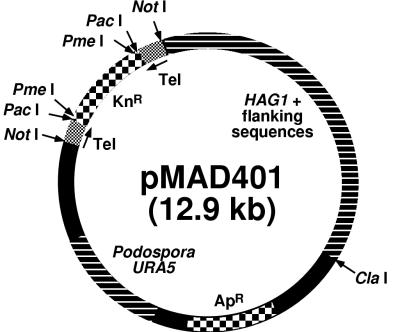
Plasmid pH is a pBluescript SK(−) derivative containing the H. capsulatum G217B H antigen gene HAG1 (∼3.8 kb), more than 1 kb of 5′ and 3′ flanking sequences from the genomic locus, and the ampicillin resistance gene for selection in E. coli (4). To construct plasmid pMAD401 (Fig. 1), we cloned from plasmid pH a 5-kB NotI/ClaI fragment containing HAG1 and upstream and downstream H. capsulatum flanking sequences into pWU45 (12) digested with NotI and ClaI. The 6-kb pWU45 fragment contains the Podospora anserina URA5 gene for selection in H. capsulatum. The resulting 11-kB plasmid, pMAD400, was next digested with NotI, dephosphorylated with alkaline phosphatase to prevent vector religation, and ligated with a 1.9-kb NotI kanamycin resistance cassette (16) to generate the 12.9-kb plasmid pMAD401. The kanamycin resistance cassette provides additional selection in E. coli as well as H. capsulatum telomeric sequences inserted at either end of the cassette. PacI or PmeI digestion of pMAD401 yields an 11-kb linear molecule with appropriately oriented telomeric termini that enable autonomous replication of linear plasmids in H. capsulatum (15). Plasmid pWU45, included as a control plasmid lacking HAG1, was digested with HpaI to expose telomeric termini prior to electrotransformation of H. capsulatum.
FIG. 1.
Diagram of the telomeric HAG1 gene overexpression plasmid pMAD401 used in this study. Tel, H. capsulatum telomeric sequence comprised of repeats of the hexamer GGGTTA. Other plasmid elements are described in the text.
Transformation.
H. capsulatum (16) and E. coli (13) were electrotransformed as described previously. Selection for transformation of G184ASura5-11 and G217Bura5-23 with URA5 plasmids was done by plating on HMM agarose without uracil.
DNA preparation and Southern analyses.
Plasmids were prepared from E. coli by use of an alkaline lysis miniprep protocol (20) or Maxi columns (Qiagen, Santa Clarita, Calif.) according to the manufacturer’s recommendations. Histoplasma genomic DNA was prepared by first enzymatically digesting H. capsulatum yeast pellets with Zymolyase/Novozym plus β-glucuronidase as described previously (15). H. capsulatum spheroplasts were next treated with lysis buffer (800 mM guanidine HCl, 30 mM EDTA, 30 mM Tris-HCl, 5% Tween 20, 0.5% Triton X-100 [pH 8.0]), RNase A, and proteinase K for 90 min at 50°C. Following centrifugation (10 min, 4°C, 4,000 × g), the cleared supernatants were applied to Qiagen Midi-tips and genomic DNA was isolated following the manufacturer’s yeast DNA isolation protocols. Methods for DNA electrophoresis and Southern blotting have been described previously (15); radiolabeling of DNA probes was performed by random priming. For detection of transformants carrying telomeric linear plasmids, genomic DNA from pMAD401 or pWU45 transformants was electrophoresed uncut alongside appropriately digested transforming vectors.
RESULTS AND DISCUSSION
β-Glucosidase identification by substrate gel electrophoresis.
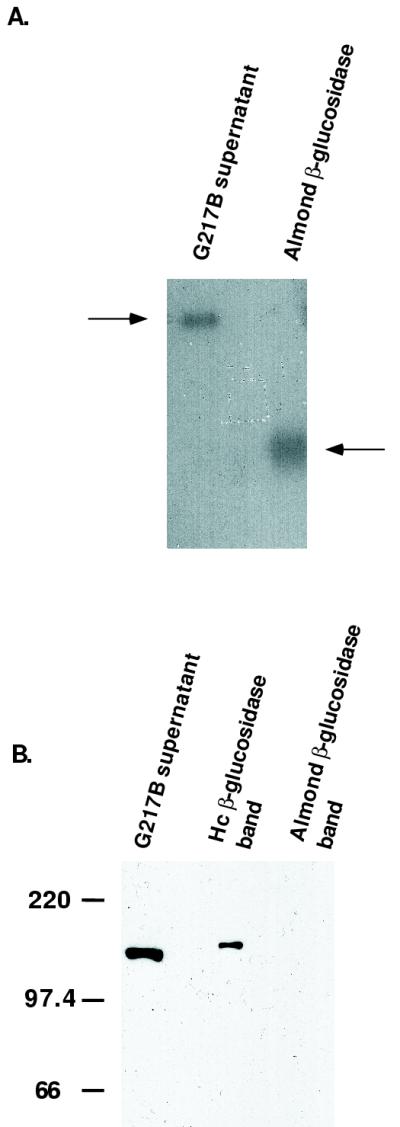
We performed substrate gel electrophoresis to visualize β-glucosidase enzyme activity in H. capsulatum culture supernatants. Areas of substrate hydrolysis in the gel were represented by yellow bands, and the G217B supernatant displayed a single band of β-glucosidase activity with a mobility similar to that of the H antigen (Fig. 2A). The band of β-glucosidase activity was excised and boiled in reducing sample buffer, and both the eluate and the gel slice were placed in a well of a new gel, reelectrophoresed, and immunoblotted with a polyclonal H antigen-specific antiserum (Fig. 2B). Immunoblotting demonstrated the presence of the H antigen within the excised band of β-glucosidase enzyme activity. The control (almond β-glucosidase) did not show serological cross-reactivity with the H antigen-specific antiserum. We observed a substantial difference in enzyme levels between an H. capsulatum RFLP class II strain (G217B) and an RFLP class III strain (G184AS) by this technique, since nearly 10-fold more total class III strain supernatant protein than class II strain supernatant protein was required to visualize any substrate hydrolysis within the gel (data not shown).
FIG. 2.
Detection of β-glucosidase activity in H. capsulatum culture supernatants and demonstration that H antigen is present within the band of enzyme activity. (A) Nonreducing, nondenaturing gel separation of G217B supernatant protein (10 μg of protein) and almond β-glucosidase (2 μg). Yellow areas of substrate hydrolysis (here converted to gray scale) are marked with arrows. (B) Immunoblot of excised bands of enzyme activity reelectrophoresed following reduction and heat denaturation. Bands were electrophoresed alongside 10 μg of G217B supernatant protein as a positive control for the H antigen. A single immunoreactive band with a mobility similar to that of the H antigen was present in H. capsulatum (Hc) β-glucosidase. The difference in migration may be an effect of the gel slice preparation technique and reelectrophoresis. Molecular size markers (in kilodaltons) are indicated on the left.
Quantification and comparison of β-glucosidase activities between H. capsulatum RFLP classes.
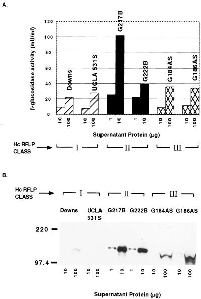
To assess potential differences in enzyme activities across H. capsulatum strains, we also quantified secreted H. capsulatum β-glucosidase activity in a colorimetric microtiter plate assay with almond β-glucosidase as a standard. We observed marked differences in the levels of secreted β-glucosidase activity between H. capsulatum RFLP class II (high levels) and H. capsulatum RFLP classes I and III (low levels) (Fig. 3A). At least 10-fold less supernatant protein from class II strains was needed to show activity equivalent to that of supernatant protein from class I or III strains. Within RFLP class II, strain G217B displayed high levels of enzyme activity, while strain G222B exhibited intermediate levels of enzyme activity (although at least 10-fold higher than those of class I and III strains).
FIG. 3.
Microtiter plate analysis of β-glucosidase enzyme activity and H antigen-specific immunoblot analysis across H. capsulatum (Hc) RFLP classes. (A) H. capsulatum β-glucosidase enzyme activity was measured against an almond β-glucosidase standard curve. The x axis illustrates amounts of supernatant protein from each H. capsulatum strain; the y axis illustrates β-glucosidase enzyme activity (values are averages from duplicate wells). Different amounts of supernatant protein were used for different strains. Similar results were obtained in three independent experiments. (B) Immunoblot analysis of H antigen expression across H. capsulatum RFLP classes with the indicated amounts of supernatant protein. Molecular size markers (in kilodaltons) are indicated on the left.
To determine if the differences in activity could be attributed to different levels of H antigen expression, we performed immunoblotting with the H antigen-specific antiserum and H. capsulatum supernatants. Figure 3B is an immunoblot examining H antigen levels in H. capsulatum strains of different RFLP classes. We found a correlation between the levels of secreted β-glucosidase activity and H protein levels in each H. capsulatum strain examined. Total supernatant protein (100 μg) from the RFLP class I strain Downs displayed a faintly immunoreactive H antigen band, while no H antigen band was detected when an equivalent amount of supernatant protein from UCLA 531S was used. Additional immunoblots of even more heavily loaded gels confirmed the production of a scant amount of H antigen by UCLA 531S (data not shown). The RFLP class III strains G184AS and G186AS displayed faint H antigen bands with 10 μg of supernatant and prominent H bands with 100 μg of supernatant. In contrast, 1 μg of supernatant from class II strains G217B and G222B exhibited strong H antigen bands. Strain G184AR (from which the variant G184AS was derived) and strain G184-HTE (a variant selected by growth in hamster tracheal epithelial cells) were also examined by β-glucosidase plate assays and H antigen immunoblotting. Each strain displayed enzyme and H antigen levels similar to those of G184AS (data not shown), although these strains differ in cell wall α-1,3-glucan expression, colony morphology, broth growth characteristics, and virulence (5, 10).
By using Western immunoblotting, we found differences in electrophoretic migration and thus the apparent size of the H antigen in different strains. For example, the G184AS protein showed a smaller apparent molecular weight than the G217B H antigen. Also, there was some discordance between Western immunoblotting and enzyme assay results for some strains. For instance, Downs and UCLA 531S showed nearly identical enzyme activities, but Downs expressed more H antigen than UCLA 531S, as detected with the H antigen-specific antiserum. It is possible that antigenic differences in the proteins from different strains cause differences in relative levels of detection by Western immunoblotting with this antiserum, which was raised against a recombinant form of the G217B protein. Alternatively, we cannot exclude the possibility that another β-glucosidase contributes to enzyme activity in some strains but is not detectable with this antiserum.
We tested the recombinant H antigen used to generate the antiserum for β-glucosidase and found no significant activity in substrate gels or the microtiter plate assay (data not shown). The native H. capsulatum H antigen is a secreted glycoprotein, with 10 predicted N-glycosylation sites. The recombinant antigen was purified from inclusion bodies in E. coli, requiring denaturation and renaturation steps. Its lack of enzymatic activity could be due to incorrect folding, lack of glycosylation, or other inappropriate posttranslational modifications in a prokaryote or could be an effect of the purification procedures.
Telomeric plasmid overexpression of the HAG1 gene.
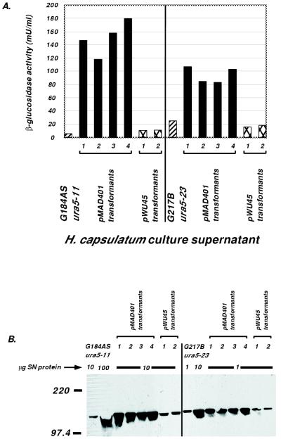
To provide further evidence for the association of β-glucosidase enzyme activity with the H antigen and to examine whether low-expression strains such as G184AS are unable to express higher levels, we used recently developed tools for the molecular genetic manipulation of H. capsulatum (16). Linear plasmids containing terminal telomeric sequences introduced by transformation replicate as multiple copies episomally (15). Plasmid pMAD401 (Fig. 1) contains the full G217B HAG1 gene sequence as well as appropriate selectable markers and telomeric sequences. This plasmid was transformed into two uracil-auxotrophic strains of H. capsulatum, G184ASura5-11 and G217Bura5-23, which display widely disparate levels of enzyme activity and H antigen production. Plasmid pWU45, lacking the HAG1 gene sequence, was used as a negative control. Following transformant isolation, genomic DNA was prepared, and the presence of monomeric, linear plasmids was confirmed by Southern analysis (data not shown). Concentrated culture supernatants from transformants and parental H. capsulatum strains were examined by both quantitative β-glucosidase microtiter plate assays and H antigen immunoblotting. Plasmid pMAD401 transformants of both uracil-auxotrophic strains of H. capsulatum, regardless of native levels of the H antigen, could effectively produce more β-glucosidase enzyme and H antigen than control pWU45 transformants, which exhibited enzyme and antigen levels consistent with those of the parental strains (Fig. 4). These results are consistent with a gene copy number effect, with protein overexpression resulting from supply of the HAG1 gene on a telomeric linear plasmid. Furthermore, we observed this phenomenon regardless of the native level of expression of the transformation recipient strain. These data indicate the absence of an inherent defect in expression by G184AS as well as a lack of negative feedback regulation for this gene, at least involving the G217B sequence supplied on the transforming plasmid.
FIG. 4.
Demonstration of HAG1 gene overexpression by increased β-glucosidase enzyme activity and H antigen expression following H. capsulatum transformation with PmeI-digested pMAD401. (A) Microtiter plate assay results for parental strains, four pMAD401 transformants, and two control pWU45 transformants for both G184ASura5-11 and G217Bura5-23 lineages. For G184ASura5-11 and its derivatives (left panel), 10 μg of supernatant protein was tested. For G217Bura5-23 and its derivatives (right panel), 1 μg of supernatant protein was tested. The average β-glucosidase activity in duplicate wells from a representative experiment is shown. Similar results were observed in two independent experiments. (B) Immunoblot analysis of the same H. capsulatum strains as those shown in panel A with the indicated amounts of supernatant (SN) protein. Molecular size markers (in kilodaltons) are indicated on the left.
Fungal β-glucosidases have been speculated to function in nutrient acquisition (by the metabolism of cellulose to acquire glucose) (17) or in cell wall remodeling events (by the breakdown of cell wall polymers and carbohydrates) (11). Relevant to the former model, Borok has speculated that the brown mycelial phenotype may be associated with growth on complex carbohydrate media requiring microbial substrate hydrolysis to obtain simple sugars, while the albino mycelial phenotype is favored when glucose is provided (1). The heavily sporulating brown phenotype and the more vegetative albino phenotype apply to mycelial cultures and not directly to the yeast morphotype that we have used, but it should be noted that both of our high-expression RFLP class II strains (G217B and G222B) are derived from brown mycelial isolates. With regard to the cell wall remodeling hypothesis, Kruse and Cole have demonstrated blocking of arthroconidium-to-spherule-phase transition in the fungal pathogen Coccidioides immitis by inhibition of β-glucosidase activity, suggesting a role for this enzyme in the hydrolysis of fungal cell walls (11). Definitive assignment of a biological role for the H antigen and determination of any effect on virulence will require targeted gene disruption and subsequent complementation to fulfill Koch’s molecular postulates. We are currently attempting HAG1 inactivation by allelic replacement. The telomeric linear plasmid pMAD401 should be useful for resupplying the gene to a disruptant. Moreover, we have provided here the first demonstration of protein overexpression based on gene copy number in H. capsulatum. Targeted hag1 mutants from different H. capsulatum strains may help explain the large differences in secreted β-glucosidase enzyme activities across H. capsulatum RFLP classes, the reasons for which are not immediately apparent. Although we have shown increased expression of the G217B HAG1 gene supplied by transformation in different strains, it remains to be determined whether differential expression of the native gene of a strain may explain the RFLP class differences that we observed. Alternatively, there may be a difference at the protein level, such as secretion, that is obscured by multicopy supply of the HAG1 gene.
ACKNOWLEDGMENTS
This work was supported by Public Health Service grant RO1 HL55949 from the National Heart, Lung, and Blood Institute.
We thank Erik Munson for assistance with the rabbit antiserum.
REFERENCES
- 1.Borok R. The mycelial status and reversibility in Histoplasma capsulatum. Sabouraudia. 1980;18:249–253. [PubMed] [Google Scholar]
- 2.Bradley G, Pine L, Reeves M W, Moss C W. Purification, composition, and serological characterization of histoplasmin H and M antigens. Infect Immun. 1974;9:870–880. doi: 10.1128/iai.9.5.870-880.1974. [DOI] [PMC free article] [PubMed] [Google Scholar]
- 3.Davis T E, Domer J E, Li Y-T. Cell wall studies of Histoplasma capsulatum and Blastomyces dermatitidis using autologous and heterologous enzymes. Infect Immun. 1977;15:978–987. doi: 10.1128/iai.15.3.978-987.1977. [DOI] [PMC free article] [PubMed] [Google Scholar]
- 4.Deepe G S, Jr, Durose G G. Immunobiological activity of recombinant H antigen from Histoplasma capsulatum. Infect Immun. 1995;63:3151–3157. doi: 10.1128/iai.63.8.3151-3157.1995. [DOI] [PMC free article] [PubMed] [Google Scholar]
- 5.Eissenberg L G, West J L, Woods J P, Goldman W E. Infection of P388D1 macrophages and respiratory epithelial cells by Histoplasma capsulatum: selection of avirulent variants and their potential role in persistent histoplasmosis. Infect Immun. 1991;59:1639–1646. doi: 10.1128/iai.59.5.1639-1646.1991. [DOI] [PMC free article] [PubMed] [Google Scholar]
- 6.Eissenberg L G, Goldman W E. Histoplasma variation and adaptive strategies for parasitism: new perspectives on histoplasmosis. Clin Microbiol Rev. 1991;4:411–421. doi: 10.1128/cmr.4.4.411. [DOI] [PMC free article] [PubMed] [Google Scholar]
- 7.Gass M, Kobayashi G S. Histoplasmosis. An illustrative case with unusual vaginal and joint involvement. Arch Dermatol. 1969;100:724–727. doi: 10.1001/archderm.100.6.724. [DOI] [PubMed] [Google Scholar]
- 8.Graybill J, Patino M, Ahrens J. In situ localization of antigens of Histoplasma capsulatum using colloidal gold immune electron microscopy. Mycopathologia. 1988;104:181–188. doi: 10.1007/BF00437434. [DOI] [PubMed] [Google Scholar]
- 9.Heiner D C. Diagnosis of histoplasmosis using precipitin reactions in agar gel. Pediatrics. 1958;22:616–627. [PubMed] [Google Scholar]
- 10.Klimpel K R, Goldman W E. Isolation and characterization of spontaneous avirulent variants of Histoplasma capsulatum. Infect Immun. 1987;55:528–533. doi: 10.1128/iai.55.3.528-533.1987. [DOI] [PMC free article] [PubMed] [Google Scholar]
- 11.Kruse D, Cole G T. A seroreactive 120-kilodalton β-1,3-glucanase of Coccidiodes immitis which may participate in spherule morphogenesis. Infect Immun. 1992;60:4350–4363. doi: 10.1128/iai.60.10.4350-4363.1992. [DOI] [PMC free article] [PubMed] [Google Scholar]
- 12.Retallack D, Heinecke E L, Gibbons R, Deepe G S, Jr, Woods J P. The URA5 gene is necessary for Histoplasma capsulatum growth during infection of mouse and human cells. Infect Immun. 1999;67:624–629. doi: 10.1128/iai.67.2.624-629.1999. [DOI] [PMC free article] [PubMed] [Google Scholar]
- 13.Sambrook J, Fritsch E F, Maniatis T. Molecular cloning: a laboratory manual. 2nd ed. Cold Spring Harbor, N.Y: Cold Spring Harbor Laboratory Press; 1989. [Google Scholar]
- 14.Vincent R D, Goewert R, Goldman W E, Kobayashi G S, Lambowitz A M, Medoff G. Classification of Histoplasma capsulatum isolates by restriction fragment polymorphisms. J Bacteriol. 1986;165:813–818. doi: 10.1128/jb.165.3.813-818.1986. [DOI] [PMC free article] [PubMed] [Google Scholar]
- 15.Woods J P, Goldman W E. In vivo generation of linear plasmids with addition of telomeric sequences by Histoplasma capsulatum. Mol Microbiol. 1992;6:3603–3610. doi: 10.1111/j.1365-2958.1992.tb01796.x. [DOI] [PubMed] [Google Scholar]
- 16.Woods J P, Heinecke E L, Goldman W E. Electrotransformation and expression of bacterial genes encoding hygromycin phosphotransferase and β-galactosidase in the pathogenic fungus Histoplasma capsulatum. Infect Immun. 1998;66:1697–1707. doi: 10.1128/iai.66.4.1697-1707.1998. [DOI] [PMC free article] [PubMed] [Google Scholar]
- 17.Woodward J, Wiseman A. Fungal and other β-d-glucosidases—their properties and applications. Enzyme Microb Technol. 1982;4:73–79. [Google Scholar]
- 18.Worsham P L, Goldman W E. Quantitative plating of Histoplasma capsulatum without addition of conditioned medium or siderophores. J Med Vet Mycol. 1988;26:137–143. [PubMed] [Google Scholar]
- 19.Worsham P L, Goldman W E. Selection and characterization of ura5 mutants of Histoplasma capsulatum. Mol Gen Genet. 1988;214:348–352. doi: 10.1007/BF00337734. [DOI] [PubMed] [Google Scholar]
- 20.Zhou C, Yang Y, Jong A Y. Mini-prep in ten minutes. BioTechniques. 1990;8:172–173. [PubMed] [Google Scholar]


