ABSTRACT
We present the case of a 4-year-old girl with tetralogy of Fallot associated with an incidentally detected isolation of the left brachiocephalic artery, with no communication between it and the pulmonary artery or ductus arteriosus. The case highlights the unusual association and hemodynamic consequences of the condition.
Keywords: Computed tomography angiography, isolation of left brachiocephalic artery, tetralogy of Fallot
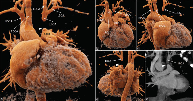
A 4-year-old girl presented with cyanosis and recurrent respiratory tract infections since infancy. Transthoracic echocardiography demonstrated features suggesting tetralogy of Fallot (TOF). The patient underwent cardiac computed tomography angiography for further evaluation of the cardiovascular morphology, which confirmed the diagnosis of TOF. A right-sided aortic arch was present, with the first and second branches from the arch being the right common carotid artery and the right subclavian artery, respectively. Interestingly, the left brachiocephalic artery was not visualized.
A blind-ending arterial stump originating from the main pulmonary trunk was seen. Another arterial channel was seen cranial to this stump, coursing in the expected location of the left brachiocephalic artery and giving rise to the left common carotid artery and the left subclavian artery [Figure 1]. Ultrasound Doppler showed retrograde flow in the left common carotid artery and antegrade flow in the left vertebral and left subclavian arteries.
Figure 1.
Volume-rendered images (a-d) and oblique coronal image (e) show a right-sided aortic arch with loss of anatomic continuity of the left brachiocephalic artery (LBCA) with aortic arch. A blind-ending arterial stump (arrowhead) is seen originating from the main pulmonary artery, with the LBCA seen cranial to this stump, giving rise to the left common carotid artery and left subclavian artery. RCCA: right common carotid artery, RBCA: right brachiocephalic artery, LCCA: Left common carotid artery, LSCA: Left subclavian artery, LBCA: Left brachiocephalic artery, MPA: Main pulmonary artery, RSCA: Right subclavian artery
Isolation of the brachiocephalic artery is rare, with only a few cases reported in the literature. Interruption of the connection of the isolated brachiocephalic artery with the pulmonary artery or ductus arteriosus is even rarer, with only one case reported previously.[1] Limb perfusion in such cases may be maintained either through collaterals from the contralateral carotid artery or subclavian artery and the descending thoracic aorta or through an intact circle of Willis through the ipsilateral common carotid artery or ipsilateral vertebral artery. In the previously reported case of “interrupted” isolated left brachiocephalic artery, the left common carotid artery and left subclavian artery were filled through the left vertebral artery, which in turn was filled by the contralateral vertebral artery.[1] In the present case, the left subclavian artery and the left vertebral artery were being filled by the left common carotid artery, which in turn was filled from the contralateral side through an intact circle of Willis.
While the conventional pulmonary steal seen in isolation of arch vessels is not seen in the presence of the above-described “interruption,” subclavian steal may still occur through the mentioned retrograde pathway and may precipitate cerebral ischemia. Therefore, along with surgical correction of TOF, reimplantation of the “interrupted” isolated left brachiocephalic artery onto the ascending aorta to reinstate the normal physiology remains the treatment of choice in the present case.
Declaration of patient consent
The authors certify that they have obtained all appropriate patient consent forms. In the form, the legal guardian has given his consent for images and other clinical information to be reported in the journal. The guardian understands that names and initials will not be published and due efforts will be made to conceal patient identity, but anonymity cannot be guaranteed.
Financial support and sponsorship
Nil.
Conflicts of interest
There are no conflicts of interest.
REFERENCE
- 1.Joseph A, Core J, Becerra JL, Kaushal RD. Right-sided aorta with complete isolation of the left innominate artery. Radiol Case Rep. 2016;11:21–4. doi: 10.1016/j.radcr.2015.11.002. [DOI] [PMC free article] [PubMed] [Google Scholar]


