Abstract
Background Knee joint osteoarthritis (OA) is among the most prevalent degenerative diseases of the joints in the body. Various scoring system exists for grading OA, such as (1) magnetic resonance imaging (MRI) Osteoarthritis Knee Score (MOAKS), (2) clinical grading by Western Ontario and McMaster Universities Arthritis Index (WOMAC), and (3) X-ray grading of the Kellgren–Lawrence grading system (K-L).
Objectives To study MRI findings and MOAKS scoring of knee OA and correlation with WOMAC and K-L scoring.
Setting and Design Cross-sectional study in hospital population.
Materials and Methods A total 40 knee OA cases underwent an MRI of the knee. MOAKS scoring was done and compared with K-L grading and WOMAC scores.
Statistical Analysis Collected data were compiled systematically and interpreted using IBM SPSS statistics software 25.0. A p -value of less than 0.05 was considered significant.
Results The mean total WOMAC score was 9. K-L grade 2 was the most prevalent X-ray grade. Bone marrow lesion (BML) and cartilage loss in MOAKS score were greater in the medial femorotibial region. A moderate positive correlation was noted between the WOMAC score and K-L grade; full-thickness articular cartilage loss score at the medial femorotibial joint (MFTJ) and WOMAC score; partial-thickness articular cartilage loss score at lateral femorotibial joint (LFTJ) and WOMAC total pain score. No correlation was found between BML and pain severity score.
Conclusion Higher WOMAC scores were associated with higher grades of K-L scoring and score of cartilage loss (partial and full thickness) of the MOAKS scoring system. The rest of the features of the MOAKS score (BML score, osteophyte, and synovitis) had no significant association with pain severity and K-L grading.
Keywords: MRI, osteoarthritis of knee, X-ray, cross-sectional study
Introduction
Osteoarthritis (OA) is a type of chronic joint disease and is among the commonest form of arthritis. Being a long-standing disease, it has a significant impact on individualistic and public health care. Its occurrence is increasing with the growing population of obese and aged persons. Due to the inadequate understanding of its etiopathogenesis and natural history, there is no effective treatment for OA. 4
OA can be categorized into primary OA, which is age related and occurs in old age, and secondary OA, which occurs due to pre-occurring diseases or posttraumatic effects.
Structural changes of OA traditionally have been assessed with radiographs, which rely on surrogate markers as indirect measures of actual pathology, like osteophytes and reduced joint space. Development of osteophytes occurs earlier than reduction of joint space. Osteophytes are the most commonly applied radiographic feature for identifying the presence of OA, while assessment of severity relies mainly on reduction of joint space narrowing and concomitant subchondral bone abnormalities. 3 5 The main disadvantages of radiography are absence of identification of soft-tissue abnormalities and its inability to detect early changes in OA.
Magnetic resonance imaging (MRI) gives an overall picture of the disease process by providing a quantitative objective evaluation of articular cartilage morphology (thickness, area, and volume) and integrity (quality). In addition, it also provides information on the periarticular soft tissue, which has been implicated in the pathogenesis of some of the clinical features of OA, including pain.
The knee joint can be considered an organ because it comprises several tissues, which are crucial for functionality of the joint. Recent research on the various scoring systems has contributed to the understanding of natural history and pathophysiology of OA and also the clinical relevance of the findings on MRI. Whole-organ evaluation of the knee and scoring of its component structure on MRI have demonstrated sensitivity, specificity, and adequate reliability, with an additional ability to signify lesion progression. Our study assessed various morphological changes associated with knee OA and their correlation with X-ray grades and clinical pain severity.
Materials and Methods
This cross-sectional observational study was conducted in a tertiary-level referral hospital in central India for 1.5 years after approval from the institute's ethics committee. Patients with primary OA of the knee, referred from the orthopaedics outpatient department (OPD), were included in the research. Secondary OA cases were precluded from the study.
Clinical Scoring
Western Ontario and McMaster scoring system (WOMAC) pain scoring was done using a questionnaire where patients rated the severity of the pain during various daily activities 2 ( Table 1 ).
Table 1. WOMAC pain scoring system.
| Pain when | None | Slight | Moderate | Severe | Extreme |
|---|---|---|---|---|---|
| Walking | 0 | 1 | 2 | 3 | 4 |
| Climbing stairs | 0 | 1 | 2 | 3 | 4 |
| Sleeping at night | 0 | 1 | 2 | 3 | 4 |
| Resting | 0 | 1 | 2 | 3 | 4 |
| Standing | 0 | 1 | 2 | 3 | 4 |
| Total score | Maximum score = 20, minimum score = 0 | ||||
Abbreviation: WOMAC, Western Ontario and McMaster scoring system.
X-Ray Acquisition and Kellgren–Lawrence Grading
Weight-bearing anteroposterior (AP) and lateral views were obtained as a part of the routine workup for knee joint complaints. However, as per the original description of Kellgren–Lawrence (KL) grading, only AP views were evaluated for grading as follows 3 :
Grade 0 (none): no features of OA.
Grade 1 (doubtful): suspicious reduction of joint space and possible osteophytes.
Grade 2 (minimal): clearly seen osteophytes and likely reduction of joint space.
Grade 3 (moderate): many osteophytes, explicit narrowing of joint space, early sclerosis, and likely deformity of bone ends.
Grade 4 (severe): osteophytes of considerable size, significant narrowing of joint space, marked sclerosis, and certain deformity of bone ends.
MRI Acquisition and Osteoarthritis Knee Score
MRI was done on a 3-T MRI system (Discovery MR750w, GE Medical Systems) using a dedicated extremity coil. The center of the was positioned at the lower margin of the patella, while the knee was kept in natural extension. Proton density fat-saturated (PDFS) turbo spin echo sequences (repetition time [TR]/echo time [TE]: 4,500/30; slice thickness: 3.0/0.5; matrix: 320 × 320; field of view [FOV]: 150 × 140) in coronal, sagittal, and axial planes; T1-weighted turbo spin echo (TR/TE: 650/25 milliseconds) in coronal plane; and 3D-Multiple Echo Recombined Gradient Echo (MERGE) (TR/TE: 32/13 milliseconds; slice thickness: 1.0/0.5 mm; FOV: 17 × 15 cm; matrix: 288 × 288) in coronal plane were acquired.
For scoring as per the MRI Osteoarthritis Knee Score (MOAKS) system, 1 the studied MRI features included bone marrow lesions (BMLs) and cysts, articular cartilage, osteophytes, synovitis-effusion, meniscal extrusion and morphology, ligaments and tendons, intra-articular loose bodies, and periarticular features (inflammation of bursae such as pes anserine bursa, prepatellar bursa and infrapatellar bursa, abnormal signals along iliotibial band [ITB] signal, presence of cysts such as ganglion cyst and popliteal cyst). All of these characteristics were evaluated and scored separately.
The knee joint was subdivided into 14 subarticular regions to score articular cartilage and BMLs. The subspinous region was also included for assessment of BMLs:
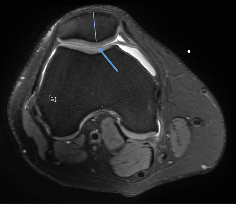
The patella was categorized into medial and lateral parts by an AP line lateral to the patellar crista, which was included in the medial patellar subdivision ( Fig. 1 ).
The femur was subdivided into six subregions: medial central femur, medial posterior femur, lateral central femur, lateral posterior femur, medial trochlea, and lateral trochlea.
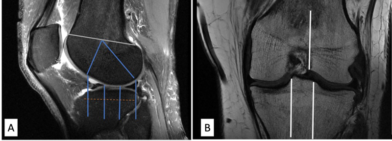
The tibia was further subdivided into six articular cartilage–covered regions, by dividing each tibial plateau into three equal parts (after excluding the osteophytes) in the central, anterior, and posterior divisions ( Fig. 2 ). The midline subspinous subregion is not covered by cartilage.
Fig. 1.

Subdivision of the patella bone. Axial proton density fat-saturated (PDFS) image depicts the division into medial and lateral patella as divided in the MRI Osteoarthritis Knee Score (MOAKS) system. The apex of the patella is a part of the medial subregion ( arrow ).
Fig. 2.

( A ) Sagittal Proton Density Fat saturated image showing subdivision of the Femur and tibia into Central (C), trochlea (T) and posterior (P) regions. ( B ) Coronal T1 weighted image showing subdivision of the femur and patella in the coronal plane.
Scoring of Each MRI Feature
Each MRI characteristic was scored separately. BMLs and articular cartilage were scored according to the subregions described earlier.
-
BMLs and cysts: BMLs include ill-defined areas of abnormal signals (hyperintense on T2/PDFS images, hypointense on T1WI) in the trabecular bone, in association with well defined fluid intensity cystic lesions in the subchondral regions (subarticular cysts).
BMLs and cysts were scored in the 15 subdivisions described earlier. For the size of BML, a single grade inclusive of all BMLs was obtained ( Table 2 ). The BML scores were combined to get a score for each patellofemoral compartment, medial femorotibial compartment, and lateral femorotibial compartment for better and easier correlation with different parameters.
Articular cartilage: Any cartilage loss (partial and full thickness) is graded separately in each articular subregion as a percentage of the surface area to the surface of the individual subregion and percentage full-thickness loss ( Table 3 ). Full- and partial-thickness articular loss grades were added to get a final score for each patellofemoral compartment, medial femorotibial compartment, and lateral femorotibial compartment for better and easier correlation with different parameters.
Osteophytes: Osteophytes were assessed in 12 locations ( Table 4 ) and categorized according to the size of the osteophyte. Absent osteophytes were scored as 0, small osteophytes as 1, medium-sized osteophytes as 2, and large-sized osteophytes as 3. Osteophytes scored were combined to get a cumulative score for the lateral femur, medial femur, and patella for a more straightforward correlation with different parameters.
-
Synovitis-effusion and Hoffa's synovitis: Hoffa's fat pad abnormal signal has been considered to represent a supplementary marker for chronic mild synovitis. It is best defined as diffuse T2/PDFS hyperintense signal within the fat pad.
Scoring is done on the basis of the size of signal abnormality: no abnormal signals are scored as 0; small size as 1, moderately sized as 2, and large sized as 3.
Size of effusion-synovitis:
Physiological amount: 0.
Small—continuous fluid in the retropatellar space: 1.
Medium—fluid with slightly convex margins of the suprapatellar bursa: 2.
Large—fluid along with distension of the joint capsule: 3.
-
Meniscus: Displacement of the meniscus (extrusion or subluxation) and morphological changes such as tear or substance loss are known to precipitate cartilage loss.
Changes in position: Grading for extrusion—less than 2 mm is grade 0; 2 to 2.9 mm is grade 1; 3 to 4.9 mm is grade 2; greater than 5 mm is grade 3. For each measurement, reference was considered at the tibial plateau edge (not including any osteophytes).
Morphology: Meniscal morphological features scored included the following:
Abnormal signals (not reaching till the meniscal surface suggesting not a tear).
Vertical tear (including longitudinal and radial tears).
Radial and horizontal tear.
Complex tear.
Tear of meniscal root (posterior horn).
Maceration, partial or complete.
Parameniscal cyst.
Hypertrophy of meniscal tissue.
Ligaments and tendon: Ligament tears were recorded as either absent or present. Partial-thickness tears in the anterior cruciate ligament (ACL) and posterior cruciate ligament (PCL) were recorded as “normal” in MOAKS. Only clearly visible complete tears were recorded as present or 1. In the patellar tendon, no abnormal signals were scored as 0, while the presence of signal abnormality was scored as 1.
-
Periarticular MRI features:
Bursitis of pes anserine bursa (increased signal in this bursa): absent/present.
Abnormal signals in ITB: absent/present.
Popliteal cyst: absent/present.
Infrapatellar bursitis: present/absent.
Prepatellar bursitis: absent/present.
Ganglion cyst: absent/present.
Loose bodies: present/absent.
Table 2. Scores of bone marrow lesions (BML) in the MOAKS system.
| Size of the overall BML by percentage of volume of the subregion | Score |
|---|---|
| None | 0 |
| <33 | 1 |
| 33–66 | 2 |
| >66 | 3 |
Abbreviation: MOAKS, MRI Osteoarthritis Knee Score.
Table 3. Articular cartilage loss scoring in the MOAKS system.
| Size of any cartilage loss (including partial- and full-thickness loss) as a % of surface area cartilage of individual region | % Full-thickness cartilage loss of the region |
|---|---|
| 0: none | 0: none |
| 1: <10% | 1: <10% |
| 2: 10–75% | 2: 10–75% |
| 3: >75% | 3: >75% |
Abbreviation: MOAKS, MRI Osteoarthritis Knee Score.
Table 4. Twelve locations of osteophytes scoring in knee joint.
| Osteophyte location | |
|---|---|
| Anterior femur (trochlea) | Medial Lateral |
| Posterior femur | Medial Lateral |
| Central femur | Medial Lateral |
| Patella | Superior Inferior Medial Lateral |
| Tibia | Medial Lateral |
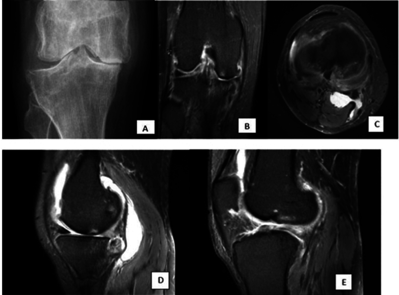
A representative case is depicted in Fig. 3A - E , showing X-ray Knee with K-L Grading and MRI features of Osteoarthritis.
Fig. 3.

(Representative case). A 39-year-old woman presented with bilateral knee pain for the past 2 to 3 years. Her Western Ontario and McMaster scoring system (WOMAC) total pain score was 12/20. ( A ) Posteroanterior (PA) X-ray shows severe reduction of medial joint space with large osteophytes, subchondral sclerosis, and subchondral cysts suggesting Kellgren–Lawrence grading system (K-L) grade 4. ( B ) Coronal proton density fat-saturated (PDFS) image shows full-thickness cartilage loss in the medial femoral and tibial articular surface with large osteophytes and severe narrowing of the joint space. ( C ) Axial PDFS image shows Baker's cyst between the tendons of semimembranosus and medial head of the gastrocnemius. ( D ) Sagittal PDFS image shows complete maceration of the posterior horn of the medial meniscus. ( E ) Sagittal PDFS image reveals complete anterior cruciate ligament (ACL) tear.
Statistical Analysis
The collected data were compiled systematically. A master table was prepared, including the clinical pain scoring, X-ray grading, and MRI findings and their scoring according to the subdivisions. The data were analyzed on version 25.0 of IBM SPSS statistics software. The processed data were presented as separate tables and graphs.
Statistics were done in two steps:
Descriptive analysis for frequency, mean, and median.
Comparative analysis.
Data were compared by applying specific statistical tests to determine the statistical significance of the comparison. A p -value of less than 0.05 was considered as significant.
Results
Our study evaluated 40 knees of 17 (42%) males and 23 (58%) females. The age of the patients ranged from 39 to 74 years, with the mean age being 51.95 years. Various studies have also observed that the prevalence of OA increases with increased age. 6 7 8 9
Clinical Scoring
The total WOMAC score for pain grading varied from 3 to 14; the mean score was 9.03 and the median score was 9. The severity of the pain was higher in exertional work like climbing stairs and walking than during resting and sleeping at night.
Radiographic Grading
Most of the patients were of K-L grades 2 and 3, comprising 21 (52.5%) and 14 (35%) patients of the total study population, while 4 (10%) of them had K-L grade 4 and 1 (2.5%) of them had K-L grade 1.
MRI Findings and Scoring
The MOAKS scoring system was used to score the findings in OA of the knee according to the subdivisions.
The mean combined BML scores were found to be higher in the medial femorotibial joint (MFTJ; 2.38 ± 1.98), followed by the patellofemoral joint (PFJ; 1.88 ± 1.30) and least in the lateral femorotibial joint (LFTJ; 1.07 ± 1.07). The pathophysiology of BML occurrence due to loss of protection offered by the overlying cartilage explains the distribution of the BML in the knee.
The mean full-thickness cartilage loss scores were highest in the MFTJ (3.58 ± 3.33), followed by the PFJ (2.25 ± 1.82) and least in the LFTJ (0.55 ± 1.41), whereas the mean partial-thickness scores were in the following order: MFTJ (5.80 ± 2.24) > LFTJ (4.32 ± 1.73) > PFJ (3.28 ± 1.71). Increased stress and load bearing on the medial joint during weight-bearing activities explain the predominant cartilage loss in the MFTJ.
There were higher osteophyte scores in the femur (mean score: 6.9) than in the patella (mean score: 4.5) and tibia (mean score: 2.4). These variations in the mean score might be attributed to the variations in the compartmental subdivisions where the femur was divided into six subdivisions, the patella into four subdivisions, and the tibia into two subdivisions.
Out of the 40 patients, Hoffa's synovitis was present in 37 patients, and all 40 of them had joint effusion. Grade 1 and 2 Hoffa's synovitis was the most common, occurring in 20 (50%) and 15 (37.5%) of the total patients, while 2 (5%) of them had grade 3 synovitis. In synovitis-effusion score, mild (grade 1) and moderate (grade 2) joint effusion was the most prevalent, occurring in 20 (50%) and 18 (45%) of the total patients, while 2 (5%) of them had gross (grade 3) joint effusion.
Among the cases with extrusion of the meniscus, the most commonly and most severely involved meniscus was the medial meniscus and the most common direction of extrusion was the medial side (85%), followed by the anterior side (37.5%). Fourteen of 15 anteriorly extruded medial meniscus knees also had medial extrusion of higher grade or at least of similar grade. In contrast, one of the knees had only anterior extrusion of grade 1 and no medial extrusion.
Among the 40 knees scanned, some abnormality was noted in the medial meniscus in 37 of them, whereas 23 had some abnormality in the lateral meniscus. Intrasubstance PDFS hyperintense signal was the most prevalent abnormality in both the menisci, that is, 18 (45%) of the 40 knees had medial meniscal intrasubstance signal, whereas 22 (55%) knees had intrasubstance signal in their lateral meniscus. Partial maceration and meniscal hypertrophy (22.5 and 10%, respectively) were the next most prevalent abnormalities of medial meniscus.
Among tendon and ligament pathologies, only 2 knees had complete tear of the ACL among the 40 knees scanned. None of the knees had PCL tear or patellar tendon signal.
Regarding periarticular features, the popliteal cyst was noted among 7 (17.5%) of the knees, and pes anserine bursitis was noted in 2 (5%) of the 40 knees.
In our study, nine (22.5%) of the total participants had the presence of loose bodies.
Discussion
Imaging interpretation has a vital role in the diagnosis and assessment of OA. X-ray imaging has been widely used in the assessment of OA and grading (most commonly used K-L grade) for severity of OA based on osteophytes and degree of joint space reduction. MRI, in contrast to radiography, can evaluate these pathological changes owing to its excellent soft-tissue resolution and can be used to assess the progression of the pathologies by serial follow-ups, owing to its lack of ionizing radiation. Many semiquantitative scoring systems have been published in the literature; among them, the MOAKS scoring system, which is used in our study, has been the most refined and widely accepted scoring system.
In OA, due to progressive thinning and erosion of articular cartilage, the subchondral bone gets predisposed to damage. Repetitive microtrauma at the subfracture level causes patchy sclerosis and osteophytes and these in turn reduce bone elasticity resulting in increased transfer of loads to the cartilage and further damage of the cartilage. In consequence, the deficiency of cartilage tissue reduces the stress-absorbing ability of knee, and friction during joint motion cannot be mitigated any longer. This increased stress is directly transmitted to the subchondral region, which results in bone thickening and bone marrow edema in the subchondral region. The degraded cartilage and bone and soluble matrix proteins incite an inflammatory reaction and causes synovitis. Osteophyte formation is thought to be a compensatory mechanism, in an effort to stabilize the joint.
The subchondral bone has a rich sensory innervation and because of the fact that the subchondral lesions are a common pathology in OA patients, a lot of recent researches have focused on further exploring this coexisting relationship of subchondral marrow edema and pain in OA patients. The BMLs, as detected on MRI, have shown an association with histopathological changes of OA, such as fibrosis, reconstruction, and remodeling of bone trabeculae and bone necrosis.
The mean combined BML scores were higher in the MFTJ, then in PFJ, and least in LFTJ. In a study by Stefanik et al, 10 970 knees were assessed, of which the isolated PFJ was more commonly involved than isolated FTJ. Even in mixed PFJ and FTJ involvement, more severe lesions were found in the PFJ than in the FTJ.
The mean full-thickness articular cartilage scores were the highest in the MFTJ, then in the patellofemoral compartment, and least in the LFTJ. In contrast, the mean partial-thickness scores were the highest in the MFTJ. However, Stefanik et al 10 and Runhaar et al 11 showed a predominant involvement of the PFJ than the MFTJ. 10 11
In our study, the femur had higher osteophyte scores, followed by the patella and the tibia. In a study by Runhaar et al, 11 the baseline prevalence of osteophytes was higher in the PFJ (60%) than in the MFTJ (32%) or the LFTJ (17%).
Among the extrusions of the menisci, the most commonly and most severely involved menisci were the medial meniscus in our study. Medial extrusion of the medial meniscus was the most common with anterior extrusion being the second most common. Medial extrusion of the medial meniscus was also noted to be the most common extrusion in patients with knee OA in a study by Kaneko et al, 12 in which out of 1,191 patients, 73.5% of the patients had medial meniscus extrusion.
Among the 40 knees scanned, some abnormality was noted in the medial meniscus in 37 of them, whereas 23 had some abnormality in the lateral meniscus. Foreman et al 13 observed that meniscal abnormalities, especially extrusion and tears at the root, were found to be predisposing factors for accelerated development of knee OA and hence identifying these findings early in the course of the disease helps in early recognition of at-risk patients. This may result in early initiation of preventive measures, particularly addressing these risk factors such as reconstruction and reparative knee surgeries.
A strong positive correlation was observed between WOMAC pain score and X-ray grading. Neogi et al 14 observed similar findings, where patients with higher grades of K-L classification were associated with more frequent and more severe knee pain.
Our study noted no statistically significant correlation between MOAKS BML scores at the PFJ, MFTJ, and LFTJ and WOMAC total score. Some of the previous researches also showed no correlation between BMLs and pain severity, 15 16 while some of the other studies showed a significant association between pain severity and volume of BMLs. 17 18 19 The reason for the discordant findings is unclear though it may, in part, be attributed to differences in the populations studied and the small volume of the study sample. This may also partly be due to the fact that the abundant nerve endings are mostly present at the subchondral region, while the MOAKS BML scores take into account the volume of the BML, which may extend well beyond the subchondral region.
A statistically significant moderately positive correlation between full-thickness articular cartilage loss score at the MFTJ and WOMAC total pain score was noted. A moderately positive correlation was also noted between the partial-thickness articular cartilage loss score at the LFTJ and the WOMAC total pain score.
The WOMAC total pain score and articular cartilage loss at PFJ showed no correlation. Many previous researchers have found a positive correlation between cartilage loss and pain severity, 19 20 21 22 while some of them showed no such correlation. 15 16
The pain severity score was higher in the presence of osteophyte compared to its absence in all the subregions. However, no correlation was found between the total WOMAC pain and osteophyte scores in any subregion. Few previous studies have shown a positive correlation between osteophyte and pain severity, 16 18 19 22 while others showed no such correlation. 15 23 Total score of WOMAC grading was more in the presence of synovitis, meniscal extrusion, and meniscal abnormalities, without, however, any statistically significant correlation.
The full-thickness articular cartilage loss score at the MFTJ and PFJ showed a moderate positive correlation with BML score at the MFTJ and the PFJ, respectively. No correlation was noted between full-thickness articular cartilage loss at the LFTJ and BML score at the LFTJ. In some previous studies, similar findings were reported where BMLs were associated with cartilage defects. 21 24 25 26
While only AP radiographs have been classically used for K-L grading, lateral views also provide valuable insights into joint morphology, alignment, and pathology. One of the key advantages of lateral knee radiographs is their ability to visualize the joint space width from a different angle. This additional perspective aids in the detection of joint space narrowing and widening, which are indicative of various degenerative or inflammatory conditions affecting the knee joint. 27 Furthermore, lateral radiographs provide detailed visualization of the femoral condyles, tibial plateau, and patella from the side. This enables clinicians to evaluate the congruence of articular surfaces and detect abnormalities such as osteophytes, subchondral sclerosis, or joint effusion, all of which can impact joint space integrity. 28
In a lateral X-ray of the knee, the femur and tibia bones are superimposed on each other. This superimposition can make it difficult to clearly distinguish between the bones and accurately assess the joint space between them. As a result, the degree of joint space narrowing, which indicates cartilage loss (and potentially “bone on bone” contact), may not be accurately measured.
Conclusion
Recent studies, particularly those involving MRI in knee OA, have provided insights into the relationship between different morphological changes in soft-tissue structures of the knee and periarticular structures, which can be identified by imaging and symptoms (stiffness and pain) and natural course of the disease. Semiquantitative studies such as MOAKS have tried to divide the knee into subdivisions and score different morphological features of OA of the knee in these subdivisions. Our study was mainly aimed at identifying various characteristics of OA of the knee on MRI and correlating these imaging findings with the knee pain.
The prevalence of BML and articular cartilage loss was higher in the MFTJ, followed by the PFJ and the LFTJ.
The WOMAC pain score showed a positive correlation with K-L grading, that is, higher WOMAC pain scores were associated with advanced X-ray grades. No correlation was found between MOAKS BML scores at the PFJ, MFTJ, and LFTJ and total WOMAC score. The full-thickness articular cartilage loss score at the MFTJ showed moderate positive correlation with the WOMAC total pain score. Moderate positive correlation was also found between the partial-thickness articular cartilage loss score at the LFTJ and the WOMAC total pain score. No significant correlation was observed between the total WOMAC pain score and osteophyte score at any subregion and between the total WOMAC pain score and synovitis-effusion grading. Higher incidence and scores of BML were associated with higher degrees of full-thickness articular cartilage loss scores at the PFJ and the MFTJ; no such correlation was noted at the LFTJ.
Funding Statement
Funding None.
Conflict of Interest None declared.
Note
This work is attributed to the Department of Radiodiagnosis, All India Institute of Medical Sciences, Bhopal, Madhya Pradesh, India.
Authors' Contributions
A.M.H. contributed to the design, definition of intellectual content, literature search, clinical studies, data acquisition, data analysis, statistical analysis, and manuscript preparation. R.M. contributed to the concepts, design, definition of intellectual content, data analysis, manuscript preparation, manuscript editing, and manuscript review. R.S. contributed to the concepts, literature search, data analysis, manuscript editing, and manuscript review. J.S. contributed to the concepts, design, definition of intellectual content, literature search, clinical studies, data acquisition, data analysis, statistical analysis, manuscript preparation, manuscript editing, and manuscript review, and is the guarantor.
References
- 1.Hunter D J, Guermazi A, Lo G H et al. Evolution of semi-quantitative whole joint assessment of knee OA: MOAKS (MRI osteoarthritis knee score) Osteoarthritis Cartilage. 2011;19(08):990–1002. doi: 10.1016/j.joca.2011.05.004. [DOI] [PMC free article] [PubMed] [Google Scholar]
- 2.Riddle D L, Perera R A. The WOMAC pain scale and crosstalk from co-occurring pain sites in people with knee pain: a causal modeling study. Phys Ther. 2020;100(10):1872–1881. doi: 10.1093/ptj/pzaa098. [DOI] [PMC free article] [PubMed] [Google Scholar]
- 3.Kellgren J H, Lawrence J S. Radiological assessment of osteo-arthrosis. Ann Rheum Dis. 1957;16(04):494–502. doi: 10.1136/ard.16.4.494. [DOI] [PMC free article] [PubMed] [Google Scholar]
- 4.Roemer F W, Crema M D, Trattnig S, Guermazi A. Advances in imaging of osteoarthritis and cartilage. Radiology. 2011;260(02):332–354. doi: 10.1148/radiol.11101359. [DOI] [PubMed] [Google Scholar]
- 5.Eyre D R. Collagens and cartilage matrix homeostasis. Clin Orthop Relat Res. 2004;427:S118–S122. doi: 10.1097/01.blo.0000144855.48640.b9. [DOI] [PubMed] [Google Scholar]
- 6.National Arthritis Data Workgroup . Lawrence R C, Felson D T, Helmick C G et al. Estimates of the prevalence of arthritis and other rheumatic conditions in the United States. Part II. Arthritis Rheum. 2008;58(01):26–35. doi: 10.1002/art.23176. [DOI] [PMC free article] [PubMed] [Google Scholar]
- 7.Shane Anderson A, Loeser R F. Why is osteoarthritis an age-related disease? Best Pract Res Clin Rheumatol. 2010;24(01):15–26. doi: 10.1016/j.berh.2009.08.006. [DOI] [PMC free article] [PubMed] [Google Scholar]
- 8.Arden N, Nevitt M C. Osteoarthritis: epidemiology. Best Pract Res Clin Rheumatol. 2006;20(01):3–25. doi: 10.1016/j.berh.2005.09.007. [DOI] [PubMed] [Google Scholar]
- 9.Neogi T. The epidemiology and impact of pain in osteoarthritis. Osteoarthritis Cartilage. 2013;21(09):1145–1153. doi: 10.1016/j.joca.2013.03.018. [DOI] [PMC free article] [PubMed] [Google Scholar]
- 10.Stefanik J J, Niu J, Gross K D, Roemer F W, Guermazi A, Felson D T. Using magnetic resonance imaging to determine the compartmental prevalence of knee joint structural damage. Osteoarthritis Cartilage. 2013;21(05):695–699. doi: 10.1016/j.joca.2013.02.003. [DOI] [PMC free article] [PubMed] [Google Scholar]
- 11.Runhaar J, Schiphof D, van Meer B, Reijman M, Bierma-Zeinstra S MA, Oei E HG. How to define subregional osteoarthritis progression using semi-quantitative MRI osteoarthritis knee score (MOAKS) Osteoarthritis Cartilage. 2014;22(10):1533–1536. doi: 10.1016/j.joca.2014.06.022. [DOI] [PubMed] [Google Scholar]
- 12.Kaneko H, Ishijima M, Aoki T et al. Prevalence of medial meniscus extrusion in elderly persons: the Bunkyo Health Study. Osteoarthritis Cartilage. 2020;28:S295. [Google Scholar]
- 13.Foreman S C, Liu Y, Nevitt M Cet al. Meniscal root tears and extrusion are significantly associated with the development of accelerated knee osteoarthritis: data from the osteoarthritis initiative Cartilage 202113(1_suppl):239S–248S. [DOI] [PMC free article] [PubMed] [Google Scholar]
- 14.Neogi T, Felson D, Niu J et al. Association between radiographic features of knee osteoarthritis and pain: results from two cohort studies. BMJ. 2009;339:b2844. doi: 10.1136/bmj.b2844. [DOI] [PMC free article] [PubMed] [Google Scholar]
- 15.Ai F, Yu C, Zhang W, Morelli J N, Kacher D, Li X. MR imaging of knee osteoarthritis and correlation of findings with reported patient pain. J Huazhong Univ Sci Technolog Med Sci. 2010;30(02):248–254. doi: 10.1007/s11596-010-0223-0. [DOI] [PubMed] [Google Scholar]
- 16.Kornaat P R, Bloem J L, Ceulemans R YT et al. Osteoarthritis of the knee: association between clinical features and MR imaging findings. Radiology. 2006;239(03):811–817. doi: 10.1148/radiol.2393050253. [DOI] [PubMed] [Google Scholar]
- 17.Carotti M, Salaffi F, Di Carlo M, Giovagnoni A. Relationship between magnetic resonance imaging findings, radiological grading, psychological distress and pain in patients with symptomatic knee osteoarthritis. Radiol Med. 2017;122(12):934–943. doi: 10.1007/s11547-017-0799-6. [DOI] [PubMed] [Google Scholar]
- 18.Torres L, Dunlop D D, Peterfy C et al. The relationship between specific tissue lesions and pain severity in persons with knee osteoarthritis. Osteoarthritis Cartilage. 2006;14(10):1033–1040. doi: 10.1016/j.joca.2006.03.015. [DOI] [PubMed] [Google Scholar]
- 19.Hayes C W, Jamadar D A, Welch G W et al. Osteoarthritis of the knee: comparison of MR imaging findings with radiographic severity measurements and pain in middle-aged women. Radiology. 2005;237(03):998–1007. doi: 10.1148/radiol.2373041989. [DOI] [PubMed] [Google Scholar]
- 20.Wluka A E, Wolfe R, Stuckey S, Cicuttini F M. How does tibial cartilage volume relate to symptoms in subjects with knee osteoarthritis? Ann Rheum Dis. 2004;63(03):264–268. doi: 10.1136/ard/2003.007666. [DOI] [PMC free article] [PubMed] [Google Scholar]
- 21.Sowers M F, Hayes C, Jamadar D et al. Magnetic resonance-detected subchondral bone marrow and cartilage defect characteristics associated with pain and X-ray-defined knee osteoarthritis. Osteoarthritis Cartilage. 2003;11(06):387–393. doi: 10.1016/s1063-4584(03)00080-3. [DOI] [PubMed] [Google Scholar]
- 22.Sayre E C, Guermazi A, Esdaile J M et al. Associations between MRI features versus knee pain severity and progression: data from the Vancouver Longitudinal Study of Early Knee Osteoarthritis. PLoS One. 2017;12(05):e0176833. doi: 10.1371/journal.pone.0176833. [DOI] [PMC free article] [PubMed] [Google Scholar]
- 23.Sengupta M, Zhang Y Q, Niu J B et al. High signal in knee osteophytes is not associated with knee pain. Osteoarthritis Cartilage. 2006;14(05):413–417. doi: 10.1016/j.joca.2005.11.012. [DOI] [PubMed] [Google Scholar]
- 24.Baranyay F J, Wang Y, Wluka A E et al. Association of bone marrow lesions with knee structures and risk factors for bone marrow lesions in the knees of clinically healthy, community-based adults. Semin Arthritis Rheum. 2007;37(02):112–118. doi: 10.1016/j.semarthrit.2007.01.008. [DOI] [PubMed] [Google Scholar]
- 25.Freund E. The pathological significance of intra-articular pressure. Edinburgh Med J. 1940;47(03):192–203. [PMC free article] [PubMed] [Google Scholar]
- 26.Rhaney K, Lamb D W. The cysts of osteoarthritis of the hip: a radiological and pathological study. J Bone Joint Surg Br. 1955;37-B(04):663–675. doi: 10.1302/0301-620X.37B4.663. [DOI] [PubMed] [Google Scholar]
- 27.Hunter D J, Lo G H, Gale D, Grainger A J, Guermazi A, Conaghan P G. The reliability of a new scoring system for knee osteoarthritis MRI and the validity of bone marrow lesion assessment: BLOKS (Boston Leeds Osteoarthritis Knee Score) Ann Rheum Dis. 2008;67(02):206–211. doi: 10.1136/ard.2006.066183. [DOI] [PubMed] [Google Scholar]
- 28.Matsumoto K, Hukuda S, Ishizawa M et al. Experimental knee joint osteoarthritis in dogs. A comparison of the Pond-Nuki and medial meniscectomized models. Clin Orthop Relat Res. 1999;367:234–245. [Google Scholar]


