Abstract
Background
Recently, intestinal ultrasound (IUS) scores such as International Bowel Ultrasound Segmental Activity Score (IBUS-SAS) and Simple Ultrasound Score for Crohn’s Disease (SUS-CD) have been established to evaluate disease activity in Crohn’s disease (CD), but these require further external validation. This study thus aimed to compare recent IUS scores in patients with colonic or small intestinal CD in order to objectively assess their value and appropriate application.
Methods
This retrospective study consecutively enrolled data of patients with CD from October 2020 to November 2022. The endoscopic and ultrasound images were collected, and the affected segments were rescored according to endoscopic scores [Simple Endoscopic Score for Crohn’s Disease (SES-CD), Rutgeerts score for patients who have undergone surgery, and the Lewis score for CD of the small intestine]; IUS scores were also collected, including the IBUS-SAS, Ultrasound Consolidated Score (UCS), SUS-CD, Simple Ultrasound Score (Simple US Score), and Bowel Ultrasound Score (BUSS). Subsequently, the correlation of IUS scores with endoscopic scores and the identification of disease activity was calculated. The Spearman rank correlation coefficient was used to calculate the correlation of parameters, and the Kruskal-Wallis test was used to compare different groups. Receiver operating characteristic (ROC) curve analysis was performed to evaluate the diagnostic efficiency of each score, and corresponding area under the curve (AUC), cutoff values, sensitivity, specificity, and 95% confidence intervals (CIs) were calculated.
Results
A total of 203 patients were included in this study. All scores correlated well with endoscopic scores and showed the ability to identify colonic CD activity with high sensitivity and specificity. Among all the scores, IBUS-SAS had the highest value overall and for colonic CD, with sensitivity of 92.7% and a specificity of 91.4% in identifying endoscopic activity and a sensitivity of 95.0% and a specificity of 88.2% in identifying severe endoscopic activity. In small intestinal CD, the UCS showed the highest correlation with endoscopic score, with a relative coefficient of 0.708. The corresponding cutoff values for identifying endoscopic activity and severe activity were also calculated.
Conclusions
Consistent with endoscopy, IUS scores are accurate in retrospective activity evaluation of CD, and suitable scores can be chosen according to the given circumstances.
Keywords: Intestinal ultrasound score (IUS score), Crohn’s disease (CD), activity assessment, endoscopic score
Introduction
Crohn’s disease (CD), as one of the inflammatory bowel diseases, is a chronic, progressive, and debilitating condition that can affect any segment of the gastrointestinal tract. It is characterized by transmural inflammation and relapsing-remitting activity, necessitating frequent evaluations throughout the disease course (1). Accurate, serial, and objective assessment of disease activity plays a crucial role in the management of CD (2). In active cases, activity assessment guides therapeutic decision-making and helps assess treatment response, while proactive monitoring during remission aims to identify early signs of disease recurrence (3). Moreover, since symptom severity does not always correlate with the extent of disease, the treatment goal for CD has shifted from symptom control to attaining mucosal or transmural cure (4). Therefore, in alignment with the treat-to-target therapeutic approach, objective and precise evaluation of bowel damage and activity has become increasingly essential (5).
Currently, several modalities are considered valuable for CD activity assessment, with endoscopy being the preferred, gold standard method, especially colonoscopy for colonic CD. Endoscopy allows for the direct visualization of characteristic mucosal changes associated with CD, such as ulcerations and cobblestone appearance (6). However, endoscopy may be limited in cases of stricturing and does not provide information regarding extraluminal manifestations. Therefore, perienteric assessment is important, as peri-intestinal signs, including inflamed fat, have been increasingly associated with various pathological changes and prognoses (7). Cross-sectional imaging is advantageous for extraintestinal evaluation, and the European Crohn’s and Colitis Organization and European Society of Gastroenterology and Abdominal Radiology (ECCO-ESGAR) Guideline has recommended cross-sectional imaging in the complementary phenotype and complication assessment of CD (3). Commonly used cross-sectional imaging modalities include computed tomography (CT), magnetic resonance imaging (MRI), and intestinal ultrasound (IUS), all of which exhibit comparable diagnostic efficacy in monitoring both colonic and small intestinal CD (8,9). Considering the need for repeated examinations during the disease course, MRI and IUS are preferred as first-line choices, with CT reserved for emergency situations due to radiation concerns (7,10). The selection between MRI and IUS depends on the specific clinical scenario, as both modalities appear to provide equivalent value in CD monitoring (3).
IUS, as a non-invasive and convenient modality, also offers accurate assessment of CD activity, with reported sensitivities ranging from 75% to 90% and specificities ranging from 75% to 100% (4). IUS also provides high accuracy in detecting CD complications such as stenosis, fistula, or abscess, with reported sensitivities ranging from 77% to 100% and specificities ranging from 90% to 96% (11). Furthermore, IUS examinations are better tolerated by patients compared to other imaging modalities, and monitoring with IUS can enhance shared understanding, thereby increasing patients’ confidence in disease management (12,13). In order to facilitate broader adoption in routine clinical practice, there is a need for robust and validated IUS scoring systems that can accurately evaluate disease activity and treatment response (4,14). Several IUS scores have been developed in recent years (15-19), and while some of these scores have demonstrated reproducibility in certain studies, they remain controversial (20,21). Therefore, a consensus regarding IUS scores has not yet been reached, and further external and comprehensive validation is warranted (22). Although prospective validation is considered more persuasive than retrospective validation, the latter offers advantages in terms of completeness of information and larger sample sizes. Given the prolonged disease course of those with CD, repeated comparisons with established examinations are crucial for follow-up monitoring. Consequently, a valid scoring system should demonstrate accuracy in both the prospective and retrospective settings. Some scoring systems require gastrointestinal ultrasound experience, which also hinders the more widespread use of IUS scores. Moreover, it is difficult for both gastroenterologists and ultrasound physicians to select an appropriate score according to the individual patient’s situation. In addition, application of IUS scores in small intestinal CD is still scarce. Few validations have been conducted in Chinese cohorts, and whether this population differs in any meaningful way remains unknown, hindering the application of IUS activity scores in Chinese patients. This study was conducted in the West China Hospital, a leading hospital of China Western Inflammatory Bowel Disease Alliance, and aimed to retrospectively and comprehensively compare recent IUS scores in a large cohort of patients with CD (including colonic and small intestinal CD) from Southwest China. Stringent criteria were employed in order to objectively assess the value IUS scores and their appropriate application for clinical practice in China. We present this article in accordance with the STROBE reporting checklist (available at https://qims.amegroups.com/article/view/10.21037/qims-24-742/rc).
Methods
This retrospective cross-sectional study was conducted in accordance with the Declaration of Helsinki (as revised in 2013) and was approved by institutional ethics committee of the West China Hospital of Sichuan University (No. 359, 2020). The requirement for individual consent was waived due to the retrospective nature of the analysis.
This retrospective study consecutively collected the data of patients diagnosed with CD according to the diagnostic criteria proposed by the World Health Organization and recommended by World Gastroenterology Organization (23) at the West China Hospital of Sichuan University from October 2020 to November 2022. Clinical data, laboratory examinations, and baseline demographic data were collected. The following exclusion criteria were used to screen out unqualified data: (I) application of endoscopy over 1 month before or after IUS examination; (II) administration of treatment between endoscopy and IUS examination; (III) unrestorable endoscopic or IUS images; (IV) inability of endoscopy to examine the IUS-assessed most-affected segment due to reasons such as impassable stenosis; (V) disease involving the isolated upper gastrointestinal tract or rectum; (VI) systemic diseases such as malignant tumor or other known gastrointestinal diseases; and (VII) pregnancy or lactation. If the patients underwent multiple examinations, the latest examination meeting the criteria was used. During the rescoring process, the endoscopist and IUS physician were blinded to each other’s final results.
The endoscopy images were reread and scored by an endoscopist (J.Y.J.), who had specialized in inflammatory bowel disease for over 5 years, using the Simple Endoscopic Score for Crohn’s Disease (SES-CD), Rutgeerts score (for patients who had undergone surgery), and Lewis score (for patients with isolated small bowel involvement). For colonoscopy, four segments were evaluated: the terminal ileum, cecum and ascending colon (including the ileocecal valve), transverse colon, descending, and sigmoid colon. For enteroscopy or capsule endoscopy, the small bowel was divided into the jejunum and ileum (excluding the terminal ileum). Segmental scores were recorded, and segmental activity was defined as SES-CD ≥3 or Rutgeerts score ≥ 2b, while severe activity was defined as SES-CD ≥9 or Rutgeerts score 4 (24,25). Since the sample size of isolated small-bowel involvement was relatively small, the Lewis score was only used to calculate the correlation with IUS scores, without further division into activity or remission categories.
For IUS scores, the images were reread and remeasured by an ultrasound physician (J.Y.Z.) who specialized in CD for more than 5 years and reviewed by an experienced IUS physician (H.Z.). The bowel segment division was consistent with the endoscopy. In patients with isolated small bowel involvement, significant pathological changes (such as huge ulcers, stenosis, or a single lesion) and location helped to match the segment in both IUS and enteroscopy. If the evidence was insufficient to ensure the correspondence of two examinations, then the sample was excluded. If there was more than one segment demonstrating activity, the one with the highest ultrasound scores would be recorded. If there were no active findings on IUS, the terminal ileum would be chosen to complete the score.
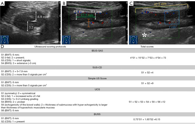
All parameters involved in the IUS scores were remeasured according to each score’s grading standard (Figure 1). The International Bowel Ultrasound Segmental Activity Score (IBUS-SAS) includes four parameters: bowel wall thickness (BWT) (mm), inflammatory fat (absent, uncertain, or present), color Doppler signal (absent, short signals, long signals inside bowel, or long signals inside and outside the bowel), and bowel wall stratification (BWS) (normal, uncertain, focal, or extensive). The Simple Ultrasound Score for Crohn’s Disease (SUS-CD) includes two parameters): BWT (<3.0, 3.0–4.9, 5.0–7.9, or ≥8.0 mm) and color Doppler signal (no or single vessel, 2–5 vessels per cm2, or >5 vessels per cm2). The Ultrasound Consolidated Score (UCS) includes six parameters: BWT (mm), symmetry (symmetrical or asymmetrical), echo of peribowel fat (not increased or increased), bowel wall vascularization (Limberg type 0–2 or Limberg type 3–4), BWS (clear, less clear, or unclear), and echo of the bowel walls (thickness of submucosa < muscularis mucosa, thickness of submucosa ≈ muscularis mucosa, or thickness of submucosa > muscularis mucosa). The Simple Ultrasound Score (Simple US Score) includes two parameters: BWT (mm) and color Doppler signal (absent, 1–2 vessels per cm2, 3–5 vessels per cm2, or >5 vessels per cm2). The Bowel Ultrasound Score (BUSS) includes two parameters: BWT (mm) and color Doppler signal (absent or present). The scoring formulas for all IUS scores are listed below, and the detailed grading protocols can be found in the corresponding literature (15-19).
Figure 1.
Practical example of IUS score remeasurement and scoring details. (A) Transverse section of affected bowel segment. (B,C) Longitudinal section of the affected bowel segment in color Doppler mode. (D) The details of score calculation according to each scoring protocol. White two-way arrows: BWT measurements in two different sections; green two-way arrows: distance between two measuring sites, with >10 mm indicating qualification; yellow line segment: length of the segmental BWS loss; orange line segment: the ratio of the thickness of mucosa (hypoechoic) and submucosa (hyperechoic); blue line square: area of 1 cm2 for semiquantitative evaluation of bowel wall blood signal; asterisk: inflamed fat wrapping. IBUS-SAS, International Bowel Ultrasound Segmental Activity Score; BWT, bowel wall thickness; i-fat, inflammatory fat; CDS, color Doppler signal; BWS, bowel wall stratification; SUS-CD, Simple Ultrasound Score for Crohn’s Disease; Simple US Score, Simple Ultrasound Score; UCS, Ultrasound Consolidated Score; BUSS, Bowel Ultrasound Score; IUS, intestinal ultrasound.
| [1] |
| [2] |
| [3] |
| [4] |
| [5] |
Statistical analysis was performed using SPSS 27 (IBM Corp., Armonk, NY, USA). The Shapiro-Wilk test was used to determine normality. Normally distributed data are expressed as the mean ± standard deviation, and nonnormally distributed data are expressed as the median. Categorical data are expressed as frequency (percentage). The Spearman correlation coefficient was used to calculate the correlation of parameters, and the Kruskal-Wallis test was used for the comparison of different groups. Receiver operating characteristic (ROC) curve analysis was performed to evaluate the diagnostic efficiency of each score, and the corresponding area under the curve (AUC), cutoff values, sensitivity, specificity, and 95% confidence intervals (CIs) were calculated. Two-sided probability values <0.05 were considered statistically significant.
Results
Overall data
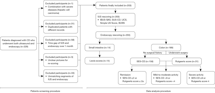
Initially, 329 patients met the inclusion criteria, and according to the exclusion criteria, but 33 were excluded for nonmatching segments between IUS and endoscopy, 58 for a time interval between IUS and endoscopy exceeding 1 month, 31 for being duplicate patients with different records, 3 for having unclear pictures for scoring, and 1 for having complication with other severe diseases (hepatic cell carcinoma). Finally, 203 patients in total were included in this study, and the sample sizes vary due to different subgrouping criterion in the subsequent results (Figure 2). The baseline demographic data and endoscopic and IUS scores are shown in Table 1. Among the enrolled patients, 126 (62.1%) were male and 77 (37.9%) were female, the mean age was 28 years, and with median disease duration was 36 months. There were 92 (45.3%) patients who had received biologics therapy and 31 (15.3%) who had undergone surgery due to CD complications such as obstruction or perforation. In the Montreal classification, patients in our study were mostly A2 (n=150, 73.9%), L3 (n=122, 64.6%), and B1 (n=87, 42.9%). The most commonly affected segment on IUS was the cecum and ascending colon (53, 26.1%). Additionally, 17 patients (8.4%) had ileocolonic anastomosis affected after surgery, and 50 patients (24.6%) showed no active findings.
Figure 2.
Flowchart of the patient screening and data analysis procedure. CD, Crohn’s disease; IUS, intestinal ultrasound; IBUS-SAS, International Bowel Ultrasound Segmental Activity Score; SUS-CD, Simple Ultrasound Score for Crohn’s Disease; UCS, Ultrasound Consolidated Score; Simple US Score, Simple Ultrasound Score; BUSS, Bowel Ultrasound Score; SES-CD, Simple Endoscopic Score for Crohn’s Disease.
Table 1. Baseline demographic data.
| Parameter | Data |
|---|---|
| Gender | |
| Male | 126 (62.1) |
| Female | 77 (37.9) |
| Age (years) | 28 [14–67] |
| Disease duration (months) | 36 [1–300] |
| Biologics therapy | 92 (45.3) |
| Surgery history | 31 (15.3) |
| Isolated small bowel affection | 14 (6.9) |
| CDAI | 232.93±127.7 [26.28–548.19] |
| CRP (mg/L) | 9.4 [1–149] |
| Montreal classification | |
| A (n=203) | |
| 1 (age under 17 years) | 34 (16.7) |
| 2 (age 17–40 years) | 150 (73.9) |
| 3 (age over 40 years) | 19 (9.4) |
| L (n=189)† | |
| 1 (ileum) | 17 (9.0) |
| 2 (colon) | 50 (26.5) |
| 3 (ileocolon) | 122 (64.6) |
| B (n=203) | |
| 1 (nonstricturing or penetrating) | 87 (42.9) |
| 2 (stricturing) | 57 (28.1) |
| 3 (penetrating) | 44 (21.7) |
| 2 + 3 (stricturing and penetrating) | 15 (7.4) |
| Endoscopic scores (n=203) | |
| SES-CD (n=158) | 3 [0–12] |
| Rutgeerts score (n=31) | 1 [0–4] |
| Lewis (n=14) | 1,559 [0–3,721] |
| Endoscopic activity (n=189)†,‡ | |
| Remission | 93 (49.2) |
| Mild-to-moderate activity | 76 (40.2) |
| Severe activity | 20 (10.6) |
| Ultrasound scores (n=203) | |
| IBUS-SAS | 30 [8–95] |
| SUS-CD | 3 [0–5] |
| Simple US Score | 6 [2–14] |
| UCS | 6 [2–18] |
| BUSS | 5.4 [1.5–10.65] |
| The most-affected segment in ultrasound | |
| Small bowel | 14 (6.9) |
| Terminal ileum | 22 (10.8) |
| Cecum and ascending colon | 53 (26.1) |
| Transverse colon | 5 (2.5) |
| Descending and sigmoid colon | 42 (20.7) |
| Ileocolonic anastomosis | 17 (8.4) |
| No active finding in ultrasound | 50 (24.6) |
Data are presented as n (%), median [range], or mean ± standard deviation [range]. †, the sample size excludes patients with isolated small bowel involvement (n=14); ‡, the endoscopic activity was defined by SES-CD and Rutgeerts score but not the Lewis score. CDAI, Crohn’s Disease Activity Index; CRP, C-reactive protein; SES-CD, Simple Endoscopic Score for Crohn’s Disease; IBUS-SAS, International Bowel Ultrasound Segmental Activity Score; SUS-CD, Simple Ultrasound Score for Crohn’s Disease; Simple US Score, Simple Ultrasound Score; UCS, Ultrasound Consolidated Score; BUSS, Bowel Ultrasound Score.
Correlation overview
The correlations of IUS scores are shown in Table 2, with all scores being highly correlated with one another. The correlation of IUS scores and endoscopic scores, Crohn’s Disease Activity Index (CDAI) and C-reactive protein (CRP) are shown in Table 3. All IUS scores were highly and positively correlated with SES-CD, with correlation coefficients (r) ranging from 0.822 to 0.891, and IBUS-SAS demonstrated the highest correlation with SES-CD (r=0.891). As for the Rutgeerts score, all IUS scores showed moderate correlation, with the IBUS-SAS again having the highest correlation (r=0.610). For the Lewis score, SUS-CD and BUSS were not significantly correlated (P>0.05), while the IBUS-SAS, Simple US Score, and UCS showed moderate correlation, with UCS being the highest (r=0.708). All scores showed correlation with CDAI and CRP, with the IBUS-SAS exhibiting the highest for both (rCDAI=0.590; rCRP=0.688). Overall, the IBUS-SAS had highest correlation but was slightly inferior to UCS in its correlation with the Lewis scores of patients with isolated small bowel involvement.
Table 2. Intracorrelation of IUS scores.
| Score | IBUS-SAS | SUS-CD | Simple US Score | UCS | |||||||
|---|---|---|---|---|---|---|---|---|---|---|---|
| r | P | r | P | r | P | r | P | ||||
| SUS-CD | 0.928 | <0.001 | |||||||||
| Simple US Score | 0.946 | <0.001 | 0.976 | <0.001 | |||||||
| UCS | 0.920 | <0.001 | 0.923 | <0.001 | 0.955 | <0.001 | |||||
| BUSS | 0.930 | <0.001 | 0.925 | <0.001 | 0.962 | <0.001 | 0.974 | <0.001 | |||
IUS, intestinal ultrasound; IBUS-SAS, International Bowel Ultrasound Segmental Activity Score; SUS-CD, Simple Ultrasound Score for Crohn’s Disease; Simple US Score, Simple Ultrasound Score; UCS, Ultrasound Consolidated Score; BUSS, Bowel Ultrasound Score.
Table 3. Score correlation overview.
| Score | SES-CD (n=158) | Rutgeerts (n=31) | Lewis (n=14) | CDAI (n=203) | CRP (n=203) | |||||||||
|---|---|---|---|---|---|---|---|---|---|---|---|---|---|---|
| r | P | r | P | r | P | r | P | r | P | |||||
| IBUS-SAS | 0.891 | <0.001 | 0.610 | <0.001 | 0.610 | <0.05 | 0.590 | <0.001 | 0.688 | <0.001 | ||||
| SUS-CD | 0.822 | <0.001 | 0.564 | <0.001 | 0.405 | 0.151 | 0.373 | <0.05 | 0.620 | <0.001 | ||||
| Simple US Score | 0.828 | <0.001 | 0.555 | <0.05 | 0.539 | <0.05 | 0.419 | <0.05 | 0.673 | <0.001 | ||||
| UCS | 0.844 | <0.001 | 0.545 | <0.05 | 0.708 | <0.05 | 0.419 | <0.05 | 0.645 | <0.001 | ||||
| BUSS | 0.831 | <0.001 | 0.583 | <0.001 | 0.521 | 0.056 | 0.425 | <0.001 | 0.668 | <0.001 | ||||
SES-CD, Simple Endoscopic Score for Crohn’s Disease; CDAI, Crohn’s Disease Activity Index; CRP, C-reactive protein; IBUS-SAS, International Bowel Ultrasound Segmental Activity Score; SUS-CD, Simple Ultrasound Score for Crohn’s Disease; Simple US Score, Simple Ultrasound Score; UCS, Ultrasound Consolidated Score; BUSS, Bowel Ultrasound Score.
Activity evaluation
Fourteen patients with isolated small bowel affection were excluded from activity evaluation due to the small sample size of each grade according to the Lewis score. They were also not included in the Montreal location classification, as it was not applicable (26). The endoscopic activity was defined based on SES-CD and Rutgeerts scores. Remission was defined as SES-CD ≤2 or Rutgeerts score ≤ 2a, mild-to-moderate activity as SES-CD <9 or Rutgeerts score <4, and severe activity as SES-CD ≥9 or Rutgeerts score 4. Additionally, 94 patients (49.2%) were in remission, 76 patients (40.2%) had mild-to-moderate activity, and 20 patients (10.6%) exhibited severe activity.
Considering that the definition of endoscopic activity was different in the original article of SUS-CD (remission, SES-CD ≤2; mild activity, SES-CD <7; moderate activity, SES-CD ≥7) (16), we also regrouped SUS-CD according to its original standard. As the original endoscopic activity classification of SUS-CD only depends on SES-CD, the sample size was smaller in this classification than it was in others (158 vs. 189).
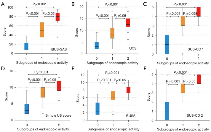
Results of the Kruskal-Wallis test showed that all IUS scores, except SUS-CD, were significantly different between the three activity-status groups (P<0.05), as shown in Figure 3. There was no significant difference between mild-to-moderate and severe activity groups in SUS-CD scoring (P=0.058). However, after regrouping was conducted according to its original classification, the SUS-CD scores were significantly different between all three activity statuses (P<0.05 in all paired tests) (Figure 3F).
Figure 3.
Box diagrams of IUS score distribution in the different endoscopic activity groups. Group 0: remission; group 1: mild-to-moderate activity; group 2: severe activity. P1–3 represents the probability values between the corresponding groups. IBUS-SAS, International Bowel Ultrasound Segmental Activity Score; UCS, Ultrasound Consolidated Score; SUS-CD, Simple Ultrasound Score for Crohn’s Disease; Simple US Score, Simple Ultrasound Score; BUSS, Bowel Ultrasound Score; IUS, intestinal ultrasound.
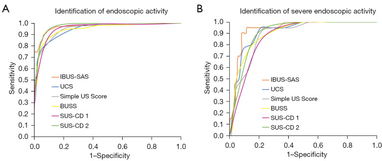
The ROC curves of all IUS scores are shown in Figure 4, and the corresponding diagnostic efficiencies and cutoff values are presented in Table 4. Similarly, both classifications of activity were evaluated in SUS-CD. In identifying endoscopic activity, all IUS scores yielded a high AUC, ranging from 0.941 to 0.970, suggesting good accuracy. Among them, IBUS-SAS had the highest AUC of 0.970, with a sensitivity and specificity of 92.7% and 91.4%, respectively. In identifying severe endoscopic activity, all scores had a slightly inferior but good performance, with the AUCs ranging from 0.863 to 0.937. Once again, IBUS-SAS exhibited the highest AUC, with a sensitivity and specificity of 95.0% and 88.2%, respectively.
Figure 4.
ROC curves of IUS scores for identifying the (A) presence and (B) severity of endoscopic activity. IBUS-SAS, International Bowel Ultrasound Segmental Activity Score; UCS, Ultrasound Consolidated Score; Simple US Score, Simple Ultrasound Score; BUSS, Bowel Ultrasound Score; SUS-CD, Simple Ultrasound Score for Crohn’s Disease; ROC, receiver operating characteristic; IUS, intestinal ultrasound.
Table 4. Diagnostic efficiency of IUS scores.
| Scores | Identification of endoscopic activity | Identification of severe endoscopic activity | |||||||
|---|---|---|---|---|---|---|---|---|---|
| AUC (95% CI) | Cutoff | Sensitivity (%) | Specificity (%) | AUC (95% CI) | Cutoff | Sensitivity (%) | Specificity (%) | ||
| IBUS-SAS | 0.970 (0.947–0.993) | 27.5 | 92.7 | 91.4 | 0.937 (0.896–0.978) | 65.5 | 95.0 | 88.2 | |
| UCS | 0.944 (0.914–0.975) | 6.5 | 79.2 | 93.5 | 0.912 (0.860–0.964) | 8.5 | 95.0 | 78.7 | |
| Simple US Score | 0.944 (0.911–0.976) | 5.5 | 95.8 | 78.5 | 0.884 (0.824–0.943) | 8.5 | 90.0 | 79.3 | |
| BUSS | 0.942 (0.909–0.975) | 5.0 | 92.7 | 82.8 | 0.898 (0.841–0.955) | 6.5 | 85.0 | 79.9 | |
| SUS-CD 1† | 0.941 (0.908–0.974) | 2.5 | 93.0 | 87.0 | 0.863 (0.801–0.924) | 3.5 | 85.0 | 78.1 | |
| SUS-CD 2† | 0.960 (0.932–0.989) | 2.5 | 94.3 | 90.1 | 0.891 (0.839–0.943) | 3.5 | 89.3 | 81.5 | |
†, SUS-CD was evaluated according to two activity standards. SUS-CD 1 was consistent with other scores in this study, while SUS-CD 2 used its original standard. IUS, intestinal ultrasound; AUC, area under the curve; CI, confidence interval; IBUS-SAS, International Bowel Ultrasound Segmental Activity Score; UCS, Ultrasound Consolidated Score; Simple US Score, Simple Ultrasound Score; BUSS, Bowel Ultrasound Score; SUS-CD, Simple Ultrasound Score for Crohn’s Disease.
The cutoff values of IUS scores indicating endoscopic activity were 27.5 for IBUS-SAS, 6.5 for UCS, 5.5 for Simple US Score, 5.0 for BUSS, and 2.5 for SUS-CD in both standards. The cutoff values indicating severe endoscopic activity were 65.5 for IBUS-SAS, 8.5 for UCS, 8.5 for Simple US Score, 6.5 for BUSS, and 3.5 for SUS-CD in both standards. Interestingly, the SUS-CD when graded according to its original standard had the same cutoff values but did show higher diagnostic efficiency.
Discussion
The development of accurate and repeatable IUS scoring is critical for broadening the use of IUS in CD. Several scores have been established in recent years, yet the retrospective comparison and validation of these scores using the same cohort has not been attempted. The importance of retrospective validation is comparable with that prospective validation in the evaluation of the clinical applicability of IUS scores. Therefore, we compared and validated several IUS scores in the same cohort of patients with CD, using strict criteria. To our knowledge, this study is the first retrospective study to include IUS parameter-based scores established within the past 5 years and the first to determine the value of these scores in assessing small intestinal CD. Our study was based at the West China Hospital, a leading hospital of the China Western Inflammatory Bowel Disease Alliance, so the CD cohort in our study could adequately represent the context of southwest China. The proportion of Montreal classifications in our study was different compared to other countries or other Chinese cohorts, especially in disease location and behavior (21,27,28), as the rate of colonic CD is higher than ileal CD among Chinese CD patients. Overall, all the scores demonstrated good potential in assessing CD activity and were comparable to one another, and thus the selection scoring system should depend on the given context.
In our study, IUS scores were found to be capable of identifying endoscopic activity, especially in colonic CD. All IUS scores exhibited a close correlation with SES-CD, a commonly used endoscopic score in CD, indicating that IUS scores are able to objectively reflect the activity of the bowel wall. The scores also demonstrated moderate-to-high correlation with the Rutgeerts score, an endoscopic score for patients who have undergone resection. Although the correlation of IUS scores with Rutgeerts score was slightly inferior to that with SES-CD, it is nonetheless encouraging that IUS scores can detect postoperative recurrence. Evaluating the activity of small intestinal CD has long been a challenge for imaging modalities including IUS, and a gold standard for quantifying the activity extent of small intestinal CD is lacking. As a result, the diagnosis and management of small intestinal CD are often delayed (29,30). The Lewis score, proposed in 2008 by Gralnek et al. and based on capsule endoscopy, was used as a temporary reference standard in this study (31). To our knowledge, few validations of IUS scores have been conducted in small bowel CD. Although ultrasound is inferior to magnetic resonance enterography in small intestinal CD, with an average accuracy of about 70% (9,32), the results in our study indicate that IUS scores are capable of evaluating the activity of small bowel CD, and the correlation of IUS scores (except SUS-CD and BUSS) with Lewis score was comparable with that of the Rutgeerts score. This demonstrated accuracy of IUS scores in the context of small bowel CD in our study is close to that for detecting postoperative recurrence, with the latter being confirmed by previous studies and guidelines (3,33). However, further validation is needed to determine the cutoff values of IUS scores in small bowel CD.
We also confirmed a correlation between IUS scores and other indices such as CRP and CDAI, which is consistent with previously published research (34). CRP and CDAI are important supplementary parameters in CD management, but they are less sensitive for indicating activity. CDAI is based on symptoms, which correlate poorly with disease activity and extent (3); meanwhile, CRP can be influenced by disease location, and a negative test does not exclude the presence of a flare (35). Assessments that reveal objective evidence of bowel wall activity, such as endoscopy, are more valuable. In our study, IUS scores correlated better with endoscopy than did CRP or CDAI, suggesting that IUS is a promising tool that can satisfy the requirements of a treat-to-target strategy.
IUS scores performed well in this study, and the most significant difference among these scores was number of parameters included. It is important to note that the more parameters a scoring system has, the more experience is required, increasing operator dependency. Simple US Score, BUSS, and SUS-CD each include only two parameters, while IBUS-SAS and UCS have four and six parameters, respectively. BWT and bowel wall blood flow are the most important and most commonly studied ultrasound parameters in CD, form the foundation of the ultrasonic assessment of transmural inflammation, and can also predict response or recurrence (2,13,36). BWT and bowel wall blood flow were included in all IUS scores, and are the only two parameters in the Simple US Score, BUSS, and SUS-CD; therefore, these systems are reliable and sensitive for rapid and preliminary activity evaluations, only requiring basic IUS training. Besides BWT and bowel wall vascularization, the loss of BWS and the presence of mesenteric fat wrapping should also be reported according to ECCO-ESGAR topical review, as they can reflect transmural inflammation and may contribute to underlying pathophysiology (7,37). This is why IBUS-SAS includes two more parameters, providing excellent sensitivity and specificity. The parameters of UCS represent the greatest number of ultrasound changes in CD among the scoring systems, including the ratio of submucosa to muscularis mucosa. However, with each additional parameter, sensitivity is decreased while specificity is increased. Therefore, UCS is more suitable for scientific applications rather than routine clinical practice, requiring operators with relevant experience in IUS, but UCS can yield high accuracy in cases of small intestinal CD. Based on the overall results, IBUS-SAS appears to be the most preferable IUS score for clinical practice in Southwest China, as it strikes a balance between precision and operator dependence.
Previous validation studies for these IUS scores have been conducted, and generally, the diagnostic efficiencies of these scores and their cutoff values vary. IBUS-SAS has been the most commonly studied system, followed by SUS-CD, and the scores in our study are in accordance with those reported elsewhere (38). These two scores are the most promising for application in clinical practice based on the current evidence. However, the cutoff values for these scores vary and have not been standardized, and the values used in our study are consistent with those of some studies but inconsistent with those of others (20,21,38). There is relatively less published research on the validation of the Simple US Score, UCS, and BUSS, and the cutoff values of Simple US Score and UCS in our study are nearly identical to those of previous studies (17,18,21), while that of BUSS was not (19,21). Therefore, further investigation into appropriate cutoff values remains necessary.
Several limitations to this study should be noted. First, we employed a retrospective design using still images, and we excluded the cases with unrecognizable images, which might have led to in data collection and remeasurements. However, we demonstrated the feasibility of comparing IUS scores with previous reports if the collected images allowed for the rereading and remeasuring of parameters. This is important for the application of these scores in clinical practice. Second, we did not assess inter- or intraoperator consistency, as measurements on static images significantly reduce operator dependency and can falsely increase consistency; moreover, IUS is generally standardized and repeatable among operators with varying experience in IUS according to the current evidence (7,27). Third, the reference standard of endoscopy scores for small intestinal CD in this study was the Lewis score, which was established for capsule endoscopy (31). However, some of our small intestinal CD cases underwent enteroscopy instead, and there is currently no standardized CD score for enteroscopy. Therefore, our study, which suggested the potential value of IUS scores in small intestinal CD, only conducted exploratory research in small intestinal cases, and thus further validation is needed. Considering the need of high replication of each IUS score in this primary transverse validation, the most seriously affected segment was chosen to complete the scores strictly according to the original score protocols. In addition, we did not include subjective IUS scoring of activity. Based on this pilot study, we plan to evaluate more segments with a greater variety of comparisons in future research.
With the emerging trend of treat-to-target therapeutic strategies, developing accurate IUS scores for CD is a critical step toward developing a novel strategy and can meet the needs of precision clinical medicine (39). Indeed, IUS results can provide valuable guidance at various stages of CD management, including de-escalation or withdrawal of maintenance therapy (40). Validated IUS scores may serve as a reference and as guidance during long disease courses. The ability of IUS scores to objectively reflect bowel wall damage is comparable with that endoscopy; furthermore, IUS can demonstrate the peribowel situation and overcome certain limitations, such as stenosis. Therefore, the IUS score is an attractive alternative to endoscopic scores for CD activity evaluation and treatment response. Additionally, the IUS score can serve as a useful supplement for challenging conditions such as small bowel CD. As indicated by ECCO-ESGAR, IUS possesses the potential to be one of the core monitoring modalities in CD management, as it is non-invasive, simple to conduct, and easily interpretable (3).
Conclusions
IUS scores were closely correlated with endoscopic activity scores, indicating their ability to quantify the disease activity via ultrasound parameters in clinical practice. Furthermore, different IUS scores may be more suitable in different contexts, and the appropriate selection of the scoring system can optimize evaluation.
Supplementary
The article’s supplementary files as
Acknowledgments
The ultrasound images and results used for rescoring were all obtained previously during routine clinical practice by members in gastrointestinal ultrasound subgroup, including Ji-Gang Jing, Qiong Zhang, and Yu-Ting Wu.
Funding: This work was supported by the 1·3·5 Project for Disciplines of Excellence, West China Hospital, Sichuan University (grant No. ZYJC18037).
Ethical Statement: The authors are accountable for all aspects of the work in ensuring that questions related to the accuracy or integrity of any part of the work are appropriately investigated and resolved. This retrospective cross-sectional study was conducted in accordance with the Declaration of Helsinki (as revised in 2013) and was approved by institutional ethics committee of the West China Hospital of Sichuan University (No. 359, 2020). The requirement for individual consent was waived due to the retrospective nature of the analysis.
Footnotes
Reporting Checklist: The authors have completed the STROBE reporting checklist. Available at https://qims.amegroups.com/article/view/10.21037/qims-24-742/rc
Conflicts of Interest: All authors have completed the ICMJE uniform disclosure form (available at https://qims.amegroups.com/article/view/10.21037/qims-24-742/coif). All authors report that this work was supported by the 1·3·5 Project for Disciplines of Excellence, West China Hospital, Sichuan University (grant No. ZYJC18037). The authors have no other conflicts of interest to declare.
References
- 1.Bernstein CN, Eliakim A, Fedail S, Fried M, Gearry R, Goh KL, Hamid S, Khan AG, Khalif I, Ng SC, Ouyang Q, Rey JF, Sood A, Steinwurz F, Watermeyer G, LeMair A; Review Team. World Gastroenterology Organisation Global Guidelines Inflammatory Bowel Disease: Update August 2015. J Clin Gastroenterol 2016;50:803-18. 10.1097/MCG.0000000000000660 [DOI] [PubMed] [Google Scholar]
- 2.Wilkens R, Novak KL, Maaser C, Panaccione R, Kucharzik T. Relevance of monitoring transmural disease activity in patients with Crohn's disease: current status and future perspectives. Therap Adv Gastroenterol 2021;14:17562848211006672. 10.1177/17562848211006672 [DOI] [PMC free article] [PubMed] [Google Scholar]
- 3.Maaser C, Sturm A, Vavricka SR, Kucharzik T, Fiorino G, Annese V, et al. ECCO-ESGAR Guideline for Diagnostic Assessment in IBD Part 1: Initial diagnosis, monitoring of known IBD, detection of complications. J Crohns Colitis 2019;13:144-64. 10.1093/ecco-jcc/jjy113 [DOI] [PubMed] [Google Scholar]
- 4.Goodsall TM, Nguyen TM, Parker CE, Ma C, Andrews JM, Jairath V, Bryant RV. Systematic Review: Gastrointestinal Ultrasound Scoring Indices for Inflammatory Bowel Disease. J Crohns Colitis 2021;15:125-42. 10.1093/ecco-jcc/jjaa129 [DOI] [PubMed] [Google Scholar]
- 5.Turner D, Ricciuto A, Lewis A, D'Amico F, Dhaliwal J, Griffiths AM, et al. STRIDE-II: An Update on the Selecting Therapeutic Targets in Inflammatory Bowel Disease (STRIDE) Initiative of the International Organization for the Study of IBD (IOIBD): Determining Therapeutic Goals for Treat-to-Target strategies in IBD. Gastroenterology 2021;160:1570-83. 10.1053/j.gastro.2020.12.031 [DOI] [PubMed] [Google Scholar]
- 6.Matsuoka K, Kobayashi T, Ueno F, Matsui T, Hirai F, Inoue N, et al. Evidence-based clinical practice guidelines for inflammatory bowel disease. J Gastroenterol 2018;53:305-53. 10.1007/s00535-018-1439-1 [DOI] [PMC free article] [PubMed] [Google Scholar]
- 7.Hameed M, Taylor SA. Small bowel imaging in inflammatory bowel disease: updates for 2023. Expert Rev Gastroenterol Hepatol 2023;17:1117-34. 10.1080/17474124.2023.2274926 [DOI] [PubMed] [Google Scholar]
- 8.Rimola J, Torres J, Kumar S, Taylor SA, Kucharzik T. Recent advances in clinical practice: advances in cross-sectional imaging in inflammatory bowel disease. Gut 2022;71:2587-97. 10.1136/gutjnl-2021-326562 [DOI] [PMC free article] [PubMed] [Google Scholar]
- 9.Taylor SA, Mallett S, Bhatnagar G, Baldwin-Cleland R, Bloom S, Gupta A, et al. Diagnostic accuracy of magnetic resonance enterography and small bowel ultrasound for the extent and activity of newly diagnosed and relapsed Crohn's disease (METRIC): a multicentre trial. Lancet Gastroenterol Hepatol 2018;3:548-58. 10.1016/S2468-1253(18)30161-4 [DOI] [PMC free article] [PubMed] [Google Scholar]
- 10.Wang YD, Zhang RN, Mao R, Li XH. Inflammatory bowel disease cross-sectional imaging: What's new? United European Gastroenterol J 2022;10:1179-93. 10.1002/ueg2.12343 [DOI] [PMC free article] [PubMed] [Google Scholar]
- 11.Losurdo G, De Bellis M, Rima R, Palmisano CM, Dell'Aquila P, Iannone A, Ierardi E, Di Leo A, Principi M. Small Intestinal Contrast Ultrasonography (SICUS) in Crohn's Disease: Systematic Review and Meta-Analysis. J Clin Med 2023;12:7714. 10.3390/jcm12247714 [DOI] [PMC free article] [PubMed] [Google Scholar]
- 12.Goodsall TM, Noy R, Nguyen TM, Costello SP, Jairath V, Bryant RV. Systematic Review: Patient Perceptions of Monitoring Tools in Inflammatory Bowel Disease. J Can Assoc Gastroenterol 2021;4:e31-41. 10.1093/jcag/gwaa001 [DOI] [PMC free article] [PubMed] [Google Scholar]
- 13.Dolinger MT, Kayal M. Intestinal ultrasound as a non-invasive tool to monitor inflammatory bowel disease activity and guide clinical decision making. World J Gastroenterol 2023;29:2272-82. 10.3748/wjg.v29.i15.2272 [DOI] [PMC free article] [PubMed] [Google Scholar]
- 14.Nardone OM, Calabrese G, Testa A, Caiazzo A, Fierro G, Rispo A, Castiglione F. The Impact of Intestinal Ultrasound on the Management of Inflammatory Bowel Disease: From Established Facts Toward New Horizons. Front Med (Lausanne) 2022;9:898092. 10.3389/fmed.2022.898092 [DOI] [PMC free article] [PubMed] [Google Scholar]
- 15.Novak KL, Nylund K, Maaser C, Petersen F, Kucharzik T, Lu C, Allocca M, Maconi G, de Voogd F, Christensen B, Vaughan R, Palmela C, Carter D, Wilkens R. Expert Consensus on Optimal Acquisition and Development of the International Bowel Ultrasound Segmental Activity Score [IBUS-SAS]: A Reliability and Inter-rater Variability Study on Intestinal Ultrasonography in Crohn's Disease. J Crohns Colitis 2021;15:609-16. 10.1093/ecco-jcc/jjaa216 [DOI] [PMC free article] [PubMed] [Google Scholar]
- 16.Sævik F, Eriksen R, Eide GE, Gilja OH, Nylund K. Development and Validation of a Simple Ultrasound Activity Score for Crohn's Disease. J Crohns Colitis 2021;15:115-24. 10.1093/ecco-jcc/jjaa112 [DOI] [PMC free article] [PubMed] [Google Scholar]
- 17.Ripollés T, Poza J, Suarez Ferrer C, Martínez-Pérez MJ, Martín-Algíbez A, de Las Heras Paez B. Evaluation of Crohn's Disease Activity: Development of an Ultrasound Score in a Multicenter Study. Inflamm Bowel Dis 2021;27:145-54. 10.1093/ibd/izaa134 [DOI] [PubMed] [Google Scholar]
- 18.Liu C, Ding SS, Zhang K, Liu LN, Guo LH, Sun LP, Zhang YF, Sun XM, Ren WW, Zhao CK, Li XL, Wang Q, Xu XR, Xu HX. Correlation between ultrasound consolidated score and simple endoscopic score for determining the activity of Crohn's disease. Br J Radiol 2020;93:20190614. 10.1259/bjr.20190614 [DOI] [PMC free article] [PubMed] [Google Scholar]
- 19.Allocca M, Craviotto V, Bonovas S, Furfaro F, Zilli A, Peyrin-Biroulet L, Fiorino G, Danese S. Predictive Value of Bowel Ultrasound in Crohn's Disease: A 12-Month Prospective Study. Clin Gastroenterol Hepatol 2022;20:e723-40. 10.1016/j.cgh.2021.04.029 [DOI] [PubMed] [Google Scholar]
- 20.Freitas M, de Castro FD, Macedo Silva V, Arieira C, Cúrdia Gonçalves T, Leite S, Moreira MJ, Cotter J. Ultrasonographic scores for ileal Crohn's disease assessment: Better, worse or the same as contrast-enhanced ultrasound? BMC Gastroenterol 2022;22:252. 10.1186/s12876-022-02326-6 [DOI] [PMC free article] [PubMed] [Google Scholar]
- 21.Dragoni G, Gottin M, Innocenti T, Lynch EN, Bagnoli S, Macrì G, Bonanomi AG, Orlandini B, Rogai F, Milani S, Galli A, Milla M, Biagini MR. Correlation of Ultrasound Scores with Endoscopic Activity in Crohn's Disease: A Prospective Exploratory Study. J Crohns Colitis 2023;17:1387-94. 10.1093/ecco-jcc/jjad068 [DOI] [PubMed] [Google Scholar]
- 22.Barchi A, D'Amico F, Zilli A, Furfaro F, Parigi TL, Fiorino G, Peyrin-Biroulet L, Danese S, Dal Buono A, Allocca M. Recent advances in the use of ultrasound in Crohn's disease. Expert Rev Med Devices 2023;20:1119-29. 10.1080/17434440.2023.2283166 [DOI] [PubMed] [Google Scholar]
- 23.Bernstein CN, Fried M, Krabshuis JH, Cohen H, Eliakim R, Fedail S, Gearry R, Goh KL, Hamid S, Khan AG, LeMair AW, Malfertheiner, Ouyang Q, Rey JF, Sood A, Steinwurz F, Thomsen OO, Thomson A, Watermeyer G. World Gastroenterology Organization Practice Guidelines for the diagnosis and management of IBD in 2010. Inflamm Bowel Dis 2010;16:112-24. 10.1002/ibd.21048 [DOI] [PubMed] [Google Scholar]
- 24.Daperno M, D'Haens G, Van Assche G, Baert F, Bulois P, Maunoury V, Sostegni R, Rocca R, Pera A, Gevers A, Mary JY, Colombel JF, Rutgeerts P. Development and validation of a new, simplified endoscopic activity score for Crohn's disease: the SES-CD. Gastrointest Endosc 2004;60:505-12. 10.1016/S0016-5107(04)01878-4 [DOI] [PubMed] [Google Scholar]
- 25.Rutgeerts P, Geboes K, Vantrappen G, Beyls J, Kerremans R, Hiele M. Predictability of the postoperative course of Crohn's disease. Gastroenterology 1990;99:956-63. 10.1016/0016-5085(90)90613-6 [DOI] [PubMed] [Google Scholar]
- 26.Satsangi J, Silverberg MS, Vermeire S, Colombel JF. The Montreal classification of inflammatory bowel disease: controversies, consensus, and implications. Gut 2006;55:749-53. 10.1136/gut.2005.082909 [DOI] [PMC free article] [PubMed] [Google Scholar]
- 27.Smith RL, Taylor KM, Friedman AB, Su HY, Con D, Gibson PR. Interrater reliability of the assessment of disease activity by gastrointestinal ultrasound in a prospective cohort of patients with inflammatory bowel disease. Eur J Gastroenterol Hepatol 2021;33:1280-7. 10.1097/MEG.0000000000002253 [DOI] [PubMed] [Google Scholar]
- 28.Zhou Q, Zhu Q, Liu W, Li W, Ma L, Xiao M, Liu J, Yang H, Qian J. New score models for assessing disease activity in Crohn's disease based on bowel ultrasound and biomarkers: Ideal surrogates for endoscopy or imaging. Clin Transl Sci 2023;16:1639-52. 10.1111/cts.13575 [DOI] [PMC free article] [PubMed] [Google Scholar]
- 29.Kucharzik T, Maaser C. Intestinal ultrasound and management of small bowel Crohn's disease. Therap Adv Gastroenterol 2018;11:1756284818771367. 10.1177/1756284818771367 [DOI] [PMC free article] [PubMed] [Google Scholar]
- 30.Bollegala N, Griller N, Bannerman H, Habal M, Nguyen GC. Ultrasound vs Endoscopy, Surgery, or Pathology for the Diagnosis of Small Bowel Crohn's Disease and its Complications. Inflamm Bowel Dis 2019;25:1313-38. 10.1093/ibd/izy392 [DOI] [PubMed] [Google Scholar]
- 31.Gralnek IM, Defranchis R, Seidman E, Leighton JA, Legnani P, Lewis BS. Development of a capsule endoscopy scoring index for small bowel mucosal inflammatory change. Aliment Pharmacol Ther 2008;27:146-54. 10.1111/j.1365-2036.2007.03556.x [DOI] [PubMed] [Google Scholar]
- 32.Allocca M, Fiorino G, Bonifacio C, Furfaro F, Gilardi D, Argollo M, Peyrin-Biroulet L, Danese S. Comparative Accuracy of Bowel Ultrasound Versus Magnetic Resonance Enterography in Combination With Colonoscopy in Assessing Crohn's Disease and Guiding Clinical Decision-making. J Crohns Colitis 2018;12:1280-7. 10.1093/ecco-jcc/jjy093 [DOI] [PubMed] [Google Scholar]
- 33.Rispo A, Imperatore N, Testa A, Nardone OM, Luglio G, Caporaso N, Castiglione F. Diagnostic Accuracy of Ultrasonography in the Detection of Postsurgical Recurrence in Crohn's Disease: A Systematic Review with Meta-analysis. Inflamm Bowel Dis 2018;24:977-88. 10.1093/ibd/izy012 [DOI] [PubMed] [Google Scholar]
- 34.You MW, Moon SK, Lee YD, Oh SJ, Park SJ, Lee CK. Assessing Active Bowel Inflammation in Crohn's Disease Using Intestinal Ultrasound: Correlation With Fecal Calprotectin. J Ultrasound Med 2023;42:2791-802. 10.1002/jum.16317 [DOI] [PubMed] [Google Scholar]
- 35.Florin TH, Paterson EW, Fowler EV, Radford-Smith GL. Clinically active Crohn's disease in the presence of a low C-reactive protein. Scand J Gastroenterol 2006;41:306-11. 10.1080/00365520500217118 [DOI] [PubMed] [Google Scholar]
- 36.de Voogd F, Bots S, Gecse K, Gilja OH, D'Haens G, Nylund K. Intestinal Ultrasound Early on in Treatment Follow-up Predicts Endoscopic Response to Anti-TNFα Treatment in Crohn's Disease. J Crohns Colitis 2022;16:1598-608. 10.1093/ecco-jcc/jjac072 [DOI] [PMC free article] [PubMed] [Google Scholar]
- 37.Kucharzik T, Tielbeek J, Carter D, Taylor SA, Tolan D, Wilkens R, Bryant RV, Hoeffel C, De Kock I, Maaser C, Maconi G, Novak K, Rafaelsen SR, Scharitzer M, Spinelli A, Rimola J. ECCO-ESGAR Topical Review on Optimizing Reporting for Cross-Sectional Imaging in Inflammatory Bowel Disease. J Crohns Colitis 2022;16:523-43. 10.1093/ecco-jcc/jjab180 [DOI] [PubMed] [Google Scholar]
- 38.Wang L, Xu C, Zhang Y, Jiang W, Ma J, Zhang H. External validation and comparison of simple ultrasound activity score and international bowel ultrasound segmental activity score for Crohn's disease. Scand J Gastroenterol 2023;58:883-9. 10.1080/00365521.2023.2181038 [DOI] [PubMed] [Google Scholar]
- 39.Zorzi F, Rubin DT, Cleveland NK, Monteleone G, Calabrese E. Ultrasonographic Transmural Healing in Crohn's Disease. Am J Gastroenterol 2023;118:961-9. 10.14309/ajg.0000000000002265 [DOI] [PubMed] [Google Scholar]
- 40.Saleh A, Abraham BP. Utility of Intestinal Ultrasound in Clinical Decision-Making for Inflammatory Bowel Disease. Crohns Colitis 360 2023;5:otad027. 10.1093/crocol/otad027 [DOI] [PMC free article] [PubMed] [Google Scholar]
Associated Data
This section collects any data citations, data availability statements, or supplementary materials included in this article.
Supplementary Materials
The article’s supplementary files as


