Endoscopic ultrasound-guided fine-needle biopsy (EUS-FNB) is useful for the diagnosis of retroperitoneal lesions and pancreatic diseases. However, the usefulness of EUS-FNB for tissue acquisition from retroperitoneal lesions in patients with surgically altered anatomy has not been established 1 2 3 4 5 . Herein, we report successful tissue acquisition from a recurrent lymph node lesion of a retroperitoneal paraganglioma by EUS-FNB using a forward-viewing echoendoscope (FV-EUS) in a patient who had undergone the Whipple procedure.
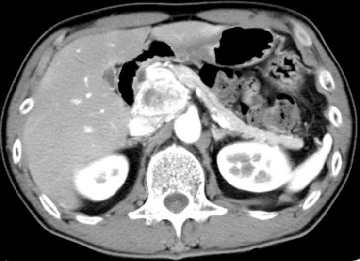
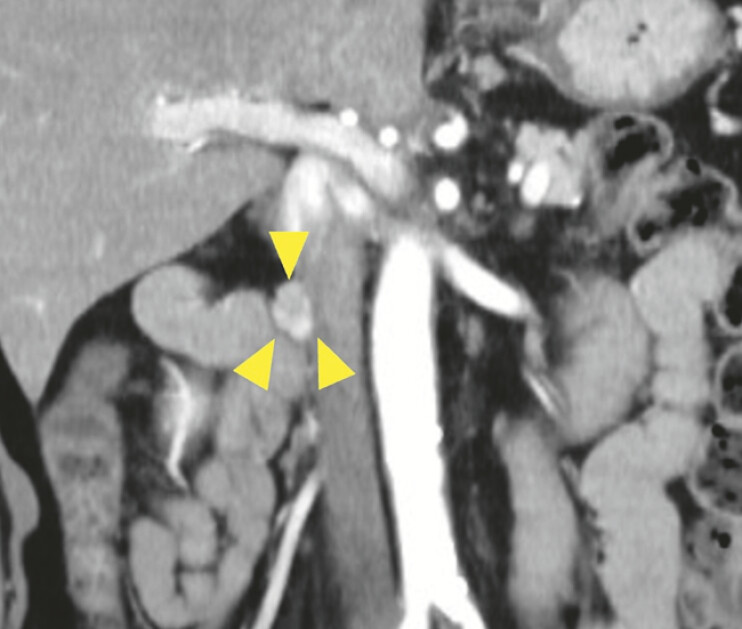
A 60-year-old man underwent the Whipple procedure for a retroperitoneal paraganglioma adjacent to the head of the pancreas ( Fig. 1 ). Follow-up computed tomography 2.5 years after surgery revealed a 15-mm swelling of the lymph node on the right side of the inferior vena cava ( Fig. 2 ). The lesion was located near the afferent loop, and we expected that it could be visualized using an FV-EUS (TGF-UC260J; Olympus, Tokyo, Japan).
Fig. 1.
Computed tomography image of a retroperitoneal paraganglioma. The tumor was located adjacent to the head of the pancreas.
Fig. 2.
Computed tomography image of a recurrent lymph node lesion (arrowheads) of the retroperitoneal paraganglioma.
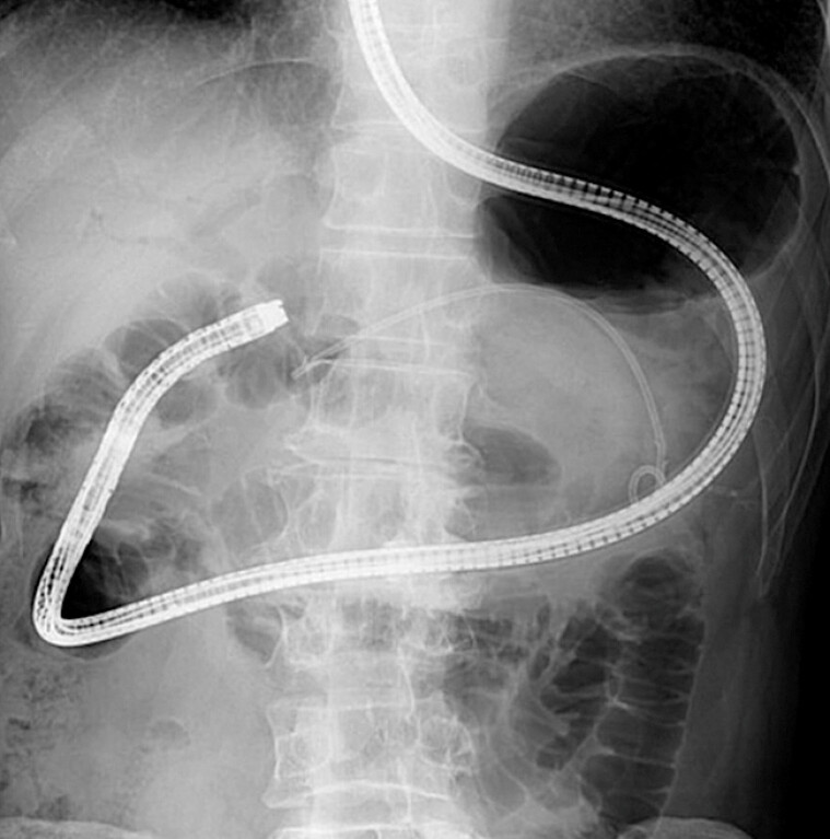
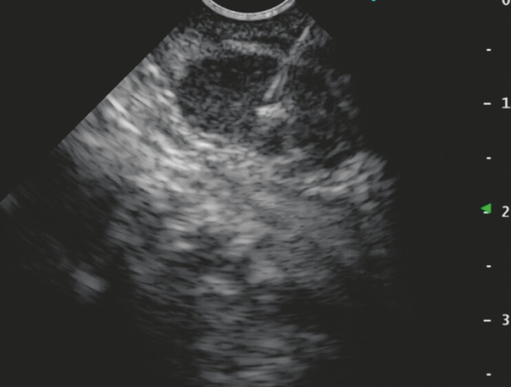
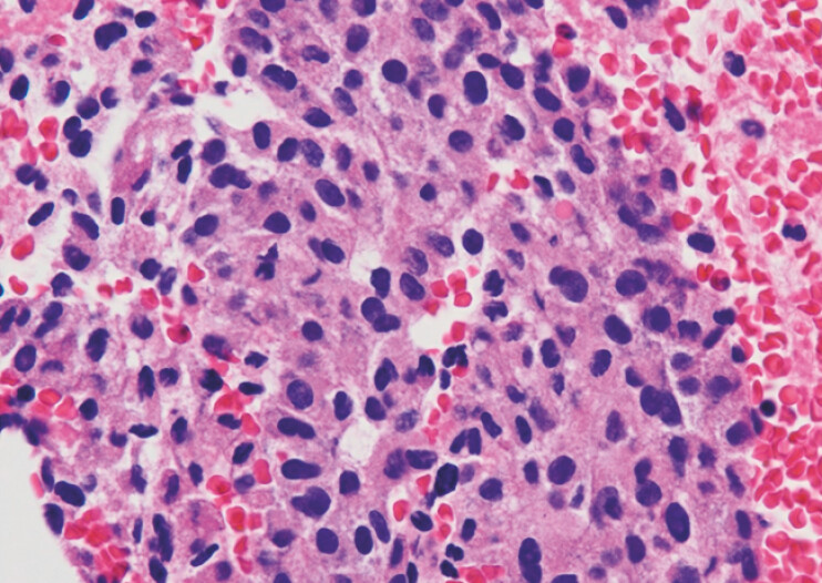
To insert the FV-EUS into the afferent loop safely, a short-type single-balloon enteroscope (SIF-H290; Olympus) was first inserted into the hepaticojejunostomy anastomosis ( Fig. 3 ), and a guidewire was placed. Then, under wire guidance, the FV-EUS was inserted up into the afferent loop, and the target lesion was visualized. EUS-FNB was performed transjejunally using a 22-gauge FNB needle ( Fig. 4 ). The histopathological diagnosis was consistent with lymph node recurrence of the retroperitoneal paraganglioma ( Fig. 5 ). Finally, open retroperitoneal tumor resection was performed, and complete resection was achieved ( Video 1 ).
Fig. 3.
A balloon enteroscope was inserted into the afferent loop.
Fig. 4.
Endoscopic ultrasound-guided fine-needle biopsy was performed using a 22-gauge needle.
Fig. 5.
The pathological specimen obtained by endoscopic ultrasound-guided fine-needle biopsy.
Successful endoscopic ultrasound-guided fine-needle biopsy of a recurrent paraganglioma using a forward-viewing echoendoscope in a patient who had undergone the Whipple procedure.
Video 1
This case demonstrates that EUS-FNB using an FV-EUS and assisted by balloon enteroscope insertion is a safe and effective method for tissue acquisition in patients with surgically altered anatomy, and can prevent adverse events, such as gastrointestinal perforation.
Endoscopy_UCTN_Code_TTT_1AS_2AC
Footnotes
Conflict of Interest The authors declare that they have no conflict of interest.
Endoscopy E-Videos https://eref.thieme.de/e-videos .
E-Videos is an open access online section of the journal Endoscopy , reporting on interesting cases and new techniques in gastroenterological endoscopy. All papers include a high-quality video and are published with a Creative Commons CC-BY license. Endoscopy E-Videos qualify for HINARI discounts and waivers and eligibility is automatically checked during the submission process. We grant 100% waivers to articles whose corresponding authors are based in Group A countries and 50% waivers to those who are based in Group B countries as classified by Research4Life (see: https://www.research4life.org/access/eligibility/ ). This section has its own submission website at https://mc.manuscriptcentral.com/e-videos .
References
- 1.Katanuma A, Hayashi T, Kin T et al. Interventional endoscopic ultrasonography in patients with surgically altered anatomy: techniques and literature review. Dig Endosc. 2020;32:263–274. doi: 10.1111/den.13567. [DOI] [PubMed] [Google Scholar]
- 2.Larghi A, Fuccio L, Chiarello G et al. Fine-needle tissue acquisition from subepithelial lesions using a forward-viewing linear echoendoscope. Endoscopy. 2014;46:39–45. doi: 10.1055/s-0033-1344895. [DOI] [PubMed] [Google Scholar]
- 3.Tanaka K, Hayashi T, Utsunomiya R et al. Endoscopic ultrasound-guided fine needle aspiration for diagnosing pancreatic mass in patients with surgically altered upper gastrointestinal anatomy. Dig Endosc. 2020;32:967–973. doi: 10.1111/den.13625. [DOI] [PubMed] [Google Scholar]
- 4.Gong TT, Zhang MM, Zou DW. EUS-FNA of a lesion in the pancreatic head using a forward-viewing echoendoscope in a patient with Billroth II gastrectomy (with video) Endosc Ultrasound. 2022;11:243–245. doi: 10.4103/EUS-D-21-00101. [DOI] [PMC free article] [PubMed] [Google Scholar]
- 5.Akdamar MK, Eltoum I, Eloubeidi MA. Retroperitoneal paraganglioma: EUS appearance and risk associated with EUS-guided FNA. Gastrointest Endosc. 2004;60:1018–1021. doi: 10.1016/s0016-5107(04)02218-7. [DOI] [PubMed] [Google Scholar]


