Abstract
Purpose:
White spot lesions (WSL) are common side-effects of orthodontic treatment with fixed multi-bracketed appliances. The aim of this review was to find all available literature and critically assess the evidence for the efficacy of bleaching as a method to treat or alleviate post-orthodontic WSLs in permanent teeth.
Materials and Methods:
Electronic databases were screened for relevant literature with the aid of predetermined search strategies. All types of studies, including randomised or nonrandomised controlled trials (RCTs or CCTs), prospective and retrospective studies, as well as in vitro studies were considered eligible for inclusion. The reference lists of all included articles were hand searched for additional studies. Two authors independently performed study selection, data extraction, and risk of bias assessment.
Results:
One RCT and 8 in vitro studies met the inclusion criteria. Seven studies were classified as having a high risk of bias while 2 in vitro studies were graded as having a moderate risk of bias. The results showed that bleaching of WSL can diminish colour disparities between carious and non-affected areas, but the certainty of the evidence was very low. The high degree of methodological heterogeneity precluded a valid interpretation of the results through pooled estimates.
Conclusions:
The findings from the present systematic review could not support or refute bleaching as an effective method for management of post-orthodontic WSLs. Because most of the studies in this field are in vitro and solid scientific evidence of low risk of bias is scare, further prospective in vivo studies are necessary.
Keywords: bleaching, enamel demineralisation, fluoride, tooth whitening, white spot lesions
Although fixed appliances have revolutionised contemporary orthodontic treatment, they are at the same time a risk factor for the integrity of tooth enamel. This is mainly attributed to plaque accumulation and the subsequent development of bacterial colonies.44 The placement of fixed orthodontic appliances interferes with standard oral hygiene procedures and causes alterations in the oral microflora by reducing pH and increasing bacterial affinity to the metallic surfaces due to electrostatic reactions.1 White spot lesions (WSL) can develop as soon as 4 weeks after placement of fixed orthodontic appliances,30 with a prevalence ranging from 5% to 97% at the time of debonding.7,17 The clinical appearance is an opaque white area of demineralised enamel that follows the shape of the bracket.32 Although WSL have the ability to remineralise after bracket removal,39 with the greatest remineralisation within the first months,21 they may in some cases still be detected up to 5 years after debonding.34
The management of WSL has traditionally focused on applying remineralising agents such as topical fluoride, amorphous calcium phosphate and self-assembling peptides.41 However, some researchers33 have warned against the use of highly concentrated fluorides, since rapid surface hypermineralisation may block deeper remineralisation of the subsurface lesion. Clinically, this may lead to a persistent intensive whitish/opaque appearance of the lesion. It is therefore suggested that slow, natural remineralisation through saliva and low-fluoride exposure may produce a more aesthetically pleasing result.26
Tooth bleaching has been suggested as an option for managing WSL. This intervention has primarily been applied to teeth with fluorosis, and a better colour match has been reported after treatment of maxillary incisors with a 35% hydrogen peroxide gel.9 Bleaching is a complex oxidation process, in which reactive forms of oxygen penetrate through the pores of enamel rods to reach the dentin.43 Reported side-effects include decreased wear resistance of enamel and dentin, increased surface roughness and decreased microhardness, as well as histomorphologic changes.6,14 Most of the literature is, however, based on in vitro studies using artificial lesions, which impedes extrapolation of data to the clinical setting. In a recent systematic review,40 no clinical trials concerning bleaching of post-orthodontic WSL fulfilled the inclusion criteria. Taking the growing aesthetic demands of orthodontic patients into account, as well as the need for minimally invasive approaches,37 a systematic review on bleaching including laboratory data was therefore indicated.
The aim of the present systematic review was to identify and critically assess the efficacy of bleaching as a method to treat or alleviate post-orthodontic WSL in permanent teeth. The primary research question was: ‘Is bleaching, alone or in combination with other methods effective in managing post-orthodontic white spots and initial caries lesions?’ The secondary question was ‘Is tooth bleaching associated with side-effects or increased risk of enamel demineralisation?’
Materials and Methods
This systematic review was conducted in accordance with The Cochrane Handbook for Systematic Reviews of Interventions.19 PICO was set as follows:
Population: patients with permanent teeth, or extracted permanent human teeth, with natural or artificial initial white spot caries lesions.
Intervention: bleaching or whitening with any chemical agent, alone or in combination with any other technique or agent such as laser treatment, fluoride applications, resin infiltration or micro-abrasion.
Control: placebo or no treatment.
Outcome: visual appearance or area of lesions after clinical scoring or fluorescence, enamel hardness and/or microradiography.
Any study design was considered eligible for inclusion in this review, including randomised controlled trials (RCTs), nonrandomised or quasi-randomised controlled trials, prospective and retrospective studies, as well as in vitro studies. Only studies with > 10 patients or teeth were considered eligible. Animal studies, case series and case reports were excluded. Studies with fluorotic or bovine teeth were excluded as well as interventions on interproximal, occlusal, and pit-and-fissure lesions.
Search Methods for Identification of Studies
For identification of relevant literature, the following electronic databases were searched through July 1st, 2018, without date restrictions: MEDLINE (via Ovid and PubMed; Appendix), EMBASE (via Ovid), the Cochrane Oral Health Group’s Trials Register and CENTRAL. Detailed search strategies were developed for each database, based on MEDLINE, but were revised for each database to take differences in controlled vocabulary and syntax rules into account. Unpublished literature was searched on ClinicalTrials.gov, the National Research Register, and Pro-Quest Dissertation Abstracts and Thesis database. There were no language restrictions. The reference lists of all eligible studies were hand searched for additional studies.
Selection of Studies
Two authors assessed the titles and abstracts of potentially eligible studies independently. When in doubt, full-text papers were ordered and evaluated by two authors. Disagreements were solved through discussion with the third author.
Data Extraction and Management
Two review authors extracted data independently and any disagreements were resolved by consulting the third review author. The following data were tabulated: author, year, and title of the study, study design, setting, inclusion criteria, type of white spot lesion (natural/artificial), participants in intervention and control groups (number, age, gender), follow-up period, main outcome assessment (method/tools), secondary outcome assessment (method/tools), results and conclusions according to the authors. If stated, sources of funding, trial registration, and publishing of the trial’s protocol were recorded in order to make a more thorough assessment of heterogeneity and the external validity of the included trials. The preferred reporting items for systematic reviews and meta-analyses31 (PRISMA) were followed. The protocol of this study was not registered in a publicly accessible database.
Assessment of Risk of Bias in Included Studies
Risk of bias in individual studies was assessed in accordance with the Cochrane Risk of Bias tool.19 For the in vitro reports, the tool was modified to the following domains: comparability of experimental conditions (selection bias), blinding of assessors (performance bias), losses or non-inclusion of specimens (attrition bias), selective reporting (reporting bias), and other bias.
An overall assessment of the risk of bias (high, moderate or low) was made for each included study; a) studies with at least one item designated to be at high risk of bias were regarded as having an overall high risk of bias; b) reports with an unclear risk of bias for one or more key domains were considered to be at moderate risk of bias; c) studies with a low risk of bias in all domains were rated as low risk of bias.
Measures of Treatment Effect
All time points during the follow-up period were recorded. Decisions on which time-of-outcome assessment to use from each study were based on the most commonly reported time point among the included studies.
Unit of Analysis and Missing Data
We anticipated that some of the included studies would present data from repeated observations on participants, which could lead to unit-of-analysis errors. In this event, the advice provided in the Cochrane Handbook for Systematic Reviews of Interventions19 was applied.
In studies where data were unclear or missing, we contacted the principal investigators or the corresponding author, or both.
Assessment of Heterogeneity and Reporting Bias
We assessed clinical and methodological heterogeneity by examining the characteristics of the studies, the similarity between the types of participants, and the interventions and outcomes as specified in inclusion criteria for considering studies for this review.
Reporting biases arise when the reporting of research findings is affected by the nature or direction of the findings themselves. In the event that more than 10 studies with a comparable outcome are included, funnel plots are constructed and analysed for asymmetry.13
Data Synthesis
We planned to conduct a meta-analysis if there were comparable studies reporting similar outcomes. If there were, risk ratios would have been combined for dichotomous data using fixed-effect models, unless there were more than 3 studies in the meta-analysis, when random-effects models would have been used.
Results
Description of Studies
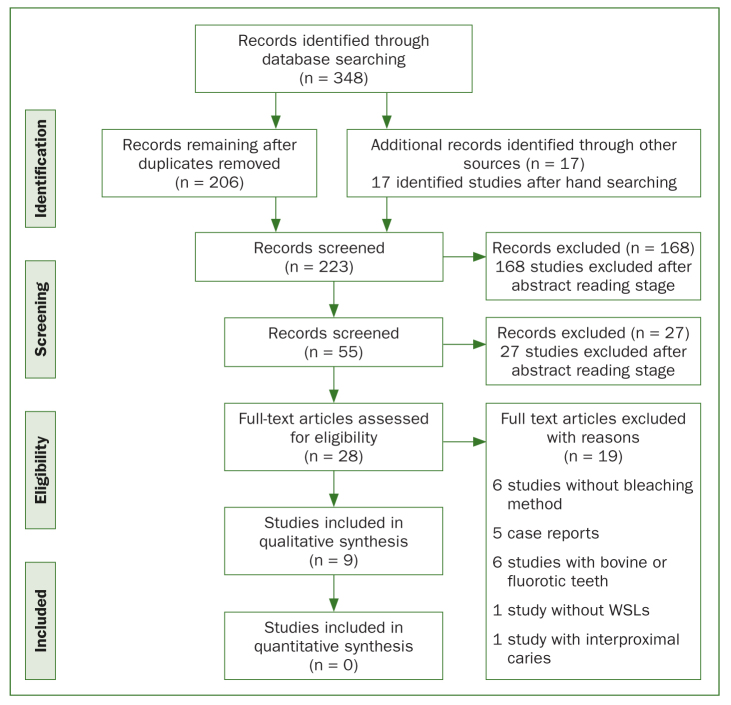
In total, 348 studies were retrieved from the electronic searches. After excluding all duplicates, abstracts, and full texts not meeting the inclusion criteria, 28 studies were found. Of these, 19 were excluded after full-text reading. Finally, 9 studies were considered eligible for inclusion in this review: one RCT24 and 8 laboratory studies.2,3,8,10,16, 23,35,38 A flow-chart of this process is presented in Fig 1. The data extracted from the included studies are shown in Tables 1a and 1b.
Fig 1.
Study flow diagram.
Table 1a.
Data extracted from included studies and study characteristics
| First author, year | Study design | Country | Primary aim | Inclusion criteria | Type of lesion | Intervention groups (HP: hydrogen peroxide; CP: carbamide peroxide) |
Participants (teeth) in intervention group | Control group | Participants (teeth) in control group |
|---|---|---|---|---|---|---|---|---|---|
| Ashnagar, 2017 | In vitro | Iran | To evaluate whether or not conventional or laser activated bleaching predispose to caries | Erupted human third molars stored in thymol solution | Artificial, pH cycling | Conventional bleaching with 40% HP for 20 minutes each time. Diode laser-assisted bleaching. Nd:YAG laser-assisted bleaching. Bleaching was followed by pH cycling |
15 molars per group | No treatment | 15 molars |
| Kim, 2016 | In vitro | South Korea | To evaluate the effect of bleaching on teeth with white spot lesions | Human maxillary premolars, sectioned into four parts | Artificial, pH cycling | Bleaching with 10% CP. Caries formation + bleaching. Caries formation + bleaching + remineralisation. Caries formation + remineralisation. |
10 enamel sections in group 1; 20 enamel sections in groups 2-4 | Only caries formation | 10 enamel sections |
| Bollineni, 2014 | In vitro | India | To evaluate the effect of adding fluoride to self-applied carbamide peroxide whitening gel on remineralisation of initial caries-like lesions | Freshly extracted third molars without defects, stored in PBS buffer | Artificial, acidified gel | Group A: demineralised + 10% CP bleaching (n=24). Group B: demineralised + 0.463% NaF + 10% CP bleaching (n=24). Group C: demineralisation only (n=24). |
24 molars, sectioned in quadrants, one quadrant per group | Group D: no treatment | N = 24 molar quadrants |
| Cavalli, 2011 | In vitro | Brazil | To evaluate the effect of adding fluoride and calcium to home-bleaching products on enamel mineral loss | Sound, extracted human third molars, stored in deionised water with thymol | Artificial | Commercial 10% CP gel, pH 6.8. Experimental 10% CP gel with 0.2% F, pH 6.8. Experimental 10% CP gel with 0.2% Ca, pH 6.5. Commercial 10% CP gel with 0.11% fluoride, pH 7.3. Commercial 10% CP gel with 0.11% fluoride, pH 6.4. |
10 enamel slabs per group | Placebo gel | 10 enamel slabs |
| Pinto, 2009 | In vitro | Brazil | To study the effect of bleaching on hardness and morphology of sound enamel and on enamel with early caries lesions | Erupted human third molars, stored in saturated thymol solution | Artificial lesions, pH-cycling (group 3-6), stored in artificial saliva (group 1,3,4) | Sound enamel bleached with CP. Sound enamel, bleached with CP and pH cycled. Carious enamel bleached with CP. Carious enamel and pH cycled. Carious enamel treated with placebo gel and pH cycled. Carious enamel, bleached with CP and pH cycled. |
10 enamel blocks per group | Group 5 | 10 enamel blocks |
| Knösel, 2007 | RCT | Germany | To evaluate the effect of external bleaching on the colour of post-orthodontic white spot lesions | Maxillary incisors and canines | Inactive WSL | Office bleaching 60 min, 14 days break, home bleaching 1 h/day for 14 days. | 10 participants | No bleaching | 9 participants |
| Alves, 2007 | In vitro | Brazil | To assess the influence of bleaching on the susceptibility of developing caries-like lesions | Human third molars | Artificial, pH cycling after bleaching | G1: home bleaching with 10% CP for 4 weeks. G2: home bleaching with 16% CP for 4 weeks. G3: in-office bleaching with 37% CP activated by a halogen light curing unit. In-office bleaching with 35% HP activated by light-emitting diode and laser energy. |
30 | G5: pH cycling, without bleaching. G6: no bleaching, no bleaching pH cycling. |
10 |
| Gladwell, 2006 | In vitro | USA | To evaluate whether a whitening system with fluoride could remineralise previously demineralised enamel | Extracted third molars, sectioned into quadrants | Artificial, with acid gel | Group A: demineralised + 10% CP gel. Group B: demineralised + 10% CP gel with fluoride. Group D: demineralised, no bleaching. |
24 enamel blocks per group | Group C: untreated, not-demineralised | 24 enamel blocks |
| Pretty, 2005 | In vitro | England | To evaluate if bleached enamel had an increased risk of either acid erosion or early caries | Human incisors | Artificial, demineralising solution | 10% CP gel. 16% CP gel. 22% CP gel. 10% CP gel with xylitol, fluoride and potassium. |
6 teeth per group | Half of each subsample was not bleached | 6 |
Table 1b.
Data extracted from included studies and study results
| First author, year | Main outcome assessment | Follow-up | Drop-outs/attrition | Main results | Author’s main conclusions |
|---|---|---|---|---|---|
| Ashnagar, 2017 | 1. Knoop microhardness 2. DIAGNOdent (DD) |
Not stated | None | All groups had a significant reduction in microhardness values but no significant differences between the groups. DD values were significantly reduced in the conventional and diode laser groups. |
Bleaching with conventional or laser-activated technologies does not make teeth vulnerable to caries development. |
| Kim, 2016 | 1. Colour (spectro-radiometer) 2. Mineral content (EPMA) 3. Knoop microhardness |
Baseline and after 14 days of bleaching | None | Bleaching of carious enamel extended whiteness without additional mineral loss. Treatment with CPP-ACP paste increased calcium, phosphate, and fluoride content in the lesion area and correlated well with microhardness. |
Bleaching reduced colour disparities between sound and carious enamel without deteriorating the chemical and mechanical properties. The remineralising agent enhanced deposition of mineral in subsurface lesions. |
| Bollineni, 2014 | 1. Colour (Vita 3D Master Shade guide) 2. Lesion depth (polarised light microscopy) |
Up to 21 days after bleaching | None | Significantly deeper lesions in groups A and C compared with Group B. Whitening data not presented. |
Fluoride added to bleaching gel showed a remineralising effect on demineralised enamel (white spot lesions). |
| Cavalli, 2011 | 1. Mineral content (FT-Raman spectroscopy) 2. Knoop microhardness 3. Lesion depth (Polarised light microscopy) |
After bleaching and after 14 days | None | Carbamide peroxide treatment decreased mineral content (subsurface mineral loss) and increased lesion depth. Inorganic deficit could be controlled by adding fluoride and calcium to the bleaching agent. |
Addition of F and Ca to home-applied bleaching agents may reduce enamel mineral loss. |
| Pinto, 2009 | 1. Knoop surface microhardness 2. Morphology (SEM) |
Not stated | None | Baseline mean microhardness values were similar in all groups. Groups exposed to enamel demineralisation (3-6) did not differ from each other and bleaching treatment reduced microhardness compared to the baseline. Changes in enamel morphology after treatments were observed for all groups. |
CP bleaching promoted mineral loss of sound enamel but did not exacerbate mineral loss of the carious enamel. |
| Knösel, 2007 | 1. Colour determination (colourimeter) 2. Patient satisfaction (questionnaire) |
28 days | None | Lightness of both sound enamel and WSLs was significantly higher after treatment. All patients in the bleaching group were satisfied with the outcome. |
External bleaching can satisfactorily camouflage post-orthodontic WSLs. |
| Alves, 2007 | 1. Visual scoring of lesions (score 0-3) by three independent examiners | Not stated | None | G1 and G2: median score 1. G3, G3 and G5: median score 2. G6: median score 0. |
Home bleaching reduced susceptibility to caries. In-office bleaching did not influence the development of caries. |
| Gladwell, 2006 | 1. Colour shade (Vita shade guide) 2. Histology (light microscopy) |
After 21 treatments | None | Bleaching gels with and without fluoride had similar whitening effect. Differences in lesion depths were significantly reduced in group B. |
Addition of fluoride to commercially available whitening gels enhance remineralisation without altering the whitening properties. |
| Pretty, 2005 | 1. Colour (Vita shade guide, Shade-Eye colourimeter) 2. QLF (erosion) 3. Transverse micro-radiography |
Not stated | None | No significant differences in whitening effects between the gels. No significant differences in mineral loss between the groups. Demineralisation increased with time with a linear relationship to bleaching time of CP exposure. |
Bleaching with carbamide peroxide gel did not increase the susceptibility of enamel to acid erosion or caries. The addition of xylitol, fluoride and potassium did not have an adverse effect on bleaching efficacy when compared to standard CP gels. |
Quality Assessment
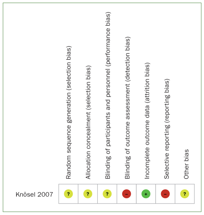
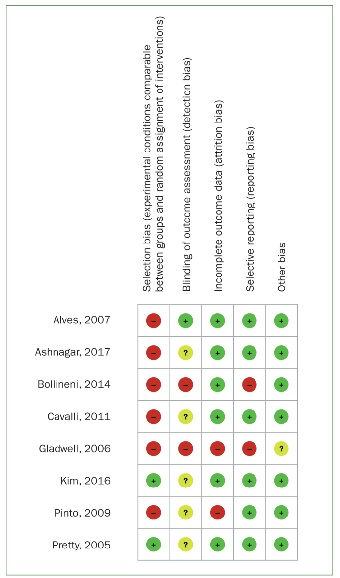
Quality assessment of the included studies is shown in Figs 2 and 3 and Tables 2a and 2b. The RCT24 was judged to be at overall high risk of bias. Six of the in vitro studies were classified as having a high risk of bias2,3,8,10,16,35 and two as having a moderate risk.23,38 Common reasons for downgrading were lack of randomisation procedures, blinding, and attrition bias.
Fig 2.

Risk of bias summary for the in vivo study.
Fig 3.

Risk of bias summary for the in vitro studies.
Table 2a.
Quality assessment of in vitro studies
| First author, year | Experimental conditions comparable between groups and random assignment of interventions (selection bias) | Blinding of assessors performing the tests (performance bias) | Losses or non-inclusion of specimens (attrition bias) | Selective reporting of results (reporting bias) | Other bias | Overall risk |
|---|---|---|---|---|---|---|
| Ashnagar, 2017 | High | Unclear | Low | Low | Low | High |
| Kim, 2016 | Low | Unclear | Low | Low | Low | Unclear |
| Bollineni, 2014 | High | High | Low | High | Low | High |
| Cavalli, 2011 | High | Unclear | Low | Low | Low | High |
| Pinto, 2009 | High | Unclear | High | Low | Low | High |
| Alves, 2007 | High | Low | Low | Low | Low | High |
| Gladwell, 2006 | High | High | High | High | Unclear | High |
| Pretty, 2005 | Low | Unclear | Low | Low | Low | Unclear |
Table 2b.
Risk of bias assessement of the in vivo study
| First author, year | Sequence generation (selection bias) | Allocation concealment (selection bias) | Blinding of participants and personnel (performance bias) | Blinding of outcome assessors (detection bias) | Incomplete outcome data (attrition bias) | Selective reporting (reporting bias) | Other sources of bias | Overall risk |
|---|---|---|---|---|---|---|---|---|
| Knösel, 2007 | Unclear | Unclear | High | High | Low | High | Unclear | High |
Descriptive Results
Clinical trial
The only clinical trial that met the inclusion criteria evaluated the effect of combined home and in-office bleaching on colour changes in 19 participants with visible inactive WSL after orthodontic treatment.24 Colour changes were registered by a colorimeter and patient satisfaction was evaluated using a questionnaire. The results, although this trial was graded as having a high risk of bias, showed that the therapeutic scheme could camouflage the WSL and that the patients were pleased with the aesthetic outcome of bleaching.
Laboratory studies
The remaining studies were performed in vitro and several concentrations, products and techniques for bleaching were applied as detailed in Table 1a. Among the outcome measures, visual examination, enamel microhardness and degree of demineralisation were the most common. The effect on colour change was addressed in three studies,8,16,23 two with high and one with moderate risk of bias, whose results indicated that bleaching reduced colour disparities between sound and carious enamel and that addition of remineralising agents did not affect the whitening properties. Studies on enamel microhardness and histology showed that the mineral loss caused by bleaching could be reduced by the presence of fluorides,8,16 calcium10 and casein phosphopeptide–amorphous calcium phosphate (CPP-ACP).23 Five studies addressed the question of whether bleaching can make teeth more vulnerable for caries development.2,3,10,35,38 Four studies at high risk of bias and one study at moderate risk reached the overall conclusion that neither home- nor in-office bleaching of permanent teeth was associated with an increased susceptibility to caries/enamel demineralisation or erosion. In two studies having high risk of bias,2,3 laser-activated bleaching did not seem to affect enamel microhardness or caries risk when compared to conventional bleaching with hydrogen or carbamide peroxide.
Qualitative synthesis of the included studies
There was considerable methodological heterogeneity across the studies with large differences in interventions, participants (type of teeth) and endpoints. Thus, a meta-analysis was not feasible for any combination of the included studies. Likewise, a funnel plot to disclose potential publication bias was not possible due to the limited number of included studies.
Discussion
Tooth bleaching has gained interest in recent decades as a key component of aesthetic dentistry. The current technologies are based on home-bleaching, either with products containing carbamide peroxide (CP) or hydrogen peroxide (HP) at concentrations between 3% and 10%, or in-office bleaching, with concentrations ranging from 15% to 38%. However, recent systematic reviews have not detected any major differences in whitening efficacy between the at-home and in-office strategies,11,12 although tray-delivered CP gels seem to perform slightly better than HP-based products.28 Tooth sensitivity and gingival pain are commonly reported side-effects of tooth bleaching procedures in adults.18,27 Thus, it is suggested that bleaching should be restricted to patients with good oral hygiene and be followed by fluoride application in order to enhance renewed mineral uptake.4
To the best of our knowledge, this is the first systematic review examining the effect of bleaching as a method for the management of post-orthodontic white spot lesions. Previous reviews on tooth bleaching implemented a more general approach,15,20,45 and focused on study design5 or adverse effects.43 Our findings were inconclusive and compromised by the limited number of studies that met the inclusion criteria, as well as the comparatively low quality of research. It is noteworthy that of the 9 papers evaluated, only one was an RCT. Furthermore, in vitro studies cannot always adequately reflect actual clinical conditions. Factors such as oral hygiene habits, diurnal alterations in saliva flow, and dietary habits are examples of parameters that are impossible to adequately simulate in the laboratory. Human saliva has been found to be less associated with enamel demineralisation than artificial saliva,5 which was supported by our observation that the studies using human saliva found post-bleaching measurements of enamel microhardness similar to those at baseline. Thus, a waiting period with natural remineralisation from saliva and self-applied fluoride toothpaste over a period of at least 3 to 6 months after debonding is advocated before any additional treatment options for WSL should be considered.36 It should also be noted that the American Academy of Pediatric Dentistry (AAPD) guidelines37 discourage the use of full-arch cosmetic bleaching for patients in the mixed dentition.
One clinical and two laboratory studies addressed the primary research question. The results showed that bleaching of WSL can diminish the colour disparities between carious and non-affected areas, but the certainty of the evidence was very low. Moreover, low-quality in vitro data indicated that the presence of fluoride or any other remineralising agent did not impair the whitening effect. The second research question could not be answered in the present review, as only in vitro studies were available. A recent meta-analysis has, however, shown that no significant changes in enamel microhardness appeared when using a 10% carbamide peroxide bleaching gel over a 21-day period.45 Although the laboratory data assessed in our study reconfirmed the fact that bleaching did not increase the risk of further demineralisation or decrease enamel microhardness, no firm conclusions can be drawn from studies with a high risk of bias. Therefore, the main conclusion of the present systematic review is that the efficacy of bleaching as a method to manage post-orthodontic white spot lesions lies in a knowledge gap. Similar conclusions were also reached by Höchli et al,20 who investigated the therapeutic and adverse effects of different interventions to treat post-orthodontic white spot lesions. According to the authors, although fluoride varnish seemed to be effective, the need for further research was pointed out.
Consequently, the need for future randomised controlled trials involving post-debonding lesions using objective endpoints such as white spot scores, light fluorescence, or impedance spectroscopy, is emphasised. Patients’ subjective perceptions should be also investigated via questionnaires, since the literature has shown that bleaching treatment produces positive changes in young participants’ oral health quality of life in terms of smiling, laughing, and showing teeth without embarrassment.25
The effects of adding fluoride and calcium to the bleaching agents were also inconclusive. The combination of carbamide peroxidase and remineralising products resulted in reduced mineral loss when applied to sound enamel,8,10,16 but this was not verified when artificial caries lesions were treated.35 The problem of extrapolating laboratory findings to clinical settings showed that adverse effects of carbamide peroxide on enamel evident in specimens bleached in vitro were not seen in situ, and that the presence of saliva could prevent the demineralising effect of bleaching gel in situ.22 Moreover, the inherently limited ability of in vitro studies to evaluate long-term results is beyond doubt; in turn, this makes the exploration of the time factor unfeasible. The rationale behind using lasers to assist bleaching is that a heat/light source may enhance the peroxide action and maximise the benefit of bleaching without requiring several lengthy sessions for the patient. With the limited information available, we found no support for adding lasers to in-office bleaching. Two systematic reviews have recently confirmed no significant differences in tooth colour change or tooth sensitivity when in-office bleaching gels with and without light were compared.29,42
As in most systematic reviews, the considerable variations in study protocols (application time, number of bleaching sessions, product concentration) and reported endpoints indicated a high degree of clinical and methodological heterogeneity, preventing a meta-analysis and making conclusions difficult. Thus, for future studies, employing standardised methodology to evaluate bleaching products would be beneficial. Nevertheless, the most striking challenge was incomplete reporting and the high risk of bias in the existing literature. In particular, a limited number of materials and undefined origin of teeth in combination with selection and performance bias frequently diminished the quality of the included studies. As shown in Tables 1a and 1b, incomplete reporting of data was also a frequent problem.
A limitation of this review is the inclusion of in vitro studies and only one clinical study. This rendered robust clinical conclusions impossible. The decision for including in vitro studies was also based on presenting all published investigations to clinicians and future researchers, irrespective of study design.
Conclusion
The findings from the present systematic review could neither support nor refute bleaching as an effective method for the management of post-orthodontic white spot lesions. The need for further prospective in vivo studies is support by the fact that most of the studies in this field are in vitro and that there is little solid scientific evidence of low risk of bias.
References
- Ahn SJ, Lee SJ, Lim BS, Nahm DS. Quantitative determination of adhesion patterns of cariogenic streptococci to various orthodontic brackets. Am J Orthod Dentofac Orthop. 2007;132:815–821. doi: 10.1016/j.ajodo.2005.09.034. [DOI] [PubMed] [Google Scholar]
- Alves EA, Alves FK, Campos Ede J, Mathias P. Susceptibility to carieslike lesions after dental bleaching with different techniques. Quintessence Int. 2007;38:e404–409. [PubMed] [Google Scholar]
- Ashnagar S, Monzavi A, Abbasi M, Aghajani M, Chiniforush N. Evaluation of the effect of different laser activated bleaching methods on enamel susceptibility to caries; an in vitro mode. J Lasers Med Sci. 2017;8(suppl 1):S62–S67. doi: 10.15171/jlms.2017.s12. [DOI] [PMC free article] [PubMed] [Google Scholar]
- Attin T, Kielbassa AM, Schwanenberg M, Hellwig E. Effect of fluoride treatment on remineralization of bleached enamel. J Oral Rehabil. 1997;24:282–286. doi: 10.1046/j.1365-2842.1997.d01-291.x. [DOI] [PubMed] [Google Scholar]
- Attin T, Schmidlin PR, Wegehaupt F, Wiegand A. Influence of study design on the impact of bleaching agents on dental enamel microhardness: a review. Dent Mater. 2009;25:143–157. doi: 10.1016/j.dental.2008.05.010. [DOI] [PubMed] [Google Scholar]
- Basting RT, Rodrigues AL, Serra MC. Micromorphology and surface roughness of sound and desmineralized enamel and dentin bleaching with a 10% carbamine peroxide bleaching agent. Am J Dent. 2007;20(2):97–102. [PubMed] [Google Scholar]
- Boersma JG, van der Veen MH, Lagerweij MD, Bokhout B, Prahl-Andersen B. Caries prevalence measured with QLF after treatment with fixed orthodontic appliances: influencing factors. Caries Res. 2005;39:41–47. doi: 10.1159/000081655. [DOI] [PubMed] [Google Scholar]
- Bollineni S, Janga RK, Venugopal L, Reddy IR, Babu PR, Kumar SS. Role of fluoridated carbamide peroxide whitening gel in the remineralization of demineralized enamel: An in vitro study. J Int Soc Prev and Comm Dent. 2014;2:117–121. doi: 10.4103/2231-0762.137638. [DOI] [PMC free article] [PubMed] [Google Scholar]
- Bussadori SK, do Rego MA, da Silva PE, Pinto MM, Pinto AC. Esthetic alternative for fluorosis blemishes with the usage of a dual bleaching system based on hydrogen peroxide at 35% J Clin Pediatr Dent. 2004;28:143–146. doi: 10.17796/jcpd.28.2.g45173lp7661g078. [DOI] [PubMed] [Google Scholar]
- Cavalli V, Rodrigues LK, Paes-Leme AF, Soares LE, Martin AA, Berger SB, Giannini M. Effects of the addition of fluoride and calcium to low-concentrated carbamide peroxide agents on the enamel surface and subsurface. Photomed Laser Surg. 2011;29:319–325. doi: 10.1089/pho.2010.2797. [DOI] [PubMed] [Google Scholar]
- de Geus JL, Wambier LM, Boing TF, Loguercio AD, Reis A. At-home bleaching with 10% vs more concentrated carbamide peroxide gels: a systematic review and meta-analysis. Oper Dent. 2018;43(4):210–222. doi: 10.2341/17-222-L. [DOI] [PubMed] [Google Scholar]
- de Geus JL, Wambier LM, Kossatz S, Loguercio AD, Reis A. At-home vs in-office bleaching: a systematic review and meta-analysis. Oper Dent. 2016;41(4):341–356. doi: 10.2341/15-287-LIT. [DOI] [PubMed] [Google Scholar]
- Egger M, Davey Smith G, Schneider M, Minder C. Bias in meta-analysis detected by a simple, graphical test. BMJ. 1997;315(7109):629–634. doi: 10.1136/bmj.315.7109.629. [DOI] [PMC free article] [PubMed] [Google Scholar]
- Ernst CP, Briseño M, Zönnchen BM. Effects of hydrogen peroxide-containing bleaching agents on the morphology of human enamel. Quintessence Int. 1996;27(1):53–56. [PubMed] [Google Scholar]
- Feliz-Matos L, Hernández L.M, Abreu N. Dental bleaching techniques; hydrogen-carbamide peroxides and light sources for activation, an update. Mini review article. Open Dent J. 2014;8:264–268. doi: 10.2174/1874210601408010264. [DOI] [PMC free article] [PubMed] [Google Scholar]
- Gladwell J, Simmons D, Wright JT. Remineralization potential of a fluoridated carbamide peroxide whitening gel. J Esthet Restor Dent. 2006;18:206–212. doi: 10.1111/j.1708-8240.2006.00021_1.x. discussion 212–213. [DOI] [PubMed] [Google Scholar]
- Gorelick L, Geiger AM, Gwinnett AJ. Incidence of white spot formation after bonding and banding. Am J Orthod. 1982;81:93–98. doi: 10.1016/0002-9416(82)90032-x. [DOI] [PubMed] [Google Scholar]
- Hasson H, Ismail AI, Neiva G. Home-based chemically-induced whitening of teeth in adults. Cochrane Database Systematic Reviews. 2006;18(4):CD006202. doi: 10.1002/14651858.CD006202. [DOI] [PubMed] [Google Scholar]
- Higgins JPT, Altman DG, Sterne JAC. Assessing risk of bias in included studies. Higgins JPT, Green S, editors. Cochrane Handbook for Systematic Reviews of Interventions. Version 5.1.0. [Internet]. The Cochrane Collaboration [updated March 2011] Available at: http://www.cochrane-handbook.org . [Google Scholar]
- Höchli D, Hersberger-Zurfluh M, Papageorgiou SN, Eliades T. Interventions for orthodontically induced white spot lesions: a systematic review and meta-analysis. Eur J Orthod. 2017;39:122–133. doi: 10.1093/ejo/cjw065. [DOI] [PubMed] [Google Scholar]
- Huang GJ, Roloff-Chiang B, Mills BE, Shalchid S, Spiekermane C, Korpak AM, Starrett JL, Greenlee GM, Drangsholt RJ, Matunas JC. Effectiveness of MI Paste Plus and PreviDent fluoride varnish for treatment of white spot lesions: A randomized controlled trial. Am J Orthod Dentofacial Orthop. 2013;143:31–41. doi: 10.1016/j.ajodo.2012.09.007. [DOI] [PMC free article] [PubMed] [Google Scholar]
- Justino LM, Tames DR, Demarco FF. In situ and in vitro effects of bleaching with carbamide peroxide on human enamel. Oper Dent. 2004;29(2):219–225. [PubMed] [Google Scholar]
- Kim Y, Son HH, Yi K, Ahn JS, Chang J. Bleaching Effects on Color, Chemical, and Mechanical Properties of White Spot Lesion. Oper Dent. 2016;41:318–26. doi: 10.2341/15-015-L. [DOI] [PubMed] [Google Scholar]
- Knösel M, Attin R, Becker K, Attin T. External bleaching effect on the color and luminosity of inactive white-spot lesions after fixed orthodontic appliances. Angle Orthod. 2007;77:646–652. doi: 10.2319/060106-224. [DOI] [PubMed] [Google Scholar]
- Kothari S, Gray A, Lyons K, Wen Tan X, Brunton PA. Vital bleaching and oral-health-related quality of life in adults: a systematic review and meta-analysis. J Dent. 2019;84:22–29. doi: 10.1016/j.jdent.2019.03.007. [DOI] [PubMed] [Google Scholar]
- Lee Linton J. Quantitative measurements of remineralization of incipient caries. Am J Orthod Dentofacial Orthop. 1996;104:590–597. doi: 10.1016/s0889-5406(96)80034-5. [DOI] [PubMed] [Google Scholar]
- Leonard RH., Jr Efficacy, longevity, side effects, and patient perceptions of night guard vital bleaching. Compend Contin Educ Dent. 1998;19:766–770, 772, 774. [PubMed] [Google Scholar]
- Luque-Martinez I, Reis A, Schroeder M, Muñoz MA, Loguercio AD, Masterson D, Maia LC. Comparison of efficacy of tray-delivered carbamide and hydrogen peroxide for at-home bleaching: a systematic review and meta-analysis. Clin Oral Investig. 2016;20:1419–1433. doi: 10.1007/s00784-016-1863-7. [DOI] [PubMed] [Google Scholar]
- Maran BM, Burey A, de Paris Matos T, Loguercio AD, Reis A. In-office dental bleaching with light vs. without light: A systematic review and meta-analysis. J Dent. 2018;70:1–13. doi: 10.1016/j.jdent.2017.11.007. [DOI] [PubMed] [Google Scholar]
- Melrose CA, Appleton J, Lovius BB. A scanning electron microscopic study of early enamel caries formed in vivo beneath orthodontic bands. Br J Orthod. 1996;23:43–47. doi: 10.1179/bjo.23.1.43. [DOI] [PubMed] [Google Scholar]
- Moher D, Liberati A, Tetzlaff J, Altman DG The PRISMA Group. Preferred Reporting Items for Systematic Reviews and Meta-Analyses: The PRISMA Statement. PLoS Med. 2009;6(7):e1000097. doi: 10.1371/journal.pmed.1000097. doi:10.1371/journal.pmed1000097. [DOI] [PMC free article] [PubMed] [Google Scholar]
- Murphy TC, Willmot DR, Rodd HD. Management of postorthodontic demineralized white lesions with microabrasion: a quantitative assessment. Am J Orthod Dentofacial Orthop. 2007;131:27–33. doi: 10.1016/j.ajodo.2005.04.041. [DOI] [PubMed] [Google Scholar]
- Ogaard B, Rolla G, Arends J. Orthodontic appliances and enamel demineralization. Part 1. Lesion development. Am J Orthod Dentofacial Orthop. 1988;94(1):68–73. doi: 10.1016/0889-5406(88)90453-2. [DOI] [PubMed] [Google Scholar]
- Ogaard B, Rolla G, Arends J, ten Cate JM. Orthodontic appliances and enamel demineralization. Part 2: prevention and treatment of lesions. Am J Orthod Dentofacial Orthop. 1988;94(2):123–128. doi: 10.1016/0889-5406(88)90360-5. [DOI] [PubMed] [Google Scholar]
- Pinto CF, Paes Leme AF, Cavalli V, Giannini M. Effect of 10% carbamide peroxide bleaching on sound and artificial enamel carious lesions. Braz Dent J. 2009;20:48–53. doi: 10.1590/s0103-64402009000100008. [DOI] [PubMed] [Google Scholar]
- Pitts N, Duckworth RM, Marsh P, Mutti B, Parnell C, Zero D. Post-brushing rinsing for the control of dental caries: exploration of the available evidence to establish what advice we should give our patients. Br Dent J. 2012;212:315–320. doi: 10.1038/sj.bdj.2012.260. [DOI] [PubMed] [Google Scholar]
- Policy on the Use of Dental Bleaching for Child. AAPD. 2018;39(6):90–92. [PubMed] [Google Scholar]
- Pretty IA, Edgar WM, Higham SM. The effect of bleaching on enamel susceptibility to acid erosion and demineralisation. Br Dent J. 2005;198:285–290. doi: 10.1038/sj.bdj.4812126. discussion 280. [DOI] [PubMed] [Google Scholar]
- Shungin D, Olsson AI, Persson M. Orthodontic treatment related white spot lesions: a 14-year prospective quantitative follow-up, including bonding material assessment. Am J Orthod Dentofacial Orthop. 2010;138(136):e1–8. doi: 10.1016/j.ajodo.2009.05.020. [DOI] [PubMed] [Google Scholar]
- Sonesson M, Bergstrand F, Gizani S, Twetman S. Management of post-orthodontic white spot lesions: an updated systematic review. Eur J Orthod. 2017;39:116–121. doi: 10.1093/ejo/cjw023. [DOI] [PubMed] [Google Scholar]
- Sonesson M, Twetman S, Bondemark L. Effectiveness of high-fluoride toothpaste on enamel demineralization during orthodontic treatment-a multicenter randomized controlled trial. Eur J Orthod. 2014;36:678–682. doi: 10.1093/ejo/cjt096. [DOI] [PubMed] [Google Scholar]
- SoutoMaior JR, de Moraes S, Lemos C, Vasconcelos BDE, Montes M, Pellizzer EP. Effectiveness of light sources on in-office dental bleaching: a systematic review and meta-analyses. Oper Dent. 2018;44:E105–E117. doi: 10.2341/17-280-L. [DOI] [PubMed] [Google Scholar]
- Tredwin CJ, Naik S, Lewis NJ, Scully C. Hydrogen peroxide tooth-whitening (bleaching) products: Review of adverse effects and safety issues. Br Dent J. 2006;200:371–376. doi: 10.1038/sj.bdj.4813423. [DOI] [PubMed] [Google Scholar]
- Zachrisson BU. Oral hygiene for orthodontic patients: current concepts and practical advice. Am J Orthod. 1974;66:487–497. doi: 10.1016/0002-9416(74)90110-9. [DOI] [PubMed] [Google Scholar]
- Zanolla J, Marques A, da Costa DC, de Souza AS, Coutinho M. Influence of tooth bleaching on dental enamel microhardness: a systematic review and meta-analysis. Austr Dent J. 2017;62:276–282. doi: 10.1111/adj.12494. [DOI] [PubMed] [Google Scholar]


