Summary:
Elbow flexion is essential for the functional use of the hand. The reconstructive procedure may also change depending on the location of the sarcoma. The nonresected muscle may alter the function of the elbow. If the proximal part of the triceps muscle remains intact, it is considered functional. Functional muscle transfer is not required in such cases. A 50-year-old patient presented with a soft-tissue defect after a wide resection of a leiomyosarcoma affecting the left elbow. The wide resection resulted in the resection of the distal third of the triceps brachii, exposing the olecranon. We performed reconstruction using a pedicled latissimus dorsi musculocutaneous flap. The flap was transferred to the elbow through a subcutaneous tunnel. Ten months after surgery, the elbow function improved. In our patient, the thoracodorsal nerve was cut to prevent mixed nerve signals. We found that patients with distal muscle defects do not require functional muscle grafting. Pedicled latissimus dorsi musculocutaneous flap transfer is a straightforward and useful procedure for reconstructing the upper arm region.
Wide muscle resection in cases of sarcomas may lead to limb dysfunction or amputation. To spare the patient’s limb function, tissue transfer is required.1,2 Muscle transfer is particularly effective for muscle reconstruction.1 After the wide resection of a leiomyosarcoma, we have observed a 1-stage reconstruction of the distal third of the triceps brachii muscle and its overlying tissue. To preserve the upper limb and elbow extension functions, we used a pedicled latissimus dorsi (LD) musculocutaneous (MC) flap, in which the muscle was noninnervated. With this flap, we performed a 1-stage reconstruction of a large defect in the triceps brachii, muscle, and overlying tissue. In our case, the proximal stump of the triceps brachii muscle was innervated. The reconstructive procedure may change depending on the defect’s location. There are no guidelines describing the relationship between the defect location and the need for functional muscle transfer. In this case report, we describe the details of our reconstruction and postoperative results.
CASE REPORT
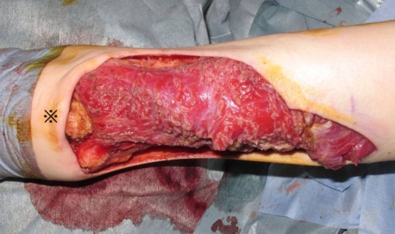
A 50-year-old man with a soft-tissue tumor in the elbow region was referred to the orthopedic surgery department of our hospital. The T2-weighted magnetic resonance imaging revealed a large solid mass. On T1-weighted imaging, the mass was isointense, nonhomogeneous, and of high intensity compared with the muscle visualized on T2-weighted imaging. A needle biopsy was performed, and the tumor was diagnosed as leiomyosarcoma. No metastases were found (T2N0M0 G2-3 Stage IIIA). A general surgeon performed a wide resection of the tumor under general anesthesia. The surgical margin was 5 cm. After tumor resection, the remaining soft tissue and skin defects measured 12 × 8 cm and 15 × 15 cm, respectively. In addition, the distal third of the triceps brachii muscle was resected, and the olecranon was exposed (Fig. 1).
Fig. 1.
Postoperative view of the elbow after tumor resection at the distal third of the triceps brachii.
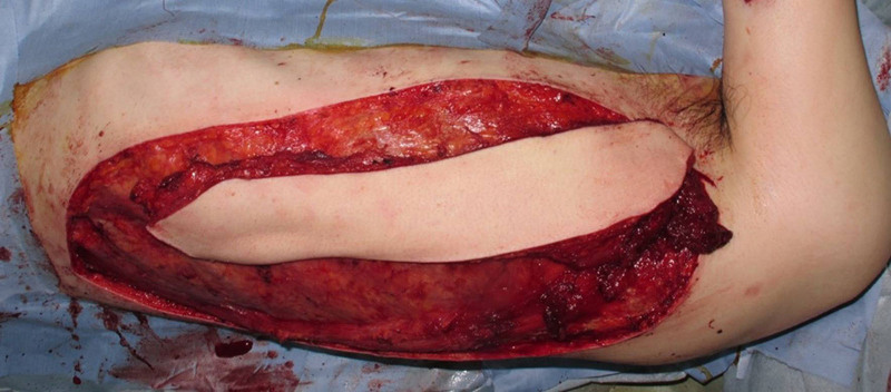
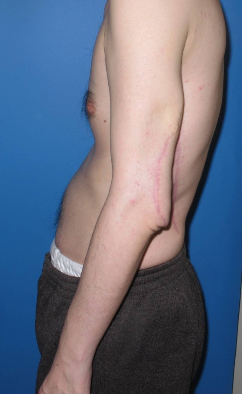
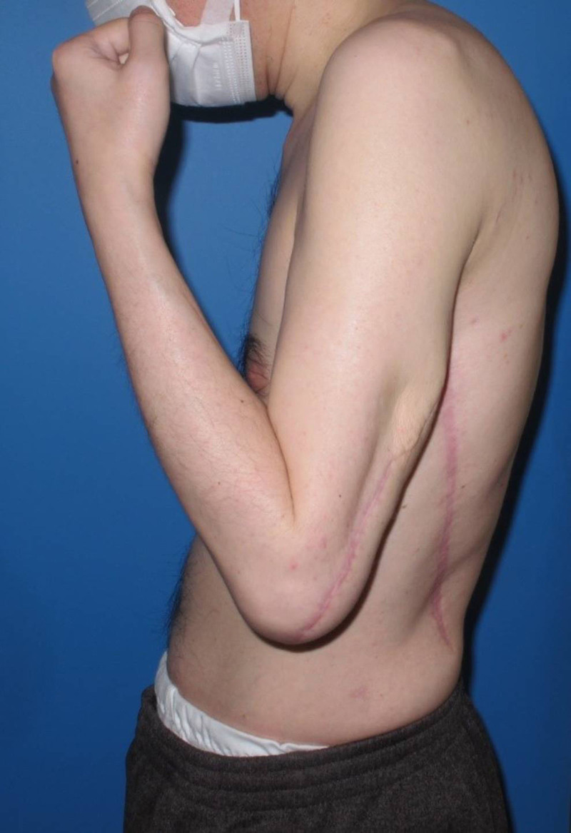
The LDMC flap was harvested from the anterior border of the muscle on the ipsilateral side. The sizes of the skin paddle and muscle belly were 25 × 7 cm and 20 × 8 cm, respectively (Fig. 2). To increase the flap’s rotation arc, the serratus branch of the thoracodorsal artery was ligated. In addition, the thoracodorsal nerve was resected. The flap was then transferred to the upper arm through a subcutaneous tunnel. The distal end of the LD muscle was fixed to the stump of the periosteum around the olecranon using a 3-0 nylon suture (Bear Medic, Tokyo, Japan), such that the olecranon was completely covered with the LD muscle. The proximal stump of the LD muscle was fixed to the proximal stump of the triceps brachii muscle using 3-0 polydioxanone (Ethicon, Baltimore, MD). After directly closing the flap donor site, a suction drain (19-Fr BLAKE, J-VAC; Ethicon, Baltimore, MD) was placed to manage the dead space at the donor site. The postoperative course was uneventful, and the patient was permitted to move his shoulder and wrist. However, the affected elbow was restrained for 3 weeks after surgery, which was followed by passive rehabilitation. Active rehabilitation began 5 weeks postoperatively. Eight weeks postoperatively, the patient regained the freedom to use his arm. Ten months after surgery, the patient could perform elbow extension and flexion (Figs. 3, 4), thus regaining the ability to use his elbow without interference in his daily life.
Fig. 2.
The harvested flap from the anterior border of the latissimus dorsi muscle.
Fig. 3.
Active extension of the elbow, demonstrating the full functional capacity of the triceps brachii muscle.
Fig. 4.
Elbow joint performing an active flexion movement.
DISCUSSION
Sarcomas are very rare malignant tumors,1–3 and only 6% of sarcomas are in the upper extremity. Wide excision of a sarcoma in the upper arm can improve patient prognosis.1 Subsequent reconstruction of the upper extremity should also consider both cosmetic and functional outcomes. If patients do not have a soft-tissue defect, elbow function may be restored by tendon transfer.4,5 Patients undergoing tumor resection with muscle and overlying soft tissue should consider musculocutaneous flap transfer. Similarly, the successful reconstruction of the upper arm from a congenital deformity or trauma using a pedicled LDMC flap has been reported.6 There have also been reports of elbow flexion restoration using the pedicled LDMC flap transfer after sarcoma resection.7–10 In the present case, we harvested the LD muscle from the muscle’s anterior border. A skin paddle was also designed along the muscle’s anterior border. The use of this flap, which maintained the direction of the muscle fibers, facilitated easy reconstruction of the defect. In our case, the patient’s distal third of the upper arm and elbow were affected. Some authors have reported the successful restoration of elbow flexion using functional muscle flap transfers.6–10 However, few reports have described the treatment of the thoracodorsal nerve. If the muscle defect involves the proximal side of the muscle or a motor nerve, tendon transfer or functional muscle transfer is necessary. However, functional muscle transfer with the motor nerve is not always required if the defect is at the distal end of the flap. This is because damage to the thoracodorsal nerve can disrupt the function of the triceps brachii muscle, which otherwise remains intact after tumor resection. In our case, the LD muscle was transferred between the proximal stump of the triceps brachii and the elbow, effectively bypassing the thoracodorsal nerve. Future studies in this area are necessary, as no report has been published on the association between the magnitude of the muscle defect and the need for muscle with motor nerve transfer.
CONCLUSIONS
After sarcoma resection, reconstruction of the upper arm defect is critical for hand function. In our case, the proximal stump of the triceps brachii muscle was functional. Nonfunctional muscle transfer prevented the disruption of triceps brachii muscle functions. A pedicled, noninnervated muscle flap is a useful surgical option for reconstructing complex muscle and soft-tissue defects on the distal side of the upper arm.
DISCLOSURE
The authors have no financial interest to declare in relation to the content of this article.
Footnotes
Published online 18 December 2024.
Disclosure statements are at the end of this article, following the correspondence information.
REFERENCES
- 1.Azurin ES, Yamamoto N, Hayashi K, et al. Clinical outcomes of soft tissue sarcoma around the elbow joint: a retrospective single institution study. Sarcoma. 2022;2022:1087726. [DOI] [PMC free article] [PubMed] [Google Scholar]
- 2.Popov P, Tukiainen E, Asko-Seljavaara S, et al. Soft-tissue sarcomas of the upper extremity: surgical treatment and outcome. Plast Reconstr Surg. 2004;113:222–230; discussion 231. [DOI] [PubMed] [Google Scholar]
- 3.Duran-Moreno J, Kontogeorgakos V, Koumarianou A. Soft tissue sarcomas of the upper extremities: maximizing treatment opportunities and outcomes. Oncol Lett. 2019;18:2179–2191. [DOI] [PMC free article] [PubMed] [Google Scholar]
- 4.Cambon-Binder A, Chammas M, Coulet B, et al. Tendon transfers to restore elbow flexion. Hand Surg Rehabil. 2022;41S:S76–S82. [DOI] [PubMed] [Google Scholar]
- 5.Weber MB, Wu KY, Spinner RJ, et al. Triceps-to-biceps tendon transfer for restoration of elbow flexion in brachial plexus injury [published online ahead of print August 2, 2023]. J Hand Surg Am. [DOI] [PubMed] [Google Scholar]
- 6.Kobayashi Y, Uchiyama Y, Yoshida S, et al. Functional reconstruction of elbow flexion with latissimus dorsi muscle rotational transfer: two case reports. J Med Case Rep. 2023;17:454. [DOI] [PMC free article] [PubMed] [Google Scholar]
- 7.Okamoto H, Kawaguchi Y, Miwa S, et al. Reverse lateral upper arm flaps for treating large soft tissue defects extending from the elbow to the forearm. Case Reports Plast Surg Hand Surg. 2022;9:145–150. [DOI] [PMC free article] [PubMed] [Google Scholar]
- 8.Zancolli E, Mitre H. Latissimus dorsi transfer to restore elbow flexion. An appraisal of eight cases. J Bone Joint Surg Am. 1973;55:1265–1275. [PubMed] [Google Scholar]
- 9.Arguello AM, Sullivan MH, Gavin L, et al. Pedicled functional latissimus flaps for reconstruction of the upper extremity following resection of soft-tissue sarcomas. Curr Oncol. 2023;30:3138–3148. [DOI] [PMC free article] [PubMed] [Google Scholar]
- 10.Ozaniak A, Hladik P, Lischke R. Successful restoration of elbow extension using the latissimus dorsi flap: case report. Plast Reconstr Surg Glob Open. 2022;10:e4121. [DOI] [PMC free article] [PubMed] [Google Scholar]


