Abstract
目的
探讨关节镜下自体髂骨植骨三滑轮四点防旋固定术治疗复发性肩关节前脱位的疗效。
方法
回顾分析2021年2月—2022年2月符合选择标准的14例复发性肩关节前脱位伴关节盂骨缺损患者临床资料,均采用关节镜下髂骨植骨三滑轮四点防旋固定术治疗。其中,男9例,女5例;年龄22~40岁,平均31.2岁。肩关节脱位4~10次,平均6.8次。初次脱位至该次就诊时间为1~6年,平均3.3年。肩关节不稳严重程度评分(ISIS)为(7.2±0.8)分,Beighton评分为(2.9±2.4)分,关节盂骨缺损宽度比例为26.64%±1.86%。手术前后采用肩关节疼痛视觉模拟评分(VAS)、美国加州大学洛杉矶分校(UCLA)评分、Constant 评分以及美国肩肘外科医师协会(ASES)评分评价肩关节疼痛及功能;肩关节CT评估移植骨块位置、愈合以及吸收重塑(关节盂面积)情况。
结果
14例患者手术均顺利完成,无严重并发症发生。术后患者均获随访,随访时间11.5~13.8个月,平均12.0个月。患者术后肩关节 VAS评分均较术前降低, UCLA评分、Constant 评分及ASES评分均提高,差异有统计学意义(P<0.05);且术后随时间延长,上述指标均进一步改善,各时间点间差异均有统计学意义(P<0.05)。影像学复查示,骨块完全填补关节盂缺损,位置良好且未发生明显移位,随时间延长骨块均愈合,并部分吸收、塑形。术后关节盂面积较术前明显增大,差异有统计学意义(P<0.05);随时间延长,关节盂面积逐渐减小,但术后各时间点间差异均无统计学意义(P>0.05)。
结论
关节镜下自体髂骨植骨固定术治疗复发性肩关节前脱位时,采用三滑轮四点防旋固定,能够有效防止骨块旋转,固定更牢靠且操作简便,可取得良好疗效。
Keywords: 复发性肩关节前脱位, 三滑轮, 四点防旋, 髂骨植骨固定术
Abstract
Objective
To explore effectiveness of arthroscopic Eden-Hybinette procedure with Triple-Pulley and four point anti-rotation fixation technique in the treatment of recurrent anterior dislocation of shoulder joint.
Methods
A clinical data of 14 patients with recurrent anterior dislocation of shoulder joint and glenoid bone defect, who were met the selective criteria and admitted between February 2021 and February 2022, was retrospectively analyzed. All patients were treated with arthroscopic Eden-Hybinette procedure and the bone blocks were fixed by using Triple-Pulley and four point anti-rotation fixation technique. There were 9 males and 5 females with an average age of 31.2 years (range, 22-40 years). Shoulder dislocation occurred 4-10 times (mean, 6.8 times). The time from the initial dislocation to the admission was 1-6 years (mean, 3.3 years). The Instability Severity Index Score (ISIS) was 7.2±0.8, the Beighton score was 2.9±2.4, and the width ratio of glenoid bone defect was 26.64%±1.86%. Pre- and post-operative visual analogue scale (VAS) score, the University of Califonia-Los Angeles (UCLA) shoulder score, Constant score, and American Association of Shoulder and Elbow Surgeons (ASES) score were used to evaluate shoulder pain and function. The position, healing, resorption, and remodeling (glenoid area) of the bone blocks were evaluated by CT of shoulder joint.
Results
All patients underwent surgery successfully without any serious complications. All patients were followed up 11.5-13.8 months (mean, 12.0 months). The VAS scores of shoulder joint after operation decreased compared to preoperative levels, while the UCLA score, Constant score, and ASES score all increased, with significant differences (P<0.05). And with the prolongation of time, the above indicators further improved, and the differences between different time points were significant (P<0.05). Imaging reexamination showed that the bone block completely filled the glenoid defect, with good position and no significant displacement. Over time, the bone block healed and partially absorbed and remodelled. The postoperative glenoid area increased significantly compared to preoperative area (P<0.05). With the prolongation of time, the glenoid area significantly decreased, but the difference was not significant between different time points (P>0.05).
Conclusion
For the recurrent anterior dislocation of shoulder joint, Triple-Pulley and four point anti-rotation fixation technique in Eden-Hybinette procedure can effectively prevent bone rotation, make fixation more reliable, and easy to operate and achieve good effectiveness.
Keywords: Recurrent anterior dislocation of shoulder joint, Triple-Pulley, four point anti-rotation, Eden-Hybinette procedure
肩关节脱位为临床常见疾病,好发于15~20岁人群,以关节前脱位为主[1]。肩关节脱位常伴随关节盂软组织损伤及骨缺损,若不进行正确处理,易转变成复发性脱位,从而导致关节盂骨缺损面积增加,进一步降低肩关节稳定性。对于复发性肩关节前脱位造成的关节盂骨缺损,目前临床常用治疗方式包括关节镜下Bankart修复术、喙突转位术(Bristow-Latarjet)以及骨移植固定术,对于骨缺损较大者主要选择后两种术式。但研究发现喙突转位术后移植骨平均吸收率达78%,可导致手术失效[2];术后并发症发生率高达15%~30%,包括螺钉松动、弯曲及断裂导致手术失效以及神经损伤等[3-5];并且相比于骨移植固定术,患者术后肩关节内旋受限更明显且功能评分更低[6-7]。骨移植固定术中移植骨块易塑形,可满足不同程度骨缺损修复需求,且术中因不需要截取喙突及联合腱,可有效避免腋神经及肩胛下肌损伤,极大降低了手术风险。因此,骨移植固定术越来越受临床重视。
自体髂骨骨移植固定术是最常用骨移植固定术式之一,临床疗效已获得广泛认可[8-9],术中常用螺钉或带袢钢板固定移植骨块。螺钉固定存在尾端撞击致软组织损伤、内植物折断或拔出、骨块骨折等问题,且相比于带袢钢板固定再手术率高[10]。带袢钢板固定虽然能避免上述问题,但抗拉力及抗旋转性能低于螺钉固定[11]。另外,螺钉及带袢钢板固定后移植骨块吸收率也达10.1%~47.6%[12-13]。针对上述情况,我们提出在关节镜下自体髂骨植骨固定术中,采用盂唇钉结合滑轮打结技术固定骨块,即“三滑轮四点防旋固定术”,于关节盂缺损上、下缘形成4个受力点(四点固定),使固定更牢靠,以解决移植骨块易旋转问题以及避免螺钉固定相关并发症。同时,该技术属于弹性固定,对移植骨块破坏少,也可避免骨块吸收。此外,相比于传统螺钉及袢钢板固定,该固定技术无需特殊骨块移植工具,手术步骤相对简便。2021年2月—2022年2月,我们采用关节镜下自体髂骨植骨三滑轮四点防旋固定术治疗14例复发性肩关节前脱位伴关节盂骨缺损患者,获得较好疗效。报告如下。
1. 临床资料
1.1. 一般资料
患者纳入标准:① 肩关节脱位达3次及以上;② 肩关节CT三维重建示关节盂骨缺损宽度比例达25%以上;③ 年龄18~40岁且有较高的运动需求。排除标准:① 合并其他损伤,如神经、血管损伤或肩袖撕裂等;② 合并其他全身性疾病无法耐受手术。
2021年2月—2022年2月,共14例患者符合选择标准纳入研究。其中,男9例,女5例;年龄22~40岁,平均31.2岁。左肩6例,右肩8例。肩关节脱位4~10次,平均6.8次。初次脱位至本次就诊时间为1~6年,平均3.3年。入院检查:Beighton评分为(2.9±2.4)分,肩关节不稳严重程度评分(ISIS)为(7.2±0.8)分,关节盂骨缺损宽度比例为26.64%±1.86%。
1.2. 手术方法
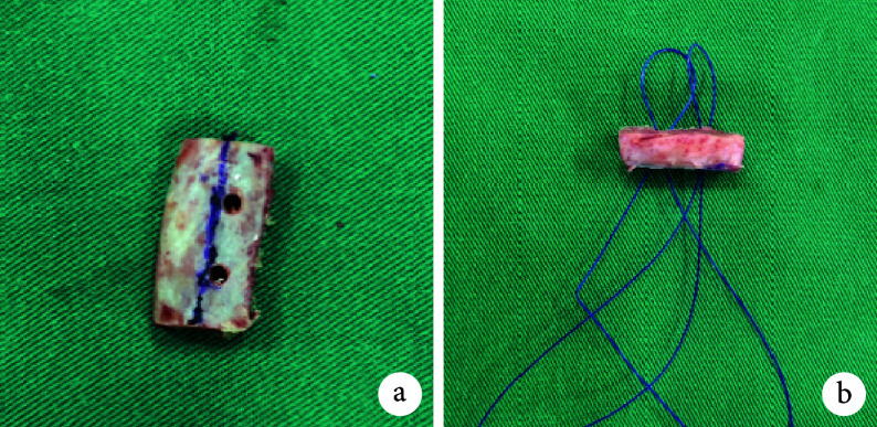
全身麻醉下,患者取健侧卧位,患肩外展30° 悬吊。首先于患侧髂前上棘后方3 cm位置作长约3 cm斜纵形切口,保留髂骨外板,分离皮下组织直达骨面。使用摆锯取长2 cm、宽1 cm、厚1 cm的长方体骨块(可根据术前影像学测量结果适当调整)。修整骨块,使骨松质面形状更贴合关节盂唇缺损面。于骨块三等分点偏离中线位置制作2个预留孔,然后引入2根对折的PDS线,用于骨块固定(图1)。
图 1.
Preparation of autologous iliac bone block
自体髂骨骨块制备
a. 制备预留孔;b. 预留孔中穿入PDS线
a. Preparation of reserved holes; b. The two PDS wires were inserted into the reserved holes
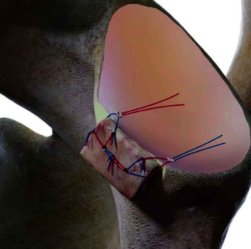
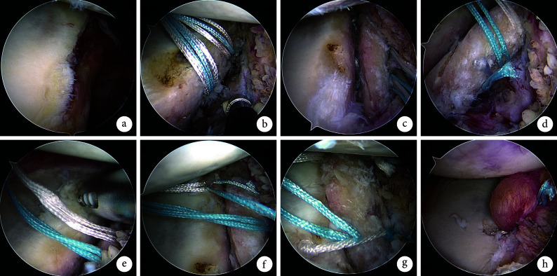
然后进行肩关节镜手术,手术入路包括标准后方入路、后上方入路、后下方入路以及前外上方入路、前方入路。首先,经后方入路进入盂肱关节,监视下建立前方入路。镜下见肱骨头后上方纵向缺损,关节盂前下缘缺损。于肱骨头后上方缺损处拧入2枚全线锚钉,并将缝线穿过冈下肌、小圆肌及软组织。对关节盂骨缺损处进行打磨至微渗血。然后,建立前外上方入路,取关节盂骨缺损中间四等分点并靠近中线内侧位置植入2枚盂唇钉。通过移植骨块PDS线将盂唇钉尾线引入预留孔中,经前方入路将去头端的10 mL注射器置于盂肱关节中。通过盂唇钉尾线及篮钳不断调整位置,使骨块与缺损处贴合。使用抓线钳将2枚盂唇钉的1根缝线进行Pulley打结,稳定骨块。经前方入路于关节盂骨缺损前上缘再拧入1枚盂唇钉,将此枚盂唇钉的1根缝线与骨块近端1枚盂唇钉剩余缝线进行Pulley打结;然后于骨缺损前下缘再拧入1枚盂唇钉,同样方法与骨块第2枚盂唇钉剩余缝线进行Pulley打结;最后将外缘2枚盂唇钉剩余2根缝线穿过盂唇组织进行Bankart修复,从而进一步稳定骨块。将肱骨头后方2枚全线锚钉打结固定,镜下见肱骨头后方与冈下肌、小圆肌及软组织完全贴合。活动肩关节见前后向稳定,缝合切口,无菌敷料包扎。见图2、3。
图 2.
Schematic diagram of arthroscopic Eden-Hybinette procedure with Triple-Pulley and four point anti-rotation fixation technique
自体髂骨植骨三滑轮四点防旋固定术示意图
图 3.
Schematic diagram of arthroscopic bone grafting and fixation
关节镜下植骨及固定操作示意图
a. 镜下见关节盂骨缺损;b. 于骨缺损中间植入2枚盂唇钉;c. 盂唇钉缝线穿过骨块预留孔并与缺损处贴合;d. 制作第1个Pulley结以稳定骨块;e. 于关节盂骨缺损前上缘拧入1枚盂唇钉;f. 于骨块外上方制作第2个Pulley结;g. 骨块外下方制作第3个Pulley结;h. 剩余缝线进行Bankart修复
a. Arthroscopic examination revealed the glenoid bone defect; b. Two glenoid anchors were inserted in the middle of the defect; c. The suture of glenoid anchor was passed through the reserved hole and adhered to the defect; d. The first Pulley knot was made to stabilize the bone block; e. A glenoid anchor was implanted at the anterior upper edge of the glenoid defect; f. The second Pulley knot was made above the outer part of the bone block; g. The third Pulley knot was made below the outer part of the bone block; h. Bankart repair with the remaining sutures
1.3. 术后处理
术后患肢佩戴肩关节外展支具,第2天开始患肢手部抓握及屈肘、屈腕等锻炼,以及患肩被动前屈上举、外展、内外旋活动,活动范围以患者能耐受疼痛为度;6周后移除支具,开始患肩主动前屈上举、外展、内外旋活动;3个月后开始患肢力量训练,包括屈肘抗阻、肩部内外旋抗阻训练等;6个月后进行患肢对抗性运动、投掷运动以及重体力活动。
1.4. 疗效评价指标
1.4.1. 临床疗效评价
术前及术后3个月、6个月、末次随访时,行患者肩关节活动时疼痛视觉模拟评分(VAS)、美国加州大学洛杉矶分校(UCLA)评分、Constant 评分以及美国肩肘外科医师协会(ASES)评分评价疗效。
1.4.2. 影像学评价
① 骨块位置:于术后即刻、6个月、末次随访时CT矢状位关节盂高度25%或50%层面,选取相对应的轴位图像进行测量。取关节盂前后缘顶点作一水平线,以肱骨头中心作同心圆。骨块位于同心圆外侧表明骨块偏外;位于水平线与同心圆之间表明位置合适;位于水平线内侧表明骨块偏内,超过内侧5 mm以上示过度偏内[10]。
② 骨块愈合:于术后6个月肩关节CT横断位观察,关节盂与骨块连接处未出现透亮区为骨块骨性愈合;透亮区长度≤5 mm为骨块纤维愈合,其中≤1 mm认为骨块稳定;透亮区>5 mm为骨块移位[11]。
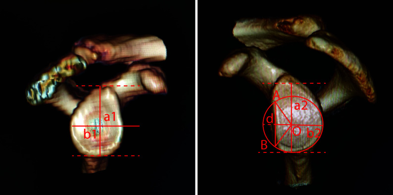
③ 骨块吸收及重塑:基于肩关节CT三维重建图像,采用Desktopview软件测算术前关节盂骨缺损宽度比例和术前及术后3个月、6个月、末次随访时关节盂面积。关节盂骨缺损宽度比例:通过关节盂切线测量健侧关节盂高度(a1)及宽度(b1),计算健侧关节盂宽高比(b1/a1);测量患侧关节盂高度(a2),根据健侧关节盂宽高比计算宽度(b2),以b2为直径、关节盂最低端切线垂直线b2/2处为圆心(O),画出患侧关节盂最适圆后,测量缺损宽度(d),计算关节盂骨缺损宽度比例(d/b2×100%),如该比例>25%则考虑骨性手术。
关节盂面积:在以b2为直径画出的患侧关节盂最适圆上,确定关节盂骨缺损与其交点(A、B),画出三角形OAB,计算三角形OAB及扇形OAB面积,两者差值即为术前关节盂骨缺损面积[14],最适圆面积与关节盂骨缺损面积差值即为术前关节盂面积。移植骨块近似长方形,按照长方形公式计算其面积,术后3个月、6个月、末次随访时关节盂面积即为移植骨块面积与术前关节盂面积之和。见图4。
图 4.
Schematic diagram of glenoid bone defect measurement
关节盂骨缺损测量示意图
1.5. 统计学方法
采用SPSS22.0统计软件进行分析。计量资料采用Shapiro-Wilk正态性检验,均服从或近似服从正态分布,数据以均数±标准差表示,手术前后比较采用重复测量方差分析,若不满足球形检验,采用Greenhouse-Geisser法进行校正,不同时间点间比较采用 Bonferroni 法。检验水准取双侧α=0.05。
2. 结果
本组手术均顺利完成,无严重并发症发生。术后患者均获随访,随访时间11.5~13.8个月,平均12.0个月。术后各时间点肩关节VAS评分均较术前降低, UCLA评分、Constant 评分及ASES评分均提高,差异有统计学意义(P<0.05);且术后随时间延长,上述指标均进一步改善,各时间点间差异均有统计学意义(P<0.05)。见表1。
表 1.
Comparison of effectiveness evaluation indicators between pre- and post-operation (n=14, x±s)
手术前后疗效评价指标比较(n=14,x±s)
| 时间 Time |
VAS评分 VAS score |
UCLA评分 UCLA score |
Constant 评分 Constant score |
ASES评分 ASES score |
关节盂面积(cm2) Glenoid area (cm2) |
|
*与术后3个月比较P<0.05,#与术后6个月比较P<0.05,△与末次随访比较P<0.05 *Compared with the value at 3 months after operation, P<0.05; #compared with the value at 6 months after operation, P<0.05; △compared with the value at last follow-up, P<0.05 | |||||
| 术前 | 6.9±0.6*#△ | 19.3±3.2*#△ | 54.4±5.1*#△ | 66.4±4.1*#△ | 10.98±0.70*#△ |
| 术后3个月 | 3.7±0.4#△ | 30.3±2.3#△ | 70.4±3.3#△ | 87.2±2.1#△ | 14.69±0.83 |
| 术后6个月 | 1.8±0.4*△ | 33.1±1.6*△ | 82.9±3.0*△ | 92.6±2.4*△ | 14.19±0.80 |
| 末次随访 | 0.7±0.4*# | 34.2±1.1*# | 89.6±2.0*# | 93.8±3.6*# | 13.65±0.98 |
| F值 | 572.443 | 61.691 | 366.761 | 384.795 | 493.944 |
| P值 | <0.001 | <0.001 | <0.001 | <0.001 | <0.001 |
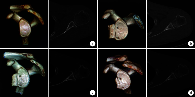
影像学复查示,术后骨块位置均未偏内或偏外,位于关节盂2:30~6:00位置且未发生明显移位。骨块完全填补关节盂骨缺损,且随时间延长逐渐愈合并达到骨性愈合,可见骨块部分吸收并塑形。术后关节盂面积较术前增大,差异有统计学意义(P<0.05);随时间延长,关节盂面积逐渐减小,但术后各时间点间差异均无统计学意义(P>0.05)。见表1及图5。
图 5.
CT three-dimensional reconstruction and axial images of a 32-year-old male patient with recurrent anterior dislocation of left shoulder
患者,男,32岁,左肩关节复发性前脱位CT三维重建及轴位图像
a. 术前关节盂骨缺损宽度比例28.3%;b. 术后即刻骨块位置良好;c. 术后6个月骨块部分吸收,整体形态及位置可;d. 术后12个月骨块吸收明显,无旋转及分离
a. Preoperative width ratio of glenoid bone defect was 28.3%; b. The good position of the bone block at immediate after operation; c. The partial absorption of the bone block was observed, and the shape and position were satisfactory at 6 months after operation; d. Significant bone resorption was observed at 12 months after operation, with no rotation or separation
3. 讨论
关节镜下骨移植固定术中,锚钉缝线固定骨块可以避免螺钉及袢钢板固定带来的问题,由于属于弹性固定,对骨块破坏少,可降低骨块吸收率[15-17]。但也有学者提出传统锚钉缝线固定存在强度不足等问题[18]。因此,我们提出自体髂骨移植术中通过植入4枚盂唇钉结合Pulley打结缝线固定提高固定牢固性。临床应用显示末次随访时,所有患者未出现严重并发症。影像学检查示骨块位置均未发生移位,骨块均达到骨性愈合并部分吸收重塑。肩关节各项疼痛及功能评分均较术前明显改善。我们总结该固定技术具有以下优势:① 植入的4枚盂唇钉于关节盂骨缺损上、下缘形成4个受力点,受力均匀,固定更牢固。② 制作3个Pulley结,其中内侧2枚盂唇钉形成的Pulley结稳定骨块,内、外侧盂唇钉形成的Pulley结防止其旋转。③ 有效避免螺钉及袢钢板固定相关并发症。④ 术中无需制备骨隧道,对关节盂破坏少。⑤ 研究表明骨移植物吸收程度与其承受应力成正相关,弹性固定应力低于坚强固定,故能有效避免骨块吸收[19]。我们提出的固定方式即属于弹性固定方式,术后关节盂面积虽随时间延长有一定减小,但各时间点间差异无统计学意义。⑥ 手术操作简便,无需其他特殊工具。
我们总结关节镜下自体髂骨植骨术中三滑轮四点防旋固定手术操作要点及注意事项:① 盂唇缺损处应充分打磨使其微渗血,并将髂骨骨块骨松质面贴合于缺损处,有利于骨块愈合。另外预留孔位置更偏于内侧,使内、外侧盂唇Pulley打结时受力面积更大以增加稳定性。② 骨块植入关节腔过程中需注意保持与关节盂骨缺损处位置一致,并始终维持内侧盂唇钉缝线张力,防止骨块翻转。③ 调整骨块在缺损处位置后,先将内侧锚钉缝线制作1个Pulley结以稳定骨块;然后每植入1枚盂唇钉便制作1个Pulley结,这样便于缝线管理及应力把握。④ 尽可能用盂唇及周围软组织对骨块进行包裹,为移植骨块提供血运,以降低骨块吸收率[20]。⑤ 固定骨块应力需适中,我们认为在固定完成后其移动度在1 mm之内为宜。⑥ 需对Bankart修复,并尽量减少对盂唇及周围软组织的破坏,最大限度保持其完整性,以进一步加强骨块稳定性。本研究中仅采用外缘2枚锚钉剩余2根缝线修复盂唇组织,我们认为2根缝线固定强度已满足需求,缝合同时可对盂唇组织形成向外提拉的作用力,在修复Bankart的同时进一步对骨块形成包裹,以加强骨块稳定性。
综上述,关节镜下自体髂骨植骨三滑轮四点防旋固定术治疗复发性肩关节前脱位,能够有效防止骨块旋转,固定更牢靠,可取得良好临床疗效,且手术操作简便。但是本研究样本量小、随访时间短,且缺少临床对照。因此,研究结论有待大样本、随机对照且长期随访明确。此外,该固定技术尚需进一步生物力学研究加以验证。
利益冲突 在课题研究和文章撰写过程中不存在利益冲突
伦理声明 研究方案经南昌市洪都中医院医学伦理委员会批准(KYKS-2021030)
作者贡献声明 周明、龚礼:研究设计、实施;侯慧铭、邹文、徐涛、胡梁深:数据收集整理及统计分析;龚礼:起草文章;范少勇:对文章的知识性内容作批评性审阅
References
- 1.Patrick CM, Snowden J, Eckhoff MD, et al Epidemiology of shoulder dislocations presenting to United States emergency departments: An updated ten-year study. World J Orthop. 2023;14(9):690–697. doi: 10.5312/wjo.v14.i9.690. [DOI] [PMC free article] [PubMed] [Google Scholar]
- 2.Provencher MT, Peebles LA, Aman ZS, et al Management of the failed Latarjet procedure: outcomes of revision surgery with fresh distal tibial allograft. Am J Sports Med. 2019;47(12):2795–2802. doi: 10.1177/0363546519871896. [DOI] [PubMed] [Google Scholar]
- 3.Cozzolino A, de Giovanni R, Malfi P, et al Arthroscopic Latarjet versus arthroscopic free bone block procedures for anterior shoulder instability: a proportional meta-analysis comparing recurrence, complication, and reoperation rates. Am J Sports Med. 2024;52(7):1865–1876. doi: 10.1177/03635465231188530. [DOI] [PubMed] [Google Scholar]
- 4.Thamrongskulsiri N, Limskul D, Tanpowpong T, et al Clinical outcomes, union rates, and complications of screw versus button fixation in the Bristow-Latarjet procedure for anterior shoulder instability: a systematic review and meta-analysis. Clin Orthop Surg. 2023;15(6):1000–1012. doi: 10.4055/cios23154. [DOI] [PMC free article] [PubMed] [Google Scholar]
- 5.Alkhelaifi K, Alzobi OZ, Mahmoud SA, et al Recurrent instability after the Latarjet procedure. J Am Acad Orthop Surg Glob Res Rev. 2023;7(12):e23. doi: 10.5435/JAAOSGlobal-D-23-00205. [DOI] [PMC free article] [PubMed] [Google Scholar]
- 6.Gilat R, Haunschild ED, Lavoie-Gagne OZ, et al Outcomes of the Latarjet procedure versus free bone block procedures for anterior shoulder instability: a systematic review and meta-analysis. Am J Sports Med. 2021;49(3):805–816. doi: 10.1177/0363546520925833. [DOI] [PubMed] [Google Scholar]
- 7.Moroder P, Schulz E, Wierer G, et al Neer Award 2019: Latarjet procedure vs. iliac crest bone graft transfer for treatment of anterior shoulder instability with glenoid bone loss: a prospective randomized trial. J Shoulder Elbow Surg. 2019;28(7):1298–1307. doi: 10.1016/j.jse.2019.03.035. [DOI] [PubMed] [Google Scholar]
- 8.Boehm E, Minkus M, Moroder P, et al Arthroscopic iliac crest bone grafting in recurrent anterior shoulder instability: minimum 5-year clinical and radiologic follow-up. Knee Surg Sports Traumatol Arthrosc. 2021;29(1):266–274. doi: 10.1007/s00167-020-05986-7. [DOI] [PMC free article] [PubMed] [Google Scholar]
- 9.Oldfield M, Burns J, Wong I Arthroscopic glenoid bone augmentation using iliac crest autograft is safe and effective for anterior shoulder instability with bone loss. Arthrosc Sports Med Rehabil. 2021;3(6):e1671–e1677. doi: 10.1016/j.asmr.2021.07.023. [DOI] [PMC free article] [PubMed] [Google Scholar]
- 10.Maguire JA, Dhillon J, Sarna N, et al Screw fixation for the Latarjet procedure may reduce risk of recurrent instability but increases reoperation rate compared to suture-button fixation: a systematic review. Arthroscopy. 2024;40(6):1882–1893. doi: 10.1016/j.arthro.2023.11.020. [DOI] [PubMed] [Google Scholar]
- 11.Reeves JM, Athwal GS, Johnson JA Double-screw and quadruple-button fixation for the glenoid: Latarjet versus bone block applications. JSES Int. 2020;4(4):780–785. doi: 10.1016/j.jseint.2020.08.015. [DOI] [PMC free article] [PubMed] [Google Scholar]
- 12.Casabianca L, Gerometta A, Massein A, et al Graft position and fusion rate following arthroscopic Latarjet. Knee Surg Sports Traumatol Arthrosc. 2016;24(2):507–512. doi: 10.1007/s00167-015-3551-6. [DOI] [PubMed] [Google Scholar]
- 13.Chakrabarti MO, Khan M Editorial Commentary: Suture button fixation for the Latarjet procedure is superior to screw fixation. Arthroscopy. 2024;40(5):1655–1657. doi: 10.1016/j.arthro.2023.11.035. [DOI] [PubMed] [Google Scholar]
- 14.Bhatia S, Saigal A, Frank RM, et al Glenoid diameter is an inaccurate method for percent glenoid bone loss quantification: analysis and techniques for improved accuracy. Arthroscopy. 2015;31(4):608–614. doi: 10.1016/j.arthro.2015.02.020. [DOI] [PubMed] [Google Scholar]
- 15.Ameziane Y, Scheibel M Arthroscopic anterior glenoid bone grafting for shoulder instability using an interconnected suture anchor technique. Arthrosc Tech. 2022;11(10):e1817–e1822. doi: 10.1016/j.eats.2022.06.019. [DOI] [PMC free article] [PubMed] [Google Scholar]
- 16.Kelly SR, Kim HM Arthroscopic bone block cerclage suture fixation of fresh distal tibial allograft for anterior glenoid reconstruction. Arthrosc Tech. 2023;12(11):e1955–e1961. doi: 10.1016/j.eats.2023.07.014. [DOI] [PMC free article] [PubMed] [Google Scholar]
- 17.Rosenow CS, Leland DP, Camp CL, et al Sutures, screws, buttons, and anchors: a review of current bone graft fixation devices for glenoid bone loss in the unstable shoulder. Curr Rev Musculoskelet Med. 2024;17(6):207–221. doi: 10.1007/s12178-024-09895-x. [DOI] [PMC free article] [PubMed] [Google Scholar]
- 18.Ritter D, Hachem AI, Scheibel M, et al Primary stability and bone contact loading evaluation of suture and screw based coracoid graft fixation for anterior glenoid bone loss. Am J Sports Med. 2023;51(11):2858–2868. doi: 10.1177/03635465231188976. [DOI] [PubMed] [Google Scholar]
- 19.Alp NB, Doğan O, Yılmaz TG, et al Understanding the causes behind coracoid graft osteolysıs in Latarjet procedure (finite element analysis and comparison of three fixation methods) Orthop Traumatol Surg Res. 2020;106(1):53–59. doi: 10.1016/j.otsr.2019.11.007. [DOI] [PubMed] [Google Scholar]
- 20.Cohen M, Zaluski AD, Siqueira GSL, et al Risk factors for coracoid graft osteolysis after the open Latarjet procedure. Rev Bras Ortop (Sao Paulo) 2020;55(5):585–590. doi: 10.1055/s-0039-1698799. [DOI] [PMC free article] [PubMed] [Google Scholar]


