Abstract
目的
对内侧髌股韧带重建术中股骨隧道Schöttle定位法研究进展进行综述,为临床应用提供参考。
方法
广泛查阅近年国内外Schöttle定位法相关研究文献,并对影响定位准确性的问题及提高定位准确性的新思路进行归纳总结。
结果
内侧髌股韧带重建术中准确定位股骨隧道很重要,隧道错位是术后并发症发生和手术失败的主要原因。Schöttle定位法是目前研究最深入、可重复性最高的股骨隧道定位方法,是被广泛应用的“金标准”。然而,影响Schöttle定位法准确性的问题仍然存在,包括膝关节内外旋、内外翻会影响定位准确性,X线片拍摄要求和解剖标志参考线绘制标准尚未明确,对于解剖结构变异患者适应性降低,以及对儿童患者缺乏进一步研究。近年来相关研究提出了有望提高Schöttle定位法定位准确性的新思路。
结论
未来研究应结合3D打印、术中导航等新技术,致力于发展个性化、智能化Schöttle定位法,进一步提高其定位准确性。
Keywords: 内侧髌股韧带, 股骨隧道, Schöttle定位法, 研究进展
Abstract
Objective
To review the research progress of Schöttle’s method in medial patellofemoral ligament reconstruction (MPFLR), and provide the latest knowledge and suggestions for surgical treatment.
Methods
The studies on Schöttle’s method at home and abroad in recent years were extensively collected, then summarized the problems affecting the accuracy of Schöttle’s method and the new ideas to improve the accuracy of localization.
Results
It’s vital to accurately locate the femoral tunnel during MPFLR. Malposition of the femoral tunnel is the main cause of postoperative complications and surgical failure. Schöttle’s method is the most well studied and most reproducible method for femoral tunnel localization, which is widely used as the “gold standard”. However, there are still problems that affect the accuracy of Schöttle’s method, including the impact of the internal/external rotation and varus/valgus of the knee on localization accuracy, unclear requirements for X-ray imaging and anatomical landmark reference line drawing standards, no suitable for patients with anatomical variations, and lack of further research on pediatric patients. In recent years, some new ideas are proposed to improve the Schöttle’s method to improve the localization accuracy.
Conclusion
Future research should combine new technologies such as three-dimensional (3D) printing and intraoperative navigation to develop personalized and intelligent Schöttle’s method, further improving their localization accuracy.
Keywords: Medial patellofemoral ligament, femoral tunnel, Schöttle’s method, research progress
内侧髌股韧带是维持膝关节屈曲稳定的基本结构[1],外伤性和急性髌骨脱位患者中90%以上合并该韧带损伤[2-5]。对于此类患者,内侧髌股韧带重建术是恢复膝关节运动、改善术后患者主观评分和减少复发性髌骨不稳定和再脱位的最合适治疗方法[6-7]。重建术中准确定位股骨隧道具有重要意义,可以恢复内侧髌股韧带的生物力学功能,包括韧带等长性、髌股关节功能[8]。如定位不准确,会导致内侧髌股关节接触压力增加、髌骨向内侧移位,进而发生疼痛和关节软骨退变,严重时会导致骨关节炎[9-10]。有研究发现股骨隧道点向任意方向错位1 mm,患者术后膝关节Kujala评分和国际膝关节文献委员会(IKDC)评分均减少0.817分[11]。
内侧髌股韧带重建术中股骨隧道的影像学定位法包括Schöttle定位法、Stephen定位法和在此基础上各种衍生定位方法。其中,Schöttle定位法研究最深入、可重复性最高,获得广泛应用,也被称为股骨隧道定位的“金标准”[12-13]。然而,Schöttle定位法仍然存在局限性,包括没有明确和充分说明X线片拍摄要求和如何绘制解剖标志参考线;膝关节内、外旋及内、外翻会影响定位准确性;对于解剖结构变异的患者适应性降低;对儿童患者缺乏进一步研究。近年来,学者们就上述局限性进行了一系列相关研究,并取得积极进展,提高了该方法适用性。现对Schöttle定位法存在问题以及有望提高定位准确性的新思路进行全面总结,以期为临床提供参考。
1. Schöttle定位法测量方法及误差范围
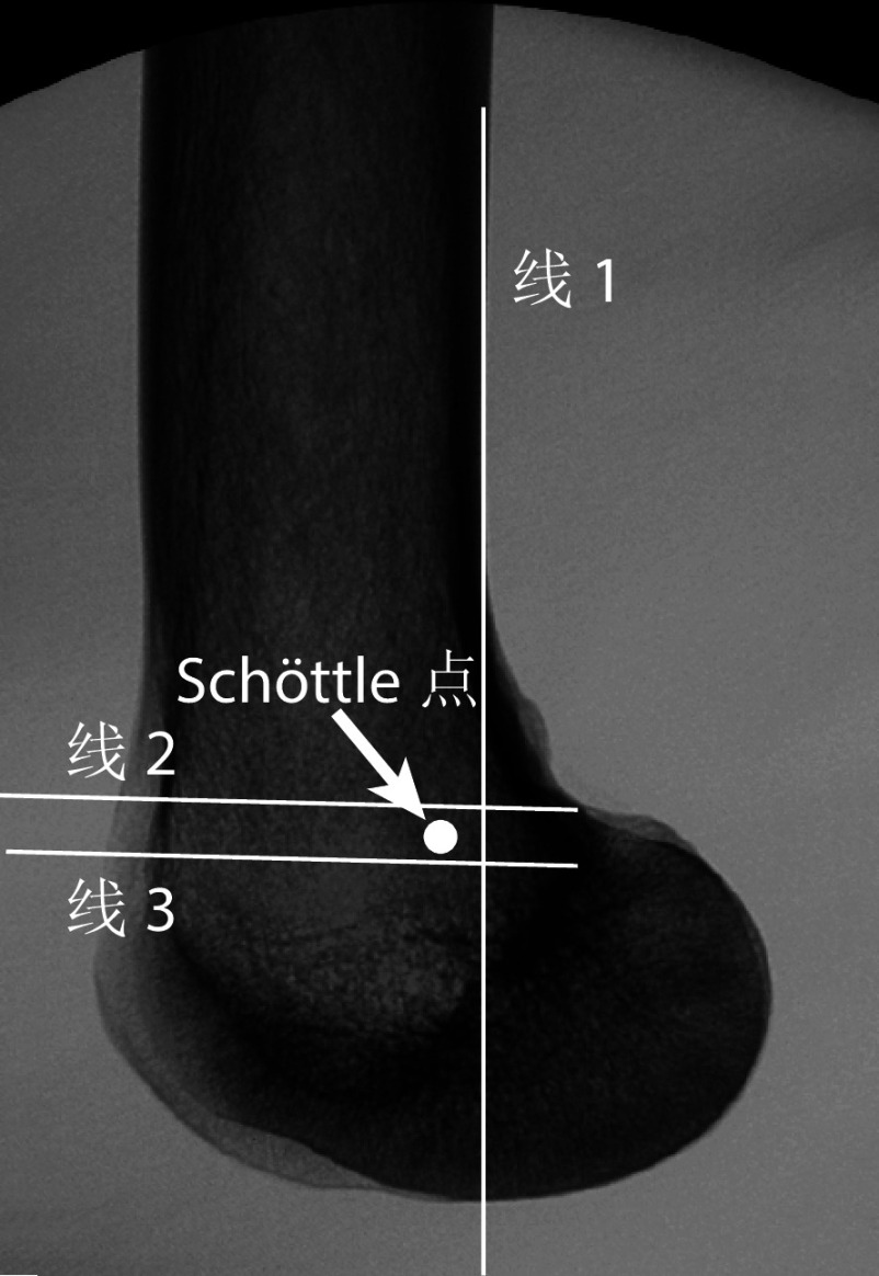
Schöttle等[14]在8个尸体膝关节标本标记内侧髌股韧带股骨止点,并拍摄纯侧位X线片(即图像中股骨内、外侧髁后端重叠),通过分析X线片标记的股骨止点在股骨前后方向和远近方向的位置,提出了“Schöttle定位法”,即在纯侧位X线片通过3条参考线定位股骨隧道,定位点(Schöttle点)位于股骨后皮质线延长线(线1)前1.3 mm、通过股骨后髁与后皮质的交界且垂直于股骨后皮质延长线的直线(线2)远端2.5 mm、过Blumensaat线后角且垂直于股骨后皮质延长线的直线(线3)近端5.5 mm。其中,线1确定了Schöttle点的前后方向位置,线2与线3确定Schöttle点的远近方向位置。见图1。
图 1.
Schematic diagram of the Schöttle’s method
Schöttle定位法示意图
在解剖研究中,内侧髌股韧带股骨止点表现为大小为3.6 cm2的区域,并非1个点,据此Schöttle等认为股骨隧道定位在一定区域内即是准确的[14]。他们发现通过Schöttle定位法确定的股骨隧道均在解剖股骨止点附近5 mm范围内。结合既往研究发现5 mm范围内的误差不会改变内侧髌股韧带的等长性[15],因此Schöttle提出以准确的解剖定位点为圆心、半径5 mm范围内的误差是可以接受的。但Redfern等[16]指出由于钻头直径在6~7 mm,已经超过Schöttle提出的5 mm范围,所以只要实际点在解剖定位点周围6~7 mm范围内即可以接受。
尽管以上研究认为股骨隧道定位在一定区域内即是准确的,但近年有研究报道仅仅5 mm错位就会破坏内侧髌股韧带等长性[17]。Stephen等[17]的生物力学试验发现Schöttle点向远端错位5 mm,膝关节屈曲过程中内侧髌股韧带长度变化达平均9.1 mm;向近端错位5 mm,韧带长度变化达平均6.4 mm。非等长变化的内侧髌股韧带会增加内侧髌股关节压力和髌骨内侧倾斜,进一步导致关节疼痛和退变[18]。另外,Stephen等[17]发现Schöttle点在前后方向上错位5 mm时,膝关节屈曲过程中没有破坏内侧髌股韧带的等长性。可见,Schöttle点在前后方向上错位造成的影响小于在远近方向上错位。因此,采用Schöttle定位法时,在股骨远近方向的错位误差越小越好,而在前后方向上5 mm内的误差被认为是可接受的。
2. Schöttle定位法存在问题及提高定位准确性的新思路
2.1. X线片拍摄要求及绘制解剖标志参考线
摄X线片时膝关节内、外旋及内、外翻会导致Schöttle点错位,分别向前、后、远、近端方向移动。此外,随着膝关节内、外旋及内、外翻程度的增加,错位程度也相应增加[19]。Ziegler等[20]发现基于纯侧位X线片确定的Schöttle点与解剖定位点间的距离平均为4.1 mm,而膝关节内、外旋及内、外翻时,这一距离会增加;并且内、外旋和内、外翻角度与Schöttle点相对于解剖定位点的距离成正比。具体来说,内、外旋及内、外翻为5° 或10° 时,Schöttle点向前、后、远、近端方向的移动距离均超过5 mm,分别达7~9 mm和10~15 mm。Balcarek等[21]也发现,膝关节内、外翻和内、外旋角度为2.5° 或5° 时,Schöttle点相对于解剖定位点向前、后、远、近端方向移动的距离分别为2.0~4.3 mm或2.6~6.8 mm。Barnett等[22]证实膝关节内、外旋程度与相应Schöttle点移位程度成正相关(P<0.0001)。
因此,需要拍摄纯侧位X线片以避免膝关节内、外旋及内、外翻导致的Schöttle点错位,但是拍摄纯侧位片也存在一些问题。在髌骨内侧韧带重建术中,医生需要多次调整C臂X线机,这一过程耗时耗力,不仅增加了辐射,而且手术时间延长可能导致感染。术中拍摄X线片时股骨内、外侧髁后端有时无法完全重叠,需要基于医生的主观判断决定Schöttle点,这也会导致定位不准确。
在股骨前后方向上,不同研究确定的Schöttle点相对于解剖定位点的距离与方位存在较大差异。例如,Redfern等[16]、Ishikawa等[23]发现Schöttle点在前后方向上分别位于解剖定位点前方2.5 mm、后方3.8 mm;Ziegler等[20]、Emre等[24]发现Schöttle点与解剖定位点的距离分别为3.2、4.1 mm。出现这一问题的原因在于Schöttle提出定位方法时没有明确定义股骨后皮质延长线。首先,股骨后皮质区域远端呈弓形轮廓,没有明确指向性,从此处绘制的延长线可能不同;其次,研究者可以随意选择延长线近端起点,存在较大主观性。上述两点最终导致Schöttle点在前后方向存在较大差异。另外,患者的长期负重活动可能导致股骨后皮质的弓形轮廓发生变化,这种情况下,股骨后皮质延长线不适合作为准确定位的参考线[17]。
基于上述分析明确和充分定义X线片拍摄要求,通过标准化来提高定位准确性,具有重要意义。Redfern等[16]与Schöttle沟通后提出了确定延长线的新标准,即基于股骨后皮质区域的最外侧轮廓确定股骨后皮质延长线,但这一新标准确定的股骨后皮质延长线仍然是可变的。
Bhimani等[25]认为不同研究者拍摄的X线片,其股骨干长度不同,导致了定位不一致。他们对9个尸体膝关节进行纯侧位X线片分析,使X线片显示的股骨干长度从1 cm逐渐增加至8 cm,每次增加1 cm,发现Schöttle点相对于股骨后皮质延长线的距离从平均1.1 cm增加至4.1 cm。通过统计分析得出,X线片可见股骨干长度(X)与Schöttle点相对于股骨后皮质延长线距离(Y)的线性回归方程为Y=−0.636X(R=0.461,R2=0.212,B=−0.636,P<0.001),其中Y正、负值分别表示Schöttle点相对于股骨后皮质延长线的方向,前方为正,进一步通过受试者工作特征曲线分析得出结论,在X线片上可见股骨干长度至少为4 cm才能确保Schöttle定位法在股骨前后方向上定位的准确性(敏感度76.7%,特异度69.0%,P<0.001)。
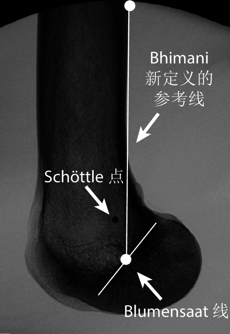
但是Bhimani等提出的上述解决方案仍不适用于一些特殊情形。例如,部分医院条件有限,仅有迷你C臂X线机,体型较大患者摄片时可能无法确保可见股骨干长度达4 cm。为此,他们提出了一种适应性更强的定位方法,重新定义了一条参考线以定位Schöttle点的股骨前后方向位置(图2)[25]。这条新参考线近端起于股骨后皮质区最近点,远端止于Blumensaat线中点,经影像学测量及统计分析证实,可见股骨干长度不会影响Schöttle点与这条参考线的距离,并且Schöttle点始终在这条参考线的后方。但是,Bhimani等未将该发现总结为临床可以直接使用的方法,也未进行临床验证,因此上述研究结论有待进一步研究。
图 2.
Schematic diagram of the reference line defined by Bhimani
Bhimani新定义参考线示意图
C臂X线机摄片时存在由内向外、由外向内两种方式,即X线发射器分别在股骨外侧与内侧。由于X线技术存在空间图像畸变问题,不同拍摄方式下股骨内、外侧髁大小存在差异,这可能影响Schöttle定位法的准确性。Korthaus等[26]评估了基于2种拍摄方式获得图像的定位准确性,结果显示不同图像确定的Schöttle点与解剖定位点的距离无显著差异。但他们的研究是基于3D打印股骨模型,解剖定位点由医生确定而非真实韧带股骨止点,并且股骨模型没有放置于C臂X线机中间位置,这些因素导致结果可信度不高。在此基础上,Cetin等[27]改进了实验,将股骨标本放置于C臂X线机中间位置并确定了相对真实的解剖定位点,发现由外向内拍摄图像的定位准确性高于由内向外拍摄,基于2种图像定位的Schöttle点与解剖定位点距离分别为(3.2±1.5)、(4.5±2.1)mm,存在显著差异;将可接受误差定义为解剖定位点周围5 mm范围,由外向内和由内向外拍摄的定位准确率分别为90.9%(40/44)和 65.9%(29/44)。
可见,通过由外向内拍摄纯侧位X线片,基于股骨后皮质区域的最外侧轮廓、确保可见股骨干长度至少为4 cm来确定股骨后皮质延长线,进而确定Schöttle点,或者通过连接股骨后皮质最近点与Blumensaat线中点的新参考线确定Schöttle点,是提高Schöttle定位法准确性的新思路。
2.2. 解剖结构变异对定位准确性的影响
Izadpanah等[28]发现严重滑车发育不良(Dejour C、D级)患者股骨内、外侧后髁完全重叠时,股骨后皮质区域远端呈宽条形而非线形,导致绘制股骨后皮质延长线具有较大的主观性,因此误差较大。他们研究证实非滑车发育不良以及Dejour C、D级滑车发育不良膝关节的Schöttle点与解剖定位点距离分别为4.3、6.7、7.3 mm。可见对于Dejour C、D级滑车发育不良的膝关节,Schöttle定位法准确性较低。
采用3D打印个性化模板定位法、术中导航定位法、通过解剖标志定位Schöttle点,有望减少解剖变异对Schöttle定位法准确性的影响。3D打印个性化模板定位法是根据CT扫描和三维重建获得的股骨立体图像,用软件将立体图像调整为纯侧位后标记Schöttle点,根据Schöttle点的定位和内侧上髁周围的骨表面设计并3D打印制备个性化导航模板。该模板可以完全贴合患者远端股骨,模板开口处即为准确的Schöttle点。Zhang等[29]发现该定位法的准确性及术后效果优于传统Schöttle定位法,可以在股骨解剖结构异常及畸形情况下较准确地定位Schöttle点。
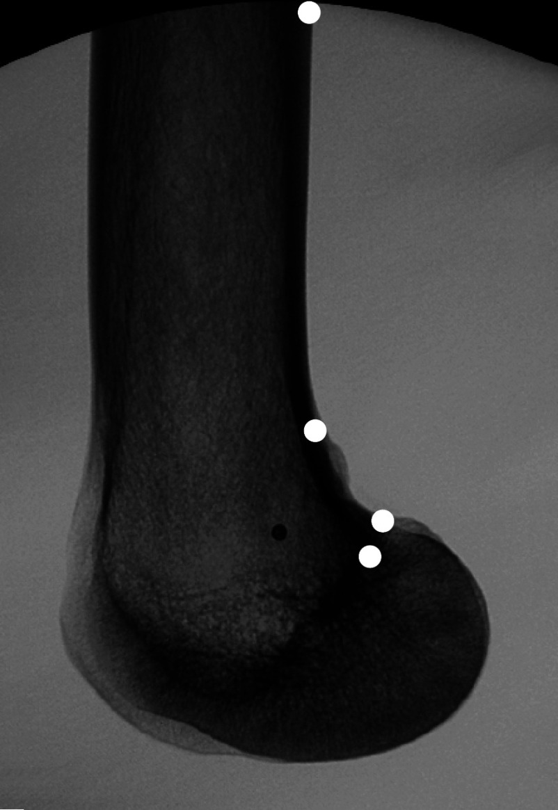
术中导航定位法是随着影像与计算机技术发展而产生的新技术,Kordon等[30]将股骨远端结构分割为多个关键点来识别Schöttle定位法所需的解剖结构(图3)。Privalov等[31]将这一方法应用于内侧髌股韧带重建中,导航软件通过识别股骨后皮质区域的远、近端点来绘制股骨后皮质延长线,通过识别股骨后内侧髁弓形轮廓与股骨干的分界点及Blumensaat线的近端点分别绘制Schöttle定位法的线2与线3,Schöttle点被标记为3条参考线之间半径为4.68 mm 的一个圆。该软件自动计算的点可实时显示在术中X线片上。并且,随着训练数据量的增加,神经网络算法可以提升其定位准确性。临床试验发现软件导航定位误差为(0.9±0.5)mm,明显小于传统Schöttle定位法的(3.0±1.4)mm。该手术导航软件不仅提高了Schöttle点定位精度,而且没有增加手术时间。张召贺等[32]通过回顾性病例对照研究也发现了类似结果。
图 3.
Schematic diagram of the software identifying key points of femur based on the Schöttle’s method
Schöttle定位法软件识别股骨结构关键点示意图
Wang等[33]发现通过解剖标志定位Schöttle点可靠性高且无需术中摄X线片。他们分析了53例患者膝关节的三维CT图像,发现Schöttle点总位于内收肌结节顶点远端8 mm处、内收肌结节后缘(即股骨内侧髁与股骨干的过渡区域边缘)前方8 mm处。经临床试验发现93.6%(44/47)患者定位准确,但对于解剖变异患者是否适用需要进一步探究。
2.3. 缺乏对儿童患者的研究
儿童患者内侧髌股韧带重建术中Schöttle定位法与成年患者不同,主要有两点原因。第一,Shea等[34]发现内侧髌股韧带的解剖起始点随着儿童年龄增长而变化,从股骨骺板远端4.7 mm处逐渐向股骨骺板近端0.8 mm处移动。第二,传统Schöttle点与股骨远端骺板非常接近,因此使用Schöttle定位法时要避免损伤骨骺,否则会影响患儿股骨生长,甚至导致畸形[35-37]。
为了达到内侧髌股韧带重建术的效果并避免损伤骺板,学者们进行了一系列研究,并提出通过控制在Schöttle点钻孔直径可以避免损伤骺板。例如,Featherall等[38]通过影像学分析发现Schöttle点始终位于股骨骨骺远端平均9.9 mm处,通过将扩孔直径控制到6 mm以下,儿童内侧髌股韧带重建时股骨骨骺是安全的。也有学者提出应将传统Schöttle点适当地向前、远端移位以避免损伤骨骺[39-41]。Metz等[42]通过49例儿童患者影像资料研究,认为将Schöttle点向股骨远端移动3 mm可以完全避免损伤骨骺。
然而,将Schöttle点向远端移位是否会改变内侧髌股韧带重建术后韧带的等长性,进一步影响膝关节活动和术后远期疗效仍不确定。Black等[43]基于8个骨骼未发育成熟的尸体膝关节标本,比较了通过Schöttle点或向远端移动8 mm行内侧髌股韧带重建术的生物力学效果。结果发现Schöttle点向远端移位后,膝关节运动学和关节面受力没有明显恶化,但内侧髌股韧带只能在膝关节屈曲0°~60° 范围内保持等长性(Schöttle点未移位情况下可在0°~100° 范围内保持等长性)。
上述研究显示控制Schöttle点钻孔直径并将其适当地向前、远端移位,有助于避免损伤儿童患者骨骺,但目前仍然无获得广泛认可和充分验证的儿童患者Schöttle定位法。
3. 总结与展望
Schöttle定位法是内侧髌股韧带重建术中股骨隧道定位的“金标准”,但其局限性不可忽视。近年来,相关研究发现了X线片拍摄、患者解剖结构变异及儿童患者解剖特异性对Schöttle定位法准确性的影响,并针对研究结果提出提高定位准确性的新思路。其中,3D打印模板、术中导航等新技术将是未来发展方向,有望用于发展个性化、智能化Schöttle定位法,进一步提高适用性。
利益冲突 在课题研究和文章撰写过程中不存在利益冲突;课题经费支持没有影响文章观点
作者贡献声明 魏雨乔:文献整理、文章撰写及文章修改;孙重霄、路凡、闫鹏安:文献收集整理及分析;夏亚一:写作思路指导及文章校对
Funding Statement
国家自然科学基金资助项目(82060405、82360436);兰州市科技计划项目(021-RC-102);甘肃省自然科学基金资助项目(22JR5RA943、22JR5RA956、23JRRA1500);兰州大学第二医院“萃英科技创新”计划(CY2021-MS-A07、CY2022-MS-A19)
National Natural Science Foundation of China (82060405, 82360436); Lanzhou Science and Technology Plan Program (2021-RC-102); Natural Science Foundation of Gansu Province (22JR5RA943, 22JR5RA956, 23JRRA1500); Cuiying Scientific and Technological Innovation Program of the Second Hospital of Lanzhou University (CY2021-MS-A07, CY2022-MS-A19)
References
- 1.Tanaka MJ Femoral origin anatomy of the medial patellofemoral complex: implications for reconstruction. Arthroscopy. 2020;36(12):3010–3015. doi: 10.1016/j.arthro.2020.06.015. [DOI] [PubMed] [Google Scholar]
- 2.张艳, 李彦林, 刘德健, 等 内侧髌股韧带重建术中股骨隧道定位点研究进展. 中国修复重建外科杂志. 2021;35(2):258–264. doi: 10.7507/1002-1892.202006059. [DOI] [PMC free article] [PubMed] [Google Scholar]
- 3.闫鹏安, 路凡, 蔡逸帆, 等 内侧髌股韧带重建术中股骨侧定位点研究进展. 中国修复重建外科杂志. 2024;38(10):1276–1282. doi: 10.7507/1002-1892.202405051. [DOI] [PMC free article] [PubMed] [Google Scholar]
- 4.吴金龙, 赵金忠 内侧髌股韧带重建的研究进展. 中国研究型医院. 2024;11(1):29–33. [Google Scholar]
- 5.黄勇, 苏帆, 陈斐, 等 内侧髌股韧带重建的股骨侧骨道预定位. 中国矫形外科杂志. 2021;29(6):540–543. [Google Scholar]
- 6.Hurley ET, Colasanti CA, Anil U, et al Management of patellar instability: a network meta-analysis of randomized control trials. Am J Sports Med. 2022;50(9):2561–2567. doi: 10.1177/03635465211020000. [DOI] [PubMed] [Google Scholar]
- 7.Erard J, Olivier J, Gunst S, et al Nonanatomical femoral tunnel positioning in isolated MPFL reconstruction is not associated with an increased risk of patellofemoral osteoarthritis after a minimum follow-up of 10 years. Knee Surg Sports Traumatol Arthrosc. 2024;32(11):2806–2817. doi: 10.1002/ksa.12264. [DOI] [PubMed] [Google Scholar]
- 8.Walsh JM, Credille K, Allahabadi S, et al. Biomechanical properties and kinematics of medial patellofemoral ligament reconstruction: a systematic review. Orthop J Sports Med, 2024, 12(6): 23259671241241537.
- 9.Velasquez Hammerle MV, Tanaka MJ Medial patellofemoral reconstruction techniques for patellar instability. Arthroscopy. 2023;39(6):1373–1375. doi: 10.1016/j.arthro.2023.01.006. [DOI] [PubMed] [Google Scholar]
- 10.Walker M, Maini L, Kay J, et al Femoral tunnel malposition is the most common indication for revision medial patellofemoral ligament reconstruction with promising early outcomes following revision reconstruction: a systematic review. Knee Surg Sports Traumatol Arthrosc. 2022;30(4):1352–1361. doi: 10.1007/s00167-021-06603-x. [DOI] [PubMed] [Google Scholar]
- 11.Maione A, Tradati D, Ferrua P, et al Accuracy of femoral tunnel positioning in medial patellofemoral ligament reconstruction: anatomic insertion leads to better clinical outcome. Knee Surg Sports Traumatol Arthrosc. 2023;31(7):2810–2817. doi: 10.1007/s00167-022-07235-5. [DOI] [PubMed] [Google Scholar]
- 12.Kaplan DJ Editorial commentary: Proper femoral tunnel placement for medial patellofemoral ligament reconstruction requires optimal radiographic technique. Arthroscopy. 2024;40(6):1867–1869. doi: 10.1016/j.arthro.2023.12.024. [DOI] [PubMed] [Google Scholar]
- 13.Zhang YQ, Zhang Z, Wu M, et al Medial patellofemoral ligament reconstruction: A review. Medicine (Baltimore) 2022;101(1):e28511. doi: 10.1097/MD.0000000000028511. [DOI] [PMC free article] [PubMed] [Google Scholar]
- 14.Schöttle PB, Schmeling A, Rosenstiel N, et al Radiographic landmarks for femoral tunnel placement in medial patellofemoral ligament reconstruction. Am J Sports Med. 2007;35(5):801–804. doi: 10.1177/0363546506296415. [DOI] [PubMed] [Google Scholar]
- 15.Smirk C, Morris H The anatomy and reconstruction of the medial patellofemoral ligament. Knee. 2003;10(3):221–227. doi: 10.1016/S0968-0160(03)00038-3. [DOI] [PubMed] [Google Scholar]
- 16.Redfern J, Kamath G, Burks R Anatomical confirmation of the use of radiographic landmarks in medial patellofemoral ligament reconstruction. Am J Sports Med. 2010;38(2):293–297. doi: 10.1177/0363546509347602. [DOI] [PubMed] [Google Scholar]
- 17.Stephen JM, Lumpaopong P, Deehan DJ, et al The medial patellofemoral ligament: location of femoral attachment and length change patterns resulting from anatomic and nonanatomic attachments. Am J Sports Med. 2012;40(8):1871–1879. doi: 10.1177/0363546512449998. [DOI] [PubMed] [Google Scholar]
- 18.Boot MR, van de Groes SAW, Dunning H, et al Length changes of the medial patellofemoral ligament during in vivo knee motion: an evaluation using dynamic computed tomography. Am J Sports Med. 2023;51(14):3724–3731. doi: 10.1177/03635465231205597. [DOI] [PMC free article] [PubMed] [Google Scholar]
- 19.Chen J, Han K, Jiang J, et al Radiographic reference points do not ensure anatomic femoral fixation sites in medial patellofemoral ligament reconstruction: a quantified anatomic localization method based on the saddle sulcus. Am J Sports Med. 2021;49(2):435–441. doi: 10.1177/0363546520972420. [DOI] [PubMed] [Google Scholar]
- 20.Ziegler CG, Fulkerson JP, Edgar C Radiographic reference points are inaccurate with and without a true lateral radiograph: the importance of anatomy in medial patellofemoral ligament reconstruction. Am J Sports Med. 2016;44(1):133–142. doi: 10.1177/0363546515611652. [DOI] [PubMed] [Google Scholar]
- 21.Balcarek P, Walde TA Accuracy of femoral tunnel placement in medial patellofemoral ligament reconstruction: the effect of a nearly true-lateral fluoroscopic view. Am J Sports Med. 2015;43(9):2228–2232. doi: 10.1177/0363546515591265. [DOI] [PubMed] [Google Scholar]
- 22.Barnett AJ, Howells NR, Burston BJ, et al Radiographic landmarks for tunnel placement in reconstruction of the medial patellofemoral ligament. Knee Surg Sports Traumatol Arthrosc. 2012;20(12):2380–2384. doi: 10.1007/s00167-011-1871-8. [DOI] [PubMed] [Google Scholar]
- 23.Ishikawa M, Hoo C, Ishifuro M, et al Application of a true lateral virtual radiograph from 3D-CT to identify the femoral reference point of the medial patellofemoral ligament. Knee Surg Sports Traumatol Arthrosc. 2021;29(11):3809–3817. doi: 10.1007/s00167-020-06403-9. [DOI] [PubMed] [Google Scholar]
- 24.Emre TY, Cetin H, Selcuk H, et al Comparison of five different fluoroscopic methods for identifying the MPFL femoral footprint. Arch Orthop Trauma Surg. 2024;144(4):1675–1684. doi: 10.1007/s00402-024-05213-9. [DOI] [PMC free article] [PubMed] [Google Scholar]
- 25.Bhimani R, Ashkani-Esfahani S, Mirochnik K, et al Radiographic landmark measurements for the femoral footprint of the medial patellofemoral complex may be affected by visible femoral shaft length on lateral knee radiographs. Arthroscopy. 2024;40(6):1858–1866. doi: 10.1016/j.arthro.2023.11.025. [DOI] [PubMed] [Google Scholar]
- 26.Korthaus A, Dust T, Berninger M, et al Influence of the fluoroscopy setting towards the patient when identifying the MPFL insertion point. Diagnostics (Basel) 2022;12(6):1427. doi: 10.3390/diagnostics12061427. [DOI] [PMC free article] [PubMed] [Google Scholar]
- 27.Cetin H, Kose O, Selcuk H, et al. Lateral to medial fluoroscopic view improves the accuracy of identifying the MPFL femoral footprint using Schottle’s technique. Knee Surg Sports Traumatol Arthrosc, 2024.
- 28.Izadpanah K, Meine H, Kubosch J, et al Fluoroscopic guided tunnel placement during medial patellofemoral ligament reconstruction is not accurate in patients with severe trochlear dysplasia. Knee Surg Sports Traumatol Arthrosc. 2020;28(3):759–766. doi: 10.1007/s00167-019-05413-6. [DOI] [PubMed] [Google Scholar]
- 29.Zhang W, Mou L, Zhang S, et al 3D-printed individualized navigation template versus the fluoroscopic guide to defining the femoral tunnel for medial patellofemoral ligament reconstruction: A retrospective study. Medicine (Baltimore) 2023;102(4):e32729. doi: 10.1097/MD.0000000000032729. [DOI] [PMC free article] [PubMed] [Google Scholar]
- 30.Kordon F, Maier A, Swartman B, et al Multi-stage platform for (semi-)automatic planning in reconstructive orthopedic surgery. J Imaging. 2022;8(4):108. doi: 10.3390/jimaging8040108. [DOI] [PMC free article] [PubMed] [Google Scholar]
- 31.Privalov M, Kordon F, Kunze H, et al Software-based method for automated intraoperative planning of Schoettle Point in surgical medial patellofemoral ligament reconstruction: A comparative validation study. Int J Med Robot. 2024;20(1):e2607. doi: 10.1002/rcs.2607. [DOI] [PubMed] [Google Scholar]
- 32.张召贺, 方禹舜, 李亚楠, 等 机器人辅助定位股骨隧道在内侧髌股韧带重建术中的应用. 中华创伤骨科杂志. 2024;26(1):19–25. doi: 10.3760/cma.j.cn115530-20230809-00058. [DOI] [Google Scholar]
- 33.Wang HJ, Song YF, Yan X, et al Using anatomic landmarks to locate Schöttle’s point was accurate without fluoroscopy during medial patellofemoral ligament reconstruction. Arthroscopy. 2021;37(6):1902–1908. doi: 10.1016/j.arthro.2021.01.041. [DOI] [PubMed] [Google Scholar]
- 34.Shea KG, Martinson WD, Cannamela PC, et al Variation in the medial patellofemoral ligament origin in the skeletally immature knee: an anatomic study. Am J Sports Med. 2018;46(2):363–369. doi: 10.1177/0363546517738002. [DOI] [PubMed] [Google Scholar]
- 35.Stavinoha TJ, Shea KG Physeal sparing approaches for MPFL reconstruction. Curr Rev Musculoskelet Med. 2023;16(12):598–606. doi: 10.1007/s12178-023-09864-w. [DOI] [PMC free article] [PubMed] [Google Scholar]
- 36.Sahin E, Tandogan R, Liebensteiner M, et al Management of patellar instability in skeletally immature patients. EFORT Open Rev. 2024;9(1):60–68. doi: 10.1530/EOR-23-0070. [DOI] [PMC free article] [PubMed] [Google Scholar]
- 37.熊藉培, 亓建洪 内侧髌股韧带重建治疗青少年髌骨脱位研究进展. 中国矫形外科杂志. 2022;30(14):1282–1286. [Google Scholar]
- 38.Featherall J, Metz AK, Froerer DL, et al The Schöttle point is consistently located distal to the medial femoral physis in pediatric patients: a digitally reconstructed radiographic study. Am J Sports Med. 2022;50(13):3565–3570. doi: 10.1177/03635465221125470. [DOI] [PubMed] [Google Scholar]
- 39.Nguyen CV, Farrow LD, Liu RW, et al Safe drilling paths in the distal femoral epiphysis for pediatric medial patellofemoral ligament reconstruction. Am J Sports Med. 2017;45(5):1085–1089. doi: 10.1177/0363546516677795. [DOI] [PubMed] [Google Scholar]
- 40.Irarrázaval S, Besa P, Fernández F, et al Anterior and distal tunnel orientation for anatomic reconstruction of the medial patellofemoral ligament is safer in patients with open growth plates. Knee Surg Sports Traumatol Arthrosc. 2021;29(6):1822–1829. doi: 10.1007/s00167-020-06229-5. [DOI] [PubMed] [Google Scholar]
- 41.Knapik DM, McCarthy CF, Drummond I, et al Optimal fluoroscopic angulation to determine intercondylar notch violation during pediatric medial patellofemoral ligament reconstruction. J Knee Surg. 2022;35(13):1440–1444. doi: 10.1055/s-0041-1724131. [DOI] [PubMed] [Google Scholar]
- 42.Metz A, Froerer D, Mortensen A, et al. Paper 20: The relationship of Schottle’s point to the medial distal femoral physis: a digitally reconstructed radiograph and 3-dimensional computed tomography study. Orthop J Sports Med, 2022, 10(7 suppl5): 2325967121S00584.
- 43.Black SR, Meyers KN, Nguyen JT, et al Comparison of ligament isometry and patellofemoral contact pressures for medial patellofemoral ligament reconstruction techniques in skeletally immature patients. Am J Sports Med. 2020;48(14):3557–3565. doi: 10.1177/0363546520966609. [DOI] [PubMed] [Google Scholar]


