. 2024 Dec 20;13:RP102222. doi: 10.7554/eLife.102222

Figure 1. Neurons expressing DNT-2 and its receptors Toll-6 and kek-6 in the adult brain.

(A, B) DNT-2A-expressing neurons (DNT-2>FlyBow1.1 in green; anti-Brp in magenta) have cell bodies in SOG and project to FLA/PRW and SMP. (C, C’, C’’) Pre-synaptic (green) and post-synaptic (magenta) terminals of DNT-2A neurons seen with DNT-2>DenMark::RFP, Dsyd1::GFP, higher magnification in (C’, C”), different specimens from (C). DNT-2A projections at SMP and PRW have both pre- and post-synaptic sites. (D) Single-neuron DNT-2A>MCFO clones. (E) DNT-2A neurons have the vesicular glutamate transporter vGlut (arrows). (F) Co-localisation between Dop2RLexA>LexAOP-CD8-GFP and DNT2Gal4>UASCD8-RFP in cell bodies of DNT-2A neurons (arrows). (G) Terminals of dopaminergic neurons (TH>mCD8GFP) abut and overlap those of DNT-2A neurons (DNT2>CD8-RFP, magenta), arrows; magnified projections on the right. (H) Illustration of neurons expressing DNT-2 (magenta) and KCs, DAN PAM and PPL1, and DAL neurons (I) Toll-6>FlyBow1.1 is expressed in Kenyon cells, PPL1, PPL2, and PAM DANs, as revealed by co-localisation with anti-TH. (J) kek-6>FlyBow1.1 co-localises with TH in MB vertical lobes, dopaminergic PALs, VUMs, PPL1, PPM2, and PPM3. SMP: superior medial protocerebrum; PRW: Prow; FLA: Flange; SOG: sub-oesophageal ganglion. Scale bars: (A, G left, I, J) 50 µm; (B, C, C”, D, G right) 30 µm (C’, E, F) 25 µm. For genotypes and sample sizes, see Supplementary file 2.

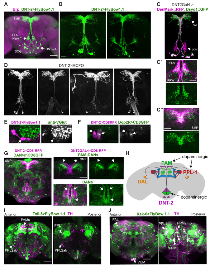
Figure 1.

Figure 1—figure supplement 1. Identification of neurotransmitter type for DNT-2A neurons.

Figure 1—figure supplement 1.

(A) All four DNT-2A neurons per hemibrain are glutamatergic as DNT-2>FlyBow1.1 reporter co-localises with anti-vGlut in these neurons as well as in their projections at SMP (arrows point at cluster of four cells). (B) All four DNT-2A neurons per hemibrain have Dop2R (arrows point to cluster, each neuron seen in magenta). (C, D) Anterior DNT-2A neurons projecting at SMP are not dopaminergic as there was no co-localisation between DNT-2>histoneYFP and anti-TH in the anterior brain (C), and there was no co-localisation in other DNT-2+ neurons in the posterior brain either (D). Higher magnification of dotted boxes on the right. (E) There was no co-localisation with Dop1R2LexA>CD8::GFP, DNT-2Gal4>CD8::RFP either. (F) DNT-2 neurons are not serotonergic as there was no co-localisation between DNT-2>FlyBow1.1 and the serotonergic neuron marker anti-5HT. (G) They are not octopaminergic as there was no overlap between TdcLexA>mCD8::GFP and DNT-2Gal4>CD8::RFP. (H) There was no overlap between DNT-2Gal4>histoneYFP and the cholinergic neuron marker anti-ChAT4b1. (I, J) Anterior DNT-2A neurons are not GABAergic, but lateral DNT-2 neurons are, as visualised with DNT-2>CD8-RFP, GADLexA>CD8-GFP. Scale bars: (A right, B, E, F, G, H, I, J) 20 µm; (A left, C,D same magnification) 50 µm. For further genotypes and sample sizes, see Supplementary file 2.
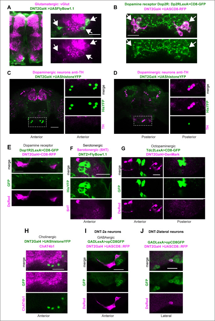
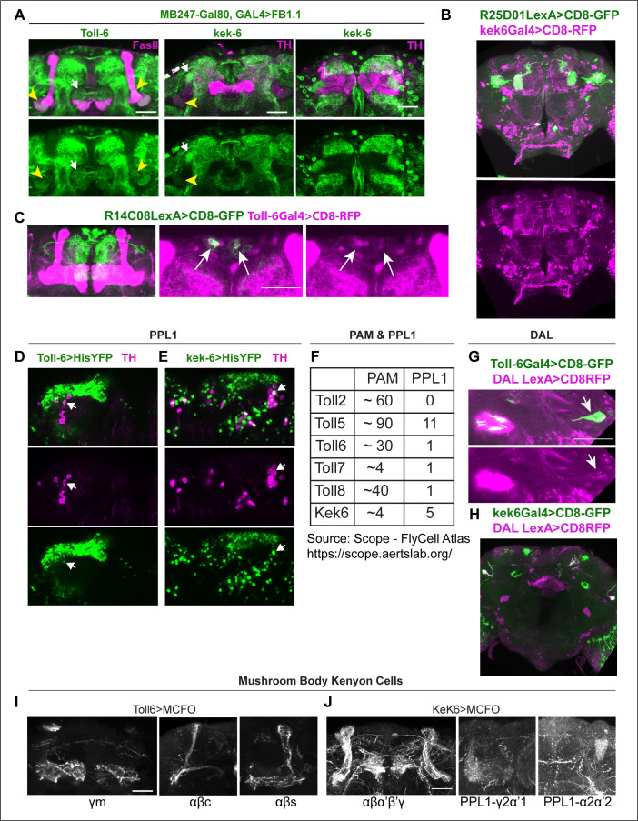
Figure 1—figure supplement 2. Identification of Toll-6 and kek-6-expressing neurons.

Figure 1—figure supplement 2.

(A)Toll-6Gal4, MB247-Gal80>UASFlyBow1.1 and kek-6Gal4, MB247-Gal80>UASFlyBow1.1 revealed Toll-6+ and Kek-6+ projections on MB γ2α’1. (B) R25D01LexA >CD8GFP, kek6-Gal4>CD8RFP overlapped in MB γ2α’1. (C) R14C08LexA >CD8GFP, Toll-6Gal4>CD8-RFP13 revealed co-localisation in MBON-M4/M6 cell bodies. (D, E) Co-localisation of Toll-6>Histone-YFP and kek-6>HistoneYFP with TH in PPL1 neurons. (F) Number of PAM and PPL1 neurons expressing Tolls and kek-6, revealed from published RNAseq data (Scope, see also Supplementary file 1). (G, H) Toll-6 and kek-6 are expressed in DAL neurons. (G) DAL neurons express Toll-6, as revealed by co-localisation between DAL-LexA (VT49239-LexA)>mCD8-GFP and Toll-6GAL4>mCD8-RFP. (H) DAL neurons express kek-6, as revealed by co-localisation between DAL-LexA (VT49239-LexA)>mCD8-GFP and kek6GAL4>mCD8RFP. (I, J) Toll-6 and kek-6 are expressed in MB neurons. (I) Toll-6>MCFO clones reveal expression at least in MB neurons γ medial (γm), αβcore (αβc), and αβsurface (αβs). (J) kek-6>MCFO clones reveal expression at least in MB Kenyon cells αβα’β’γ formed and MB γ2α’1 and α2α’2 and PPL1-γ2α’1 and PPL1-α2α’2. Scale bars: (B, H same magnification) 50 µm; (A,C,D,E,G, I, J same magnification) 30 µm. For further genotypes and sample sizes, see Supplementary file 2.