Abstract
Background
The aim of this study was to compare the effectiveness of two different vitrification methods and slow freezing in terms of the recovery of endocrine function, follicular morphology and proliferation, apoptosis of stromal cells, and angiogenesis after heterotopic transplantation of human ovarian tissue.
Methods
Ovarian tissue from young women aged 29 to 40 was subjected to two vitrification methods and one slow freezing method. The thawed ovarian tissue was then transplanted into nude mice and divided into three groups (VF1 group, VF2 group, SF group) according to the different freezing methods. Ovarian tissue samples were collected at 4 and 6 weeks post-transplantation. The recovery of ovarian function was evaluated by observing the estrous cycle and measuring estradiol levels using Elisa. Histological evaluation was performed to assess the integrity of ovarian follicles. TUNEL assay was used to detect stromal cell apoptosis, and immunohistochemistry was conducted to evaluate follicular proliferation and tissue angiogenesis.
Results
After heterotopic transplantation, mice in the experimental groups exhibited restoration of the estrous cycle. Hormone levels showed an increasing trend in the vitrification groups. At 6 weeks post-transplantation, the VF2 group had significantly higher hormone levels compared to the VF1 group and the slow freezing (SF) group (P < 0.05). At 4 weeks post-transplantation, the proportion of normal follicles was higher in the VF2 group compared to the other two groups (P > 0.05), and at 6 weeks post-transplantation, the VF2 group was significantly higher than the SF group (P < 0.05) and slightly higher than the VF1 group. Immunohistochemistry analysis indicated a higher proportion of proliferating follicles in the vitrification groups compared to the slow freezing group (P > 0.05). CD31 expression was established in all groups at 4 and 6 weeks post-transplantation, with better results in the slow freezing group compared to the vitrification group. TUNEL analysis showed that stromal cell apoptosis was higher in the SF group compared to the vitrification group at 4 weeks post-transplantation (P < 0.05), while there was no significant statistical difference among the groups at 6 weeks post-transplantation.
Conclusions
Vitrification showed better results than slow freezing, with the VF2 group performing slightly better than the VF1 group. Considering the lower economic and time costs associated with vitrification, it may be more suitable for ovarian tissue cryopreservation in major research centers in the future.
Supplementary Information
The online version contains supplementary material available at 10.1186/s12905-024-03505-1.
Keywords: Vitrification, Slow freezing, Heterotopic transplantation, Human ovarian tissue
Introduction
Since the 1970s, cancer incidence among young people has increased significantly [1]. Advances in medical technology have led to a five-year survival rate of up to 80% for these patients [2]. However, chemotherapy and radiation therapy often cause significant damage to the ovary, leading to fertility impairment and menopausal symptoms among female cancer survivors [3]. To address this, various strategies such as embryo, oocyte, or ovarian tissue cryopreservation have been advocated to preserve fertility. Among these, ovarian tissue cryopreservation stands out as the only option that can not only preserve fertility but also restore ovarian endocrine functions. Since the first successful case reported by Donnez’s team in 2004 [4], more than 200 healthy babies have been born worldwide using this method [5].
Ovarian tissue cryopreservation techniques are primarily divided into slow freezing (SF) and vitrification (VF). SF, a conventional approach, is currently recommended as the standard method for ovarian tissue cryopreservation [6]. However, ice formation during the cooling process of SF potentially damages the ovarian tissue. In contrast, VF avoids ice crystal formation due to rapid cooling rates and high concentrations of vitrification solutions [6]. Although live births through vitrification are lower than with SF, VF offers advantages such as better morphological integrity of stromal cells and vessels, shorter processing time, and inexpensive equipment. The debate on the superiority of either method is ongoing, with some studies favoring SF for follicle survival rates [7, 8] and others reporting higher interstitial cell integrity for VF [9]. Studies comparing SF and VF often differ in their findings, with some reporting no significant differences in restored ovarian tissue [10, 11]. Importantly, most studies focus on thawed ovarian tissue analysis, neglecting the long-term restoration of ovarian functions as a critical performance metric.
This study aims to comprehensively evaluate the efficacy of different cryopreservation techniques including two VF protocols and one SF protocol on estradiol production, follicle morphology and proliferation, stromal cell apoptosis, and revascularization after transplantation into Balb/c nude mice. By doing so, we aim to provide a more comprehensive understanding of the optimal cryopreservation method for preserving female fertility in cancer survivors.
Materials and methods
Collection and treatment of tissues
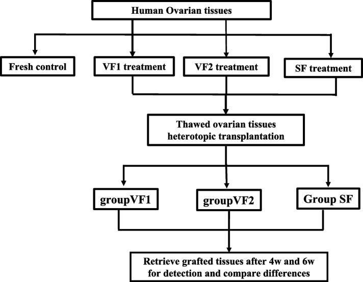
This study was approved by the Ethics Board of Peking University Shenzhen Hospital. Informed consent was obtained from three patients aged between 29 and 40 years (mean age 33 ± 6.08 years). Three patients were diagnosed with endometrioid adenocarcinoma, cervical adenocarcinoma, and non-mature teratoma of the ovary, respectively. Two underwent comprehensive staging surgery for malignant tumors, while one underwent fertility-sparing comprehensive staging surgery. Ovarian tissues were collected by the gynecologists during the surgery and placed in sterile 50 ml centrifuge tubes (Corning, USA) containing 15 ml of HEPES-buffered M199(M199, Gibco’s, Life Technologies, USA) supplemented with 10%(v/v) Serum Substitute Supplement (SSS, Irvine Scientific, Fujifilm, USA) and 100 mg/ml of streptomycin/penicillin (Gibco®, Life Technologies). The tubes were pre-warmed to 37℃. Under sterile conditions, the ovarian tissue was transferred to a Petri dish (Corning) containing the aforementioned media. A 20# scalpel was used to dissect the medulla and visible growing follicles. The ovarian fragments were cut into several pieces approximately 10mmx10mmx1-2 mm, among these, one or two pieces were fixed in 4% formaldehyde to serve as the control group, while the other ovarian tissue cubes (OTCs) were randomly frozen using different procedures (Fig. 1).
Fig. 1.
Flowchart of the study
Vitrification and warming
On the basis of our previous study [12], the following two vitrification protocols were superior in terms of normal follicle proportion, apoptosis rate and angiogenesis.
Group VF1
The vitrification protocol was performed as previously described by Amorim et al. with modifications to the equilibration time [13]. The ovarian tissues were incubated in an equilibration solution composed of 3.8% ethylene glycol (EG, Sigma-Aldrich, Merck, USA), 0.5 M sucrose, and 6% SSS with basic medium (MEM-Glumax, Gibco’s, Life Technologies, USA) for 3 min at room temperature (RT). They were then transferred to 19% EG and 0.5 M sucrose in MEM-Glumax with 6% SSS for 1 min. Finally, the tissues were incubated in a vitrification solution (38% EG, 0.5 M sucrose, MEM-Glumax with 6% SSS) for 11 min. After this procedure, the tissues were placed on a metallic grid and plunged into liquid nitrogen before being transferred to 1.8 ml cryotubes for long-term storage. For warming, the tissues were incubated in solutions with decreasing concentrations of sucrose (0.5 M, 0.25 M, 0.125 M, and 0 M) and 6% SSS in basic medium for 5 min at RT (Table S1).
Group VF2
This vitrification protocol was conducted according to Kagawa et al. with some modifications [14]. It involved a two-step protocol with ascending concentrations of EG and dimethylsulphoxide (DMSO, Sigma-Aldrich, Merck, USA) in basic medium (20% SSS and M199). The ovarian tissues were placed in 10% EG, 10% DMSO, 20% SSS in M199 for 25 min at RT. They were then exposed to a vitrification solution containing 20% EG, 20% DMSO, 0.5 M sucrose, and 20% SSS in M199 for 15 min at RT. For thawing, the tissues were submerged in a solution containing 1 M sucrose, 20% SSS, and M199 at 37℃ for 1 min. Subsequently, they were incubated in solutions with different concentrations of sucrose (0.5 M, 0 M, 0 M) and 20% SSS in M199 at RT for 5 min each step (Table S1).
Slow freezing and thawing
Group SF
Ovarian tissues were processed as described by vonWolff et al. with slight modifications (group SF) [15]. The tissues were placed in a 50 ml tube containing 20 ml of L-15 medium supplemented with 10% SSS, 10% DMSO and 0.1 M sucrose. After equilibration in the cryopreservative medium for 30 min at 4℃, the ovarian tissues were transferred to 1.8 ml cryotubes and placed in a computerized programmable freezer (CryoBioSystem, France). The cooling program involved a decrease from 2℃ to -6℃ at 2℃/min, followed by ice seeding. The cooling rate was then reduced to 0.3℃/min to reach -40℃ and 10℃/min to reach-140℃, finally the samples were stored in liquid nitrogen tank.
To initiate the thawing process, the cryopreserved cryotube vials were gently removed from liquid nitrogen and submerged in a 37℃ water bath for 2 min. Subsequently, the ovarian cubes were transferred to a petri dish containing L-15 medium enriched with 10% SSS, 1% DMSO, and 0.05 M sucrose, and incubated at 37℃ for 5 min. Following this, the specimens were rinsed twice in basic medium (L-15 medium + 10% SSS), with each wash lasting for 5 min. The tissues were then incubated in the basic medium at 37℃, prepared for transplantation (Table S1).
Transplantation to Balb/c nude mice
In terms of transplantation, our animal experiments strictly adhered to the guidelines outlined in the “Regulations of Experimental Animal Administration” issued by the State Committee of Science and Technology of the People’s Republic of China. Approval for all procedures was granted by the Animal Care and Use Committee of Peking University Shenzhen Hospital. Female Balb/c nude mice, aged 5–6 weeks, purchased from Zhuhai BesTest Bio-Tech company, China, were allowed to acclimate for a week under controlled conditions of a 28℃ temperature, a 12-h light/dark cycle, and access to a standard diet and water. Subsequently, a castration operation was performed on each mouse. All nude mice undergoing castration surgery were anesthetized with isoflurane (specification 100 ml, Zhongmu Beikang Pharmaceutical Co., Ltd., China). The induction anesthesia concentration of isoflurane was 2%-3%, and the maintenance anesthesia concentration was 1.5%-2%. Two weeks post-castration, thawed human ovarian fragments were transplanted bilaterally into the dorsolateral regions of all mice.
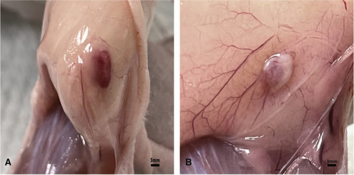
The mice were then allocated into three groups, VF1 (n = 10), VF2 (n = 10), and SF (n = 10), based on the transplanted ovarian tissues. After one week post-transplantation, mice in each group received abdominal injections of 2.0 IU human menopausal gonadotropin (HMG) every second day. Grafted tissues were retrieved for further analysis at 4 and 6 weeks post-transplantation (Fig. 2).
Fig. 2.
Ovarian tissues collected post-transplantation. A Ovarian tissue harvested 4 weeks after transplantation. Vascular coverage is observed. B Ovarian tissue harvested 6 weeks after transplantation. Uneven distribution of vascularization, with two-thirds of the ovarian tissue covered by blood vessels
Assessment of estrous cycle and estradiol concentration
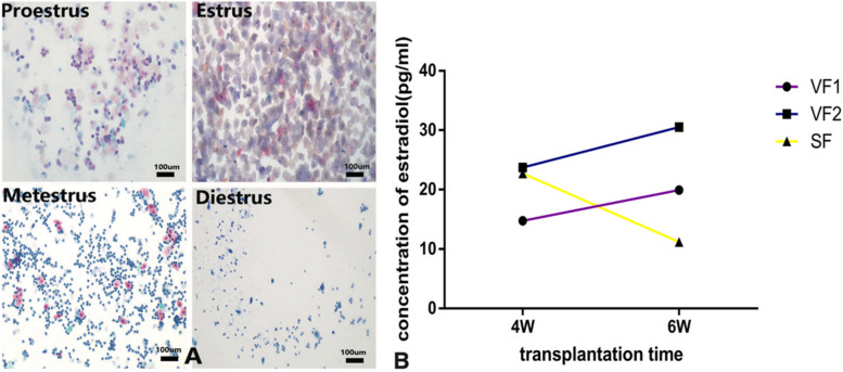
To assess the estrous cycle and estradiol concentration, vaginal smears were collected daily for 10 consecutive days, starting from the 5th day post-transplantation. These smears were fixed in 95% ethanol for 24 h and subsequently stained. With the aid of a 200 × light microscope, the estrous cycle was categorized into proestrus, estrus, metestrus, and diestrus, based on established criteria [16].
Proestrus
Characterized by a predominance of oval, medium-sized intermediate cells, with a smaller number of superficial and parabasal cells.
Estrus
The vaginal smear primarily consists of large, polygonal, exfoliated cornified cells.
Metestrus
Both large, polygonal cornified cells and polymorphonuclear leukocytes are observed.
Diestrus
The vaginal smear comprises mainly parabasal cells and numerous polymorphonuclear leukocytes.
Detection of hormone levels
To detect hormone levels, eye blood was collected from each mouse and allowed to sit at room temperature for 2 h. Following this, the samples were centrifuged at 1000 × g for 20 min and subsequently stored at -20℃ for further analysis. ELISA kit instructions (ml001962-1, Shanghai, China) were followed to determine hormone levels in the mice after transplantation.
Histological examination
Histological examination was performed on the transplanted ovarian tissue after 4 and 6 weeks of transplantation. The tissue was fixed with 4% paraformaldehyde for 48 h and subsequently dehydrated and embedded in paraffin. 4 μm-thick paraffin sections were created, and the morphology of ovarian tissue and follicles was observed using a 200 × light microscope. Every sixth section of the serially sectioned tissue was analyzed to count the number of normal and abnormal follicles, thus enabling the calculation of the proportion of normal follicles. The morphology of follicles was evaluated using the Gougeon classification criteria [17]. Normal follicles were circular or elliptical in shape, with no pyknosis in the nucleus of oocytes and granulosa cells; granulosa cells were arranged neatly, with even distribution around the oocytes, and the basement membrane was intact. Abnormal follicles were irregularly shaped, with pyknosis in the nucleus of the oocyte, irregular arrangement of granulosa cells, and detachment of the basement membrane.
Immunohistochemistry
Proliferation and angiogenesis of the transplanted tissues were assessed by examining the expression of Ki-67 and CD31. Initially, the paraffin-embedded tissue sections were deparaffinized and rehydrated. Endogenous peroxidase activity was then blocked using 3% hydrogen peroxide for 15 min at room temperature. Subsequently, the slides were subjected to antigen retrieval by heating in citrate solution at 98℃ for 25 min in a microwave oven. Following blocking, the human ovarian tissue sections were incubated overnight at 4℃ with primary antibodies Ki-67 (diluted at 1:200, Abcam, USA) and CD31 (diluted at 1:2000, Abcam, USA). After rewarming, the tissue sections were washed three times for 3 min each with PBS buffer. The secondary antibody was selected from the ready-to-use immunohistochemical ElivisionTM Super kit as the manufacturer’s instructions. Following DAB treatment, the sections were counterstained with hematoxylin, dehydrated, and mounted for microscopic evaluation.
Follicles were classified as proliferative if they exhibited at least one Ki-67-positive granulosa cell in proximity. The result was quantified as the positive rate, calculated as (number of positive follicles / total follicles) * 100%. Due to the limited number of follicles, three slices of different depths were selected from each tissue, and the total number of follicles in each slice was counted under 200 × microscope.
CD31 expression was indicative of endothelial cells within blood vessels. Thus, three randomly chosen microscopic fields were assessed from each slice to determine the number of positive vessel expressions. The expression density of positive vessels (expressed as n/mm2) was utilized to characterize angiogenesis.
Apoptosis detection (TUNEL)
Stromal cells play a crucial role in evaluating the efficacy of different protocols and as well as the follicles. Sections were deparaffinized and rehydrated following standard histologic procedures. TUNEL detection was conducted according to the instructions provided in the kit (Servicebio, Servi Biotechnology, produced in Wuhan, China). The number of positive and negative cells was observed under 200 × microscope, and four random fields were selected from each slide to calculate the positive rate. Apoptotic stromal cells were characterized by a brown-yellow stained nucleus. The apoptosis rate was calculated using the formula: (number of positive stromal cells / total stromal cell count) * 100%.
Statistical analysis
All data were presented as mean ± SD. Inter-group differences were assessed using one-way analysis of variance (ANOVA) followed by post hoc Tukey’s Honestly Significant Difference (HSD) test. Statistical significance was defined as p < 0.05. All statistical analyses were performed using SPSS 25 (IBM).
Results
Inter-group differences of estrous cycle and estradiol levels
Vaginal smears were collected from six randomly selected mice in each group starting from the sixth day post-transplantation. It was observed that 100% of the rats in groups VF1 and VF2 exhibited an intact estrous cycle. The estrus stage, characterized by the presence of fully cornified cells, appeared on average on day 8.5 and 7.6 post-transplantation for groups VF1 and VF2, respectively. In group SF, four out of six mice maintained an intact cycle, with cornified cells appearing on day 9. However, two mice lacked a discernible estrus stage.
At the 4-week post-transplantation mark, the estradiol levels of mice in groups VF2 (23.73 ± 2.67 pg/ml) and SF (22.70 ± 3.33 pg/ml) were significantly higher than those in group VF1 (14.75 ± 6.36 pg/ml) (VF2 vs VF1, p = 0.005; SF vs VF1, p = 0.008). Although not statistically significant, the estradiol levels of mice in group VF2 were slightly higher than those in group SF (p = 0.718). Furthermore, mice in the vitrification groups exhibited higher estradiol levels than SF mice after six weeks of transplantation (VF1: 19.91 ± 7.23 pg/ml; VF2: 30.52 ± 5.92 pg/ml; SF: 11.2 ± 1.68 pg/ml; VF1 vs SF, p = 0.007; VF2 vs SF, p = 0.000). Additionally, the estradiol levels of mice in group VF2 were significantly higher than those in group VF1 (p = 0.003). Further analysis revealed that the estradiol levels of mice in the vitrification groups continued to increase from the 4-week to the 6-week post-transplantation period. Conversely, SF mice exhibited a notable decrease in estradiol levels from the 4-week to the 6-week post-transplantation period (p = 0.000) (Fig. 3).
Fig. 3.
Post-transplantation estrous cycle and hormone levels. A The restored normal estrous cycle in nude mice from group VF2 after transplantation B The hormone levels in each group after transplantation
Differences of the normal follicles between the VF1, VF2 and SF group
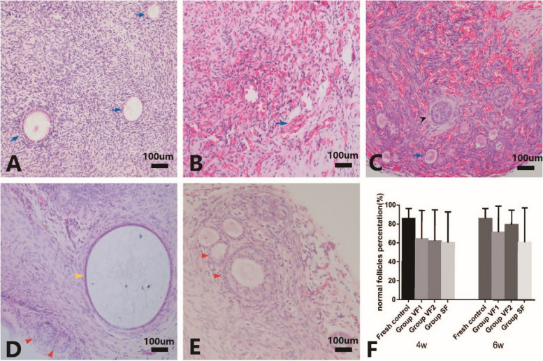
A total of 28 samples were obtained from the graft site at 4 weeks and 6 weeks post-transplantation, respectively. At the 4-week mark, the mean percentage of morphologically normal follicles was 36.81% ± 30.85%, 51.28% ± 30.89%, and 48.59% ± 34.37% in groups VF1, VF2, and SF, respectively. These rates were significantly lower than those observed in fresh ovarian tissue (92.1% ± 6.61%, p < 0.05). While the percentage of normal follicles was higher in group VF2 compared to the other two groups, the difference was not statistically significant (VF2 vs VF1, p = 0.279, VF2 vs SF, p = 0.849).
However, by the 6-week post-transplantation mark, the rate of normal follicles in ovarian tissues increased in all groups: VF1 (70.71% ± 28.84%), VF2 (85.59% ± 11.53%), and SF (55.05% ± 41.43%). Additionally, the VF2 group exhibited the highest proportion of normal follicles compared to the other two groups (VF2 vs VF1, p = 0.302; VF2 vs SF, p = 0.038). No significant difference was found between groups VF1 and SF (p = 0.196) (Fig. 4).
Fig. 4.
Inter-group differences of the normal follicles after transplantation. A Morphological examination of fresh tissue, blue arrows indicate morphologically intact primary follicles. B Abundant vascular generation observed 4 weeks after transplantation. C Multilevel follicles observed 4 weeks after transplantation, with black arrows indicating secondary follicles and blue arrows indicating primary follicles. D and E Follicles’ development in the tissue after 6 weeks of transplantation, with red arrows indicating normal secondary follicles, yellow arrow indicating apoptotic follicle. F Comparison of the proportions of normal morphological follicles among different groups
Inter-group variations for follicular proliferation and angiogenesis of transplanted ovarian tissues
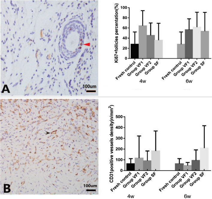
The proportion of proliferative follicles in fresh human ovarian tissue was 28.02% ± 24.03%. After 4-week transplantation, the proportion of proliferative follicles was higher than that of fresh ovarian tissue in groups VF1 (64.34% ± 29.91%, p = 0.006), VF2 (45.48% ± 24.45%, p = 0.202), and SF (35.89% ± 33.56%, p = 0.549). Group VF1 exhibited a higher proportion of proliferative follicles compared to the other two groups (VF1 vs VF2, p = 0.109; VF1 vs SF, p = 0.013), and there was no significant difference between group VF2 and SF (p = 0.549). At 6-week post-transplantation, the percentage of proliferative follicles was 56.52% ± 21.65%, 61.64% ± 29.44%, and 53.61% ± 37.23% for groups VF1, VF2, and SF, respectively. Although group VF2 had a slightly higher proportion of proliferative follicles compared to the other two groups, there was no significant difference among the three groups (VF2 vs VF1, p = 0.696; VF2 vs SF, p = 0.519). Despite slight variations in the proportion of proliferative follicles among the three groups between 4-week and 6-week post-transplantation, there were no significant differences within each group (p > 0.05).
The CD31-positive vascular density in fresh human ovarian tissue was 64.57 ± 46.94 n/mm2. At 4-week post-transplantation, this number was 116.50 ± 205.32 n/mm2, 91.40 ± 91.15 n/mm2, and 183.16 ± 185.44 n/mm2 for ovarian tissues in groups VF1, VF2, and SF, respectively. Statistically, SF exhibited a higher CD31-positive vascular density than groups VF1, VF2, and the fresh control (p = 0.094, p = 0.03, p = 0.005, respectively). There was no significant difference between the CD31-positive vascular density of groups VF1 and VF2 compared to the fresh control (VF1 vs VF2, p = 0.556;). At 6-week post-transplantation, SF still exhibited notably higher CD31-positive vascular density than groups VF1 and VF2 (VF1: 47.81 ± 27.78 n/mm2; VF2: 94.52 ± 98.09 n/mm2; SF: 209.61 ± 207.74 n/mm2; p < 0.01). The CD31-positive vascular density of the two vitrification groups did not exhibit any significant difference at 6 weeks post-transplantation, as well as at 4 weeks (VF1 vs VF2, p = 0.305;). These results indicate that the transplanted tissues showed well-established new vessels at 4 weeks post-transplantation, and there was little change within the extension of the transplantation period (Fig. 5).
Fig. 5.
Follicular proliferation and angiogenesis of transplanted ovarian tissues. A Proliferating follicles after tissue transplantation, red arrows indicate positively stained granulosa cells. Comparison of proportions of proliferating cells among different groups. B Blood vessels expressing CD31, with black arrows indicating positively stained newly formed blood vessels. Comparison of CD31-positive blood vessel density among different groups
Apoptosis of interstitial cells
At 4-week post-transplantation, the proportion of apoptotic stromal cells in ovarian tissues of the three groups was 26.61% ± 19.18%, 18.13% ± 13.34%, and 36.31% ± 13.79%, respectively. The proportion of apoptotic stromal cells in group VF2 was significantly lower than in group VF1 (p = 0.039) and group SF (p = 0.000), and group VF1 was also lower than group SF (p = 0.006). The proportion of apoptotic stromal cells showed minimal differences between the vitrification groups and the fresh control group (26.67% ± 16.28%) (VF1 vs control, p = 0.990; VF2 vs control, p = 0.080).
Although there was no statistical significance among the three groups after 6 weeks of transplantation (VF1 vs VF2, p = 0.064; VF1 vs SF, p = 0.427; VF2 vs SF, p = 0.211;), the proportion of apoptotic interstitial cells in the vitrification groups and the SF group was 24.38% ± 12.86% (VF1), 32.12% ± 14.89% (VF2), and 27.26% ± 12.09% (SF), respectively. Additionally, there were no significant differences observed between the transplanted ovarian tissues and the fresh control tissues (Fig. 6).
Fig. 6.
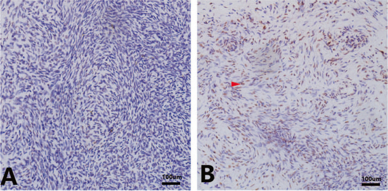
A and B Apoptotic stromal cells in fresh tissue and tissue harvested 4 weeks after transplantation, with red arrows indicating stromal cells stained brown, representing apoptotic cells
Discussion
Cryopreservation and auto-transplantation of ovarian tissues has proven to be effective methods for preserving fertility in young female cancer patients [18]. Studies have shown that approximately 95% of transplanted ovarian tissues can restore endocrine function [19], which is consistent with the findings of this study where intact estrous cycles were observed in each group. Regarding hormone levels, our results demonstrate that vitrification is more suitable than slow freezing. From 4 to 6 weeks post-transplantation, the estradiol levels in the vitrification groups showed an increasing trend, while those in the slow freezing group decreased. Suguru et al. [20] reported that slow freezing resulted in earlier estrus recovery and higher estradiol levels at 10 days after transplantation, indicating that slow freezing caused less damage to follicles compared to vitrification. However, at 20 days after transplantation, there was no significant difference in hormone levels between the two methods, and similarly, the estradiol levels in the slow freezing group decreased compared to the early stage. Based on these findings, we may speculate that slow freezing not only causes greater damage to follicles but also leads to premature recruitment of follicles in the transplanted ovarian tissue, resulting in excessive loss of follicles. The fibrous connective tissue in the ovarian cortex plays a crucial role in inhibiting the rapid growth and development of follicles [21]. Damage or destruction of this tissue, as seen in slow freezing, may result in faster depletion of follicles due to increased activation of primordial follicles after transplantation. it is known that intracellular ice crystal formation occurs during slow freezing, while the vitrification process avoids the formation of ice crystals due to the use of high concentrations of cryoprotectants and rapid cooling rates. The formation of ice crystals in slow freezing may disrupt the structure of the fibrous connective tissue in the ovarian cortex, leading to rapid recruitment and maturation of follicles, which ultimately impairs the recovery of ovarian function after transplantation. Based on these observations, we have reason to believe that vitrification is a better method than slow freezing, and further studies will be conducted to investigate the specific mechanisms involved.
In this study, the vitrification groups demonstrated certain advantages in preserving follicles and stromal cells. Consistent with other research, vitrification appeared to cause less damage to follicle integrity and reduced DNA damage in oocytes and granulosa cells compared to slow freezing [6, 22]. However, conflicting opinions exist in the literature. For instance, Ronit Abir et al. [23] did not find that vitrification was superior to slow freezing in preserving interstitial cells. Some studies even suggest that slow freezing may outperform vitrification in terms of preserving follicle numbers, follicular proliferation, and angiogenesis [8]. Abnormal morphology of primary follicles and increased sensitivity of stromal cells to ischemic damage were observed after vitrification in some studies [7], along with a higher incidence of apoptotic cells in ovarian tissue [24].
Further optimization of vitrification protocols may make it a potential alternative to slow freezing. Variations in vitrification protocols can lead to significant differences in ovarian tissue outcomes. Vitrification protocols containing dimethyl sulfoxide (DMSO) have shown higher follicle survival rates compared to protocols using a single high-concentration ethylene glycol (EG), which aligns with findings from J. Marschalek’s research in 2021 [25]. Although vitrification currently lags behind slow freezing in terms of successful deliveries, it offers advantages in terms of time and cost efficiency [10]. Continuous optimization efforts may eventually allow vitrification to replace traditional slow freezing methods.
Another key factor that determines the survival of transplanted ovarian tissue is the establishment of early vascularization. From our study, it can be observed that after four weeks of transplantation, the blood vessels of all transplanted tissues were basically established, and the vascular generation in the slow freezing group was more ideal than that in the vitrification group. There was no significant difference between the two vitrification groups. Lee et al. believed that slow freezing is significantly superior to the vitrification group in terms of angiogenesis [8]. Some studies have proposed that the expression of vascular endothelial growth factor (VEGF) and angiogenic protein 2 in ovarian tissue decreases after freezing, but there was no significant difference between the vitrification and slow freezing groups [11]. The expression can be restored after ovarian tissue transplantation. Similarly, multiple studies have concluded that there is no significant difference between slow freezing and vitrification in terms of neovascularization or gene expression of angiogenic factors [23, 26]. There are many factors that influence neovascularization in ovarian tissue, such as interventions with vitamin E [27], VEGF [28], basic Fibroblast Growth Factor( bFGF) [29], GDF9-β and adipose-derived stem cells [30, 31] before or after transplantation, which can improve the survival rate of follicles in ovarian tissue and promote neovascularization. In our study, we injected HMG intraperitoneally after ovarian tissue transplantation, and all groups showed rich vascular density. HMG has a certain synergistic effect in promoting VEGF expression in ovarian tissue, similar to the results of Wang et al. [32]. In mouse ovarian tissue autografts, new blood vessel formation can be observed as early as three days after transplantation [11], while in human ovarian tissue transplantation, new blood vessels gradually form starting from the fifth day [33]. Early ischemic injury can lead to the loss of 60–95% of follicles [34], indicating the importance of early vascular generation in reducing follicular ischemic loss and prolonging the functional lifespan of transplanted ovarian tissue. However, since our study did not include early transplantation timepoints, we cannot draw conclusions about early vascular generation. In future studies, we will incorporate artificial biomaterials to further investigate early vascularization factors in ovarian tissue transplantation, aiming to provide a basis for establishing blood vessels in ovarian tissue.
In our research, nude mice were chosen as the experimental model primarily due to their infertility, thus limiting the duration of our study. The fundamental aim of ovarian tissue cryopreservation is to preserve fertility and endocrine functions. The ability to give birth to healthy offspring and the duration of reproductive endocrine function after giving birth are important criteria for the success of ovarian tissue freezing and transplantation. Therefore, future experiments will prioritize the selection of animals with reproductive capabilities, allowing us to compare various cryopreservation protocols and monitor ovarian tissue functionality post-transplantation. Additionally, due to the challenges associated with acquiring clinical samples, it was not feasible to procure enough ovarian tissue from a single individual for transplantation in this study. So in this study, we evenly divided each participant’s ovarian samples into three groups to minimize error. Future research will utilize bovine ovarian tissue, which shares significant structural similarities with human ovarian tissue, for in vivo investigations, aiming to gather more comprehensive data.
Conclusion
In conclusion, our study evaluated the efficacy of various freezing protocols for ovarian tissue cryopreservation, focusing on tissue function restoration, follicular integrity, and vascularization. We believe that vitrification, particularly using the vitrification protocol 2, exhibits promising potential. With ongoing optimizations in vitrification techniques, it could potentially supersede traditional slow freezing methods.
Supplementary Information
Acknowledgements
Not applicable.
Abbreviations
- SF
Slow Freezing
- VF
Vitrification
- M199
HEPES-buffered M199
- OTCs
Ovarian Tissue Cubes
- EG
Ethylene Glycol
- SSS
Serum Substitute Supplement
- DMSO
Dimethylsulphoxide
- RT
Room Temperature
- HMG
Human Menopausal Gonadotropin
- PBS
Phosphate Buffer Saline
- DAB
3,3’-Diaminobenzidine
- VEGF
Vascular Endothelial Growth Factor
- bFGF
Basic Fibroblast Growth Factor
Authors’ contributions
W.R. designed this study. Z.Y. and L.Y. conducted the experiments. D.H. and L.C. helped with the experiments. Z.Y. and D.W. conducted the data analysis and drafted the manuscript. W.R. polished the manuscript. All authors reviewed the manuscript.
Funding
This study was supported by funds for Shenzhen Public Platform for Preservation of Fertility and Reproduction (XMHT20220104049), Shenzhen High-level Hospital Construction (YBH2019-260), and Peking University Shenzhen Hospital Scientific Research Fund (KYQD2021075).
Data availability
No datasets were generated or analysed during the current study.
Declarations
Ethics approval and consent to participate
This study was approved by the Ethics Board of Peking University Shenzhen Hospital.
Consent for publication
Our manuscript does not contain any individual person’s data in any form. However, three attenders provided written consents for the publication of the study.
Competing interests
The authors declare no competing interests.
Footnotes
Publisher’s Note
Springer Nature remains neutral with regard to jurisdictional claims in published maps and institutional affiliations.
Yucui Zeng and Yushan Li contributed equally to this work.
Contributor Information
Wenkui Dai, Email: daiwenkui84@gmail.com.
Ruifang Wu, Email: wurfpush@126.com.
References
- 1.Anbari F, et al. Fertility preservation strategies for cancerous women: an updated review. Turk J Obstet Gynecol. 2022;19(2):152–61. [DOI] [PMC free article] [PubMed] [Google Scholar]
- 2.Harada M, et al. Japan Society of Clinical Oncology Clinical Practice Guidelines 2017 for fertility preservation in childhood, adolescent, and young adult cancer patients: part 1. Int J Clin Oncol. 2022;27(2):265–80. [DOI] [PMC free article] [PubMed] [Google Scholar]
- 3.Gamzatova Z, et al. Autotransplantation of cryopreserved ovarian tissue–effective method of fertility preservation in cancer patients. Gynecol Endocrinol. 2014;30 Suppl 1:43–7. [DOI] [PubMed] [Google Scholar]
- 4.Donnez J, et al. Livebirth after orthotopic transplantation of cryopreserved ovarian tissue. Lancet. 2004;364(9443):1405–10. [DOI] [PubMed] [Google Scholar]
- 5.Dolmans MM, et al. Transplantation of cryopreserved ovarian tissue in a series of 285 women: a review of five leading European centers. Fertil Steril. 2021;115(5):1102–15. [DOI] [PubMed] [Google Scholar]
- 6.Mathias FJ, et al. Ovarian tissue vitrification is more efficient than slow freezing in protecting oocyte and granulosa cell DNA integrity. Syst Biol Reprod Med. 2014;60(6):317–22. [DOI] [PubMed] [Google Scholar]
- 7.Vatanparast M, et al. Evaluation of sheep ovarian tissue cryopreservation with slow freezing or vitrification after chick embryo chorioallantoic membrane transplantation. Cryobiology. 2018;81:178–84. [DOI] [PubMed] [Google Scholar]
- 8.Lee S, et al. Comparison between slow freezing and vitrification for human ovarian tissue cryopreservation and xenotransplantation. Int J Mol Sci. 2019;20(13):3346. [DOI] [PMC free article] [PubMed] [Google Scholar]
- 9.Jeong K, et al. Ovarian cryopreservation. Minerva Med. 2012;103(1):37–46. [PubMed] [Google Scholar]
- 10.Schallmoser A, et al. The effect of high-throughput vitrification of human ovarian cortex tissue on follicular viability: a promising alternative to conventional slow freezing? Arch Gynecol Obstet. 2023;307(2):591–9. [DOI] [PMC free article] [PubMed] [Google Scholar]
- 11.Choi WJ, et al. Expression of angiogenic factors in cryopreserved mouse ovaries after heterotopic autotransplantation. Obstet Gynecol Sci. 2015;58(5):391–6. [DOI] [PMC free article] [PubMed] [Google Scholar]
- 12.Li X, et al. Comparison of short-term transplantation effect of different vitrification solution on human ovarian tissue. Chin J Reprod Contracept. 2022;42(1):58–64. [Google Scholar]
- 13.Amorim CA, et al. Vitrification of human ovarian tissue: effect of different solutions and procedures. Fertil Steril. 2011;95(3):1094–7. [DOI] [PubMed] [Google Scholar]
- 14.Kagawa N, Silber S, Kuwayama M. Successful vitrification of bovine and human ovarian tissue. Reprod Biomed Online. 2009;18(4):568–77. [DOI] [PubMed] [Google Scholar]
- 15.Van der Ven H, et al. Ninety-five orthotopic transplantations in 74 women of ovarian tissue after cytotoxic treatment in a fertility preservation network: tissue activity, pregnancy and delivery rates. Hum Reprod. 2016;31(9):2031–41. [DOI] [PubMed] [Google Scholar]
- 16.Montes GS, Luque EH. Effects of ovarian steroids on vaginal smears in the rat. Acta Anat (Basel). 1988;133(3):192–9. [DOI] [PubMed] [Google Scholar]
- 17.Gougeon A, Lefèvre B, Testart J. Influence of a gonadotrophin-releasing hormone agonist and gonadotrophins on morphometric characteristics of the population of small ovarian follicles in cynomolgus monkeys (Macaca fascicularis). J Reprod Fertil. 1992;95(2):567–75. [DOI] [PubMed] [Google Scholar]
- 18.Oktay K, et al. Fertility preservation in patients with cancer: ASCO clinical practice guideline update. J Clin Oncol. 2018;36(19):1994–2001. [DOI] [PubMed] [Google Scholar]
- 19.Gellert SE, et al. Transplantation of frozen-thawed ovarian tissue: an update on worldwide activity published in peer-reviewed papers and on the Danish cohort. J Assist Reprod Genet. 2018;35(4):561–70. [DOI] [PMC free article] [PubMed] [Google Scholar]
- 20.Igarashi S, et al. Cryopreservation of ovarian tissue after pretreatment with a gonadotropin-releasing hormone agonist. Reprod Med Biol. 2010;9(4):197–203. [DOI] [PMC free article] [PubMed] [Google Scholar]
- 21.Silber SJ, et al. Cryopreservation and transplantation of ovarian tissue: results from one center in the USA. J Assist Reprod Genet. 2018;35(12):2205–13. [DOI] [PMC free article] [PubMed] [Google Scholar]
- 22.Behl S, et al. Vitrification versus slow freezing of human ovarian tissue: a systematic review and meta-analysis of histological outcomes. J Assist Reprod Genet. 2023;40(3):455–64. [DOI] [PMC free article] [PubMed] [Google Scholar]
- 23.Abir R, et al. Attempts to improve human ovarian transplantation outcomes of needle-immersed vitrification and slow-freezing by host and graft treatments. J Assist Reprod Genet. 2017;34(5):633–44. [DOI] [PMC free article] [PubMed] [Google Scholar]
- 24.Rahimi G, et al. Apoptosis in human ovarian tissue after conventional freezing or vitrification and xenotransplantation. Cryo Letters. 2009;30(4):300–9. [PubMed] [Google Scholar]
- 25.Marschalek J, et al. The effect of different vitrification protocols on cell survival in human ovarian tissue: a pilot study. J Ovarian Res. 2021;14(1):170. [DOI] [PMC free article] [PubMed] [Google Scholar]
- 26.Cho IA, et al. Angiopoietin-1 and -2 and vascular endothelial growth factor expression in ovarian grafts after cryopreservation using two methods. Clin Exp Reprod Med. 2018;45(3):143–8. [DOI] [PMC free article] [PubMed] [Google Scholar]
- 27.Nugent D, et al. Protective effect of vitamin E on ischaemia-reperfusion injury in ovarian grafts. J Reprod Fertil. 1998;114(2):341–6. [DOI] [PubMed] [Google Scholar]
- 28.Abir R, et al. Improving posttransplantation survival of human ovarian tissue by treating the host and graft. Fertil Steril. 2011;95(4):1205–10. [DOI] [PubMed] [Google Scholar]
- 29.Wang L, et al. VEGF and bFGF increase survival of xenografted human ovarian tissue in an experimental rabbit model. J Assist Reprod Genet. 2013;30(10):1301–11. [DOI] [PMC free article] [PubMed] [Google Scholar]
- 30.Manavella DD, et al. Two-step transplantation with adipose tissue-derived stem cells increases follicle survival by enhancing vascularization in xenografted frozen-thawed human ovarian tissue. Hum Reprod. 2018;33(6):1107–16. [DOI] [PubMed] [Google Scholar]
- 31.Vatanparast M, et al. GDF9-β promotes folliculogenesis in sheep ovarian transplantation onto the chick embryo chorioallantoic membrane (CAM) in cryopreservation programs. Arch Gynecol Obstet. 2018;298(3):607–15. [DOI] [PubMed] [Google Scholar]
- 32.Wang Y, et al. Effects of HMG on revascularization and follicular survival in heterotopic autotransplants of mouse ovarian tissue. Reprod Biomed Online. 2012;24(6):646–53. [DOI] [PubMed] [Google Scholar]
- 33.Van Eyck AS, et al. Electron paramagnetic resonance as a tool to evaluate human ovarian tissue reoxygenation after xenografting. Fertil Steril. 2009;92(1):374–81. [DOI] [PubMed] [Google Scholar]
- 34.Liu L, et al. Restoration of fertility by orthotopic transplantation of frozen adult mouse ovaries. Hum Reprod. 2008;23(1):122–8. [DOI] [PubMed] [Google Scholar]
Associated Data
This section collects any data citations, data availability statements, or supplementary materials included in this article.
Supplementary Materials
Data Availability Statement
No datasets were generated or analysed during the current study.


