Abstract
The authors present a case of a distal phalanx fracture secondary to a nail gun injury. The diagnosis, evaluation and emergency departmetn treatment are reviewed.
Keywords: distal phalanx fracture, tuft fracture
Introduction
The bones of the finger can be divided into three phalanges: the proximal, middle, and distal phalanx. The distal phalanx is responsible for a fingertips rounded appearance and contains a base, shaft, and tuft. The terminal extensor tendon is attached to the dorsal tubercle of the distal phalanx’s base, and the volar plate of the distal interphalangeal (DIP) joint exists on the palmar tubercle. These connections, among others, allow for the movement of the distal phalanx independent of the middle phalanx, and injury to them or the base of the distal phalanx threatens this freedom of motion. The distal phalanx is further supported by the nail bed which originates just beyond the terminal tendon.1
Injuries to the distal phalanx are generally considered less severe than those that occur in the remaining phalanges due to their ability to be treated conservatively. Intra-articular fractures pose the highest risk of a need of surgical intervention in order to ensure proper joint repair and are rare.2,3 Alternatively, extra-articular fractures, which constitute most distal phalanx fractures, can typically heal non-operatively as a result of the stability of the surrounding tissue and nail.4 These fractures most commonly affect the tuft section of the distal phalanx and are normally a result of crush trauma.5 For suspected tuft fractures, it is also important to identify whether they are simple or comminuted. Comminuted tuft fractures involve multiple breaks and a distribution of bone around the distal part of the fingertip, as opposed to simple which would consist of only one break and resembles a Seymour’s fracture.6 Most comminuted fractures can still be managed non operatively with a splint, however if the fragmentation is severe, they warrant K-wire stabilization.7
In a study from the Netherlands, bone fractures make up 12% of ED visits with hand fractures constituting 19% of those visits. 59% of those hand fractures were related to the phalanges.8 A similar study out of the Netherlands measured the economic cost of these wrist and hand injuries, totaling over $410 million dollars per year due to lost productivity.9 A study from the US on work related injuries to the upper extremities places the average expected cost at $1448 and the average days away from work at 10.10 These costs can be debilitating despite the fact that treatment for 74% of phalangeal fractures is conservative.11 While these stats vary based on geography and the political structuring of healthcare systems, it is representative of the inconvenience an individual is posed and why they may seek to dismiss seemingly minor hand or wrist injuries to avoid costs. While cost can appear to exceed the level of treatment, there is still a crucial need for diagnostic assessment of phalangeal fractures as when they are not treated sufficiently, individuals are at risk of not regaining full function, experiencing chronic pain, and deformation.12,13
Case Presentation
A 27-year-old male presented to the emergency department (ED) with a nail through his finger caused by an accident while working construction. The injury was inflicted by a pressurized nail gun resulting in a penetrating trauma. A physical examination yielded no concerns and past medical history was unremarkable. Vital signs were normal.
Upon assessment of the injury, the significant penetrative trauma to the middle finger which resulted in a distal phalanx fracture with a particular focus on the tuft region was evident. To manage the pain, the patient was given a digital block with 2% lidocaine without epinephrine. The wound was then thoroughly irrigated to remove any potentially contaminated debris. Given the nature of the trauma, a tetanus shot was given as well as one dose of antipseudomonal antibiotics as a preventative measure and the patient received a 10-day supply of oral antibiotics.
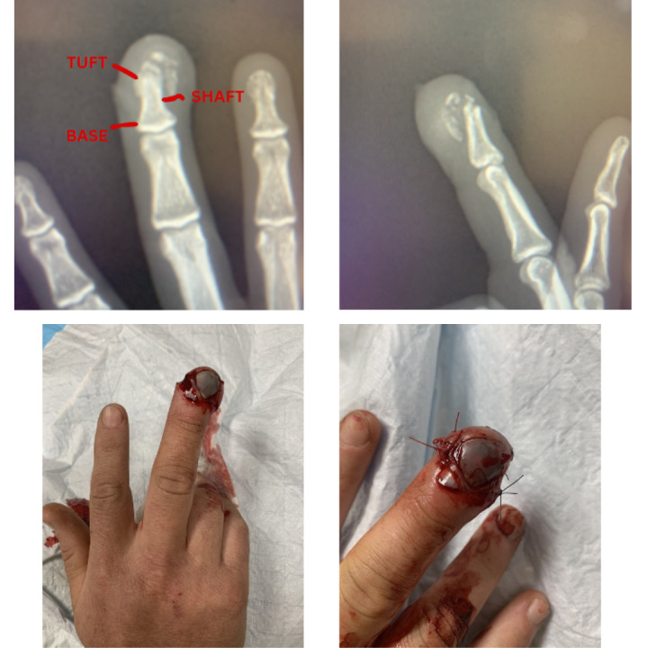
The force applied by the nail gun had caused the bone to fragment from the tuft down to the top of the shaft, indicative of a crush injury. The nail was fragmented as well as significant damage to the flesh and nerves of the fingertip, the nail bed however was intact.
Diagnostic imaging revealed a comminuted fracture of the distal phalanx (tuft fracture). Interrupted absorbable sutures were then placed on the left and right side of the finger to support healing, and the fingernail was not removed given its role as a biologic dressing [Figure 1]. Given the bone fragments were contained and the fracture was extra-articular, treatment could be fully provided within the ER and the patient was discharged.
Figure 1. Radiographs (top) and Photographs (bottom) of the phalanx injury.

Discussion
Phalanx fractures are a common reason for ED admission and their classification is necessary for determining the best method of treatment. Young adult men disproportionately represent those most affected due to occupational and behavioral risk, as well as elderly women due to fall risk.11,14,15 Of these phalanx fractures, a majority (33%) are a result of crush injuries and the distal phalanx has the highest occurrence of fracture (43%). Distal phalanx tuft fractures, in particular, account for 25% of injuries. Despite this concentration, methods of treatment vary and are best handled on the basis of factors including vitality of the nail bed and whether the wound is open or closed.
Whether the fracture is an open or closed wound, and to what extent, is a necessary element in the treatment of distal phalanx fractures. Closed wounds of the tuft lend themselves to non-operative treatment (71%) due to the support of surrounding tissue, with the binding of the middle and distal phalanges for a period of 10-14 days being sufficient for a full recovery.11,16 For open wounds, thorough irrigation and debridement is almost always necessary. Surgical intervention for open distal phalanx fractures are the same as closed with the added consideration of debridement efficacy and risk of infection.17 Treatment of these open fractures can be complicated by the detachment of the nail which can often hide an underlying phalangeal fracture as well as contribute to the instability of the distal phalanx, making radiography a necessary diagnostic step.18 Reattaching the nail when possible using methods such as figure eight suturing aids in the full recovery of the finger after a fracture.19 The nature of crush trauma might be considered a closed wound, however in this instance it typically implies a subungual hematoma which can require decompression and result in an opening of the injury.16
In any open distal phalanx fractures there is a consideration of antibiotic use. While use of antibiotics is not uncommon and is often associated with typical treatment, a case study by Metcalfe et. al asserted that prophylactic antibiotic use for the prevention of osteomyelitis showed no significance versus only irrigation and debridement.20
Complications following a distal phalanx fracture include infection, malunion, nonunion, and further nail bed damage.19 These can be avoided and ameliorated if the individual seeks immediate medical attention and the wound is properly cared for and classified at first point of contact.21
Conclusion
This case highlights treatment and diagnosis of an open distal phalanx fracture to the middle finger. Radiographs can confirm the presence of underlying fractures that could lead to future complications. Non-operative treatment was decided with sutures placed to support the tissue and recovering fracture as well as antibiotics as a precautionary measure to avoid infection.
References
- Russo F. A., Catalano L. W., III. Skeletal Trauma of the Upper Extremity. Elsevier; Phalanx fractures; pp. 611–617. [DOI] [Google Scholar]
- The upper extremity. Shaffron M. 2024Phys Assist Clin. 9(1):19–31. doi: 10.1016/j.cpha.2023.07.006. https://doi.org/10.1016/j.cpha.2023.07.006 [DOI] [Google Scholar]
- Phalanx fractures and dislocations in athletes. Chen F., Kalainov D. M. 2017Curr Rev Musculoskelet Med. 10(1):10–16. doi: 10.1007/s12178-017-9378-7. https://doi.org/10.1007/s12178-017-9378-7 [DOI] [PMC free article] [PubMed] [Google Scholar]
- McDaniel D. J., Rehman U. H. Phalanx Fractures of the Hand. StatPearls Publishing; [PubMed] [Google Scholar]
- Methods and pitfalls in treatment of fractures in the digits. Bhatt R. A., Schmidt S., Stang F. 2014Clin Plast Surg. 41(3):429–450. doi: 10.1016/j.cps.2014.03.003. https://doi.org/10.1016/j.cps.2014.03.003 [DOI] [PubMed] [Google Scholar]
- Seymour fracture: Better do not underestimate it. Perez-Lopez L. M., Parada-Avendaño I., Cabrera-Gonzalez M., Fontecha C. G. 2021Jt Dis Relat Surg. 32(3):569–574. doi: 10.52312/jdrs.2021.330. https://doi.org/10.52312/jdrs.2021.330 [DOI] [PMC free article] [PubMed] [Google Scholar]
- Morgan M. Radiopaedia.org. Radiopaedia.org; Phalangeal tuft fracture. [DOI] [Google Scholar]
- Prevalence and distribution of hand fractures. Van Onselen E. B. H., Karim R. B., Hage J. J., Ritt M. J. P. F. 2003J Hand Surg Br. 28(5):491–495. doi: 10.1016/s0266-7681(03)00103-7. https://doi.org/10.1016/s0266-7681(03)00103-7 [DOI] [PubMed] [Google Scholar]
- Healthcare costs and productivity costs of hand and wrist injuries by external cause. de Putter C. E., van Beeck E. F., Polinder S.., et al. 2016Injury. 47(7):1478–1482. doi: 10.1016/j.injury.2016.04.041. https://doi.org/10.1016/j.injury.2016.04.041 [DOI] [PubMed] [Google Scholar]
- Average lost work productivity due to non-fatal injuries by type in the USA. Peterson C., Xu L., Barnett S.B.L. 2021Inj Prev. 27(2):111–117. doi: 10.1136/injuryprev-2019-043607. https://doi.org/10.1136/injuryprev-2019-043607 [DOI] [PMC free article] [PubMed] [Google Scholar]
- Epidemiology and treatment of phalangeal fractures: conservative treatment is the predominant therapeutic concept. Kremer L., Frank J., Lustenberger T., Marzi I., Sander A.L. 2022Eur J Trauma Emerg Surg. 48(1):567–571. doi: 10.1007/s00068-020-01397-y. https://doi.org/10.1007/s00068-020-01397-y [DOI] [PMC free article] [PubMed] [Google Scholar]
- Phalangeal fracture secondary to hammering one’s finger. Ramlatchan S. R., Pomerantz L. H., Ganti L., Lee W. K., Delk G. T. 2020Cureus. 12(7):e9313. doi: 10.7759/cureus.9313. https://doi.org/10.7759/cureus.9313 [DOI] [PMC free article] [PubMed] [Google Scholar]
- Non-operative treatment of common finger injuries. Oetgen M. E., Dodds S. D. 2008Curr Rev Musculoskelet Med. 1(2):97–102. doi: 10.1007/s12178-007-9014-z. https://doi.org/10.1007/s12178-007-9014-z [DOI] [PMC free article] [PubMed] [Google Scholar]
- Incidence and demographics of hand fractures in British Columbia, Canada: a population-based study. Feehan L. M., Sheps S. B. 2006J Hand Surg Am. 31(7):1068–1074. doi: 10.1016/j.jhsa.2006.06.006. https://doi.org/10.1016/j.jhsa.2006.06.006 [DOI] [PubMed] [Google Scholar]
- The epidemiology of hand injuries in The Netherlands and Denmark. Larsen C. F., Mulder S., Johansen A. M. T., Stam C. 2004Eur J Epidemiol. 19(4):323–327. doi: 10.1023/b:ejep.0000024662.32024.e3. https://doi.org/10.1023/b:ejep.0000024662.32024.e3 [DOI] [PubMed] [Google Scholar]
- Treatment of phalangeal fractures. Carpenter S., Rohde R. S. 2013Hand Clin. 29(4):519–534. doi: 10.1016/j.hcl.2013.08.006. https://doi.org/10.1016/j.hcl.2013.08.006 [DOI] [PubMed] [Google Scholar]
- Hand fractures: Indications, the tried and true and new innovations. Cheah A.E.J., Yao J. 2016J Hand Surg Am. 41(6):712–722. doi: 10.1016/j.jhsa.2016.03.007. https://doi.org/10.1016/j.jhsa.2016.03.007 [DOI] [PubMed] [Google Scholar]
- Management of partial fingertip amputation in adults: Operative and non operative treatment. Sindhu K., DeFroda S. F., Harris A. P., Gil J. A. 2017Injury. 48(12):2643–2649. doi: 10.1016/j.injury.2017.10.042. https://doi.org/10.1016/j.injury.2017.10.042 [DOI] [PubMed] [Google Scholar]
- Treatment of distal phalanx fracture using figure-of-eight suturing of the nail. Mizanur S.K., Selvin B., Patel S., Phalak M. 2022Cureus. 14(12):e32438. doi: 10.7759/cureus.32438. https://doi.org/10.7759/cureus.32438 [DOI] [PMC free article] [PubMed] [Google Scholar]
- Prophylactic antibiotics in open distal phalanx fractures: systematic review and meta-analysis. Metcalfe D., Aquilina A. L., Hedley H. M. 2016J Hand Surg Eur Vol. 41(4):423–430. doi: 10.1177/1753193415601055. https://doi.org/10.1177/1753193415601055 [DOI] [PubMed] [Google Scholar]
- Extra-articular transverse fractures of the base of the distal phalanx (Seymour’s fracture) in children and adults. Al-Qattan M. M. 2001J Hand Surg Br. 26(3):201–206. doi: 10.1054/jhsb.2000.0549. https://doi.org/10.1054/jhsb.2000.0549 [DOI] [PubMed] [Google Scholar]


