

Abstract
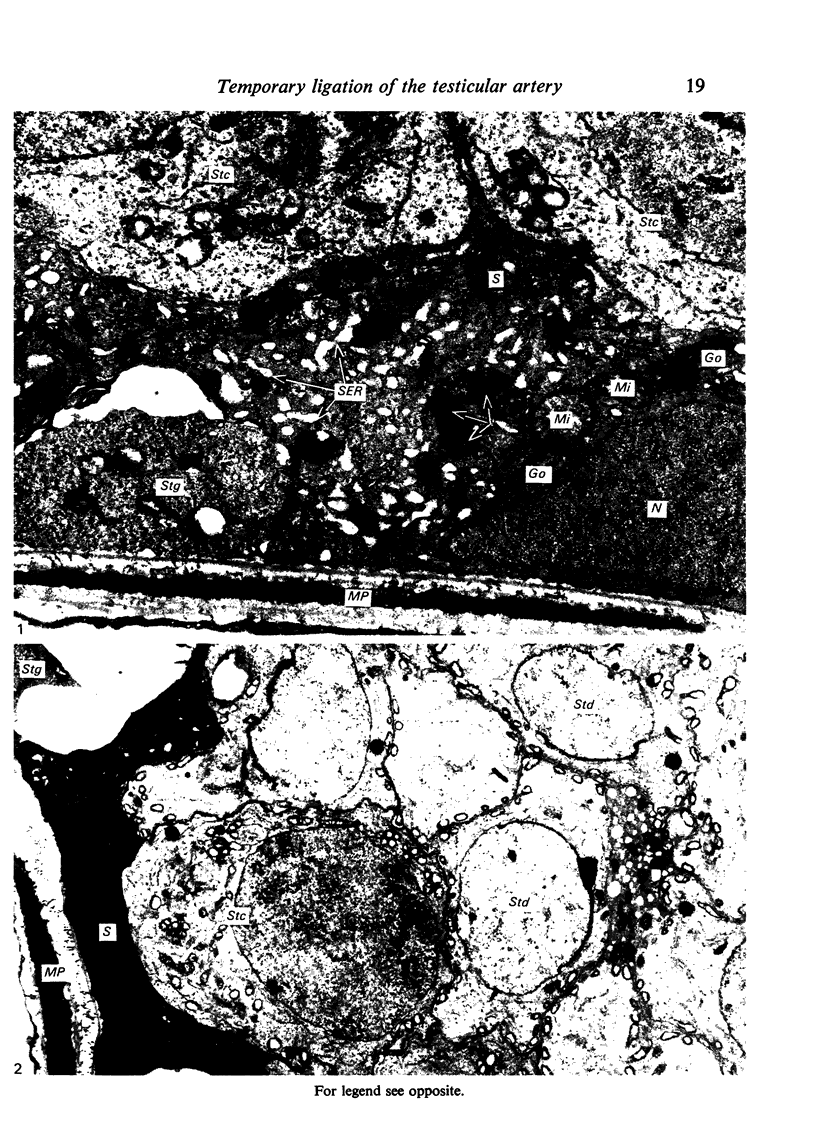
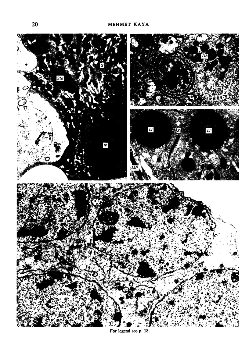
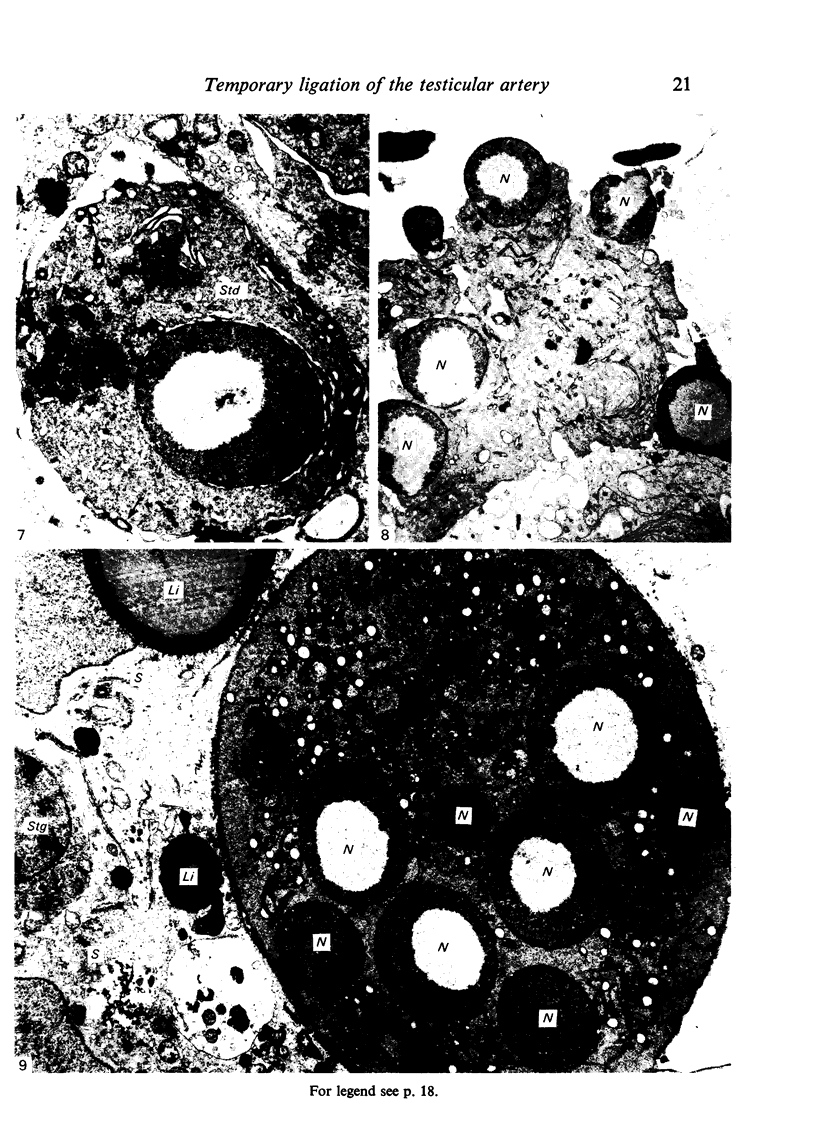
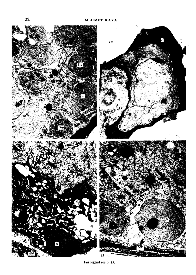
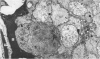
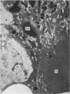
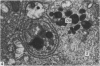
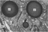
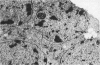
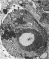
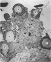
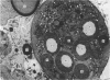
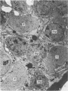
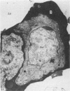
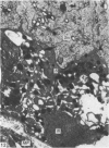
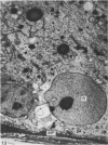
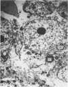
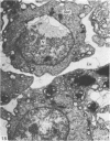
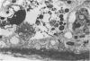
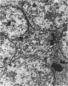
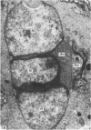
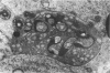
The effects of temporary ligation of the testicular artery have been analysed in rats with respect to Sertoli cells and multinucleated spermatogenic cells. The first cells to show ultrastructural changes are the Sertoli cells which progressively degenerate, leading to complete necrosis as the duration of ligation and post-ligation survival interval increases. The degree of degeneration of spermatogenic cells depends on the severity of Sertoli cell destruction. Temporary ligation of the testicular artery causes the formation of various types of multinucleated spermatogenic cells in the seminiferous epithelium. The mechanisms involved in the multinucleate formation are cell fusion, karyokinesis devoid of cytokinesis and phagocytosis. The variety of noxious agents causing formation of multinucleated spermatogenic cells in the seminiferous tubules of a number of species including man implies that the occurrence of multinucleated spermatogenic cells is not a specific response of the testis to a particular type of agent.
Full text
PDF
Images in this article
Selected References
These references are in PubMed. This may not be the complete list of references from this article.
- Anton E. Early ultrastructural changes in the rat testis after ductuli efferentes ligation. Fertil Steril. 1979 Feb;31(2):187–194. doi: 10.1016/s0015-0282(16)43821-5. [DOI] [PubMed] [Google Scholar]
- Brown P. C., Dorling J., Glynn L. E. Ultrastructural changes in experimental allergic orchitis in guinea-pigs. J Pathol. 1972 Apr;106(4):229–233. doi: 10.1002/path.1711060404. [DOI] [PubMed] [Google Scholar]
- Bryan J. H. Spermatogenesis revisited. I. On the presence of multinucleate spermatogenic cells in the seminiferous epithelium of the mouse. Z Zellforsch Mikrosk Anat. 1971;112(3):333–349. [PubMed] [Google Scholar]
- CLEGG E. J., MACMILLAN E. W. THE UPTAKE OF VITAL DYES AND PARTICULATE MATTER BY THE SERTOLI CELLS OF THE RAT TESTIS. J Anat. 1965 Apr;99:219–229. [PMC free article] [PubMed] [Google Scholar]
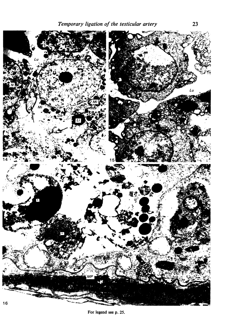
- Flickinger C., Fawcett D. W. The junctional specializations of Sertoli cells in the seminiferous epithelium. Anat Rec. 1967 Jun;158(2):207–221. doi: 10.1002/ar.1091580210. [DOI] [PubMed] [Google Scholar]
- Kanwar K. C., Bawa S. R., Singal P. K. Mode of formation of giant cells in testicular hyperthermia. Fertil Steril. 1971 Nov;22(11):778–783. doi: 10.1016/s0015-0282(16)38590-9. [DOI] [PubMed] [Google Scholar]
- Kaya M., Harrison R. G. An analysis of the effect of ischaemia on testicular ultrastructure. J Pathol. 1975 Oct;117(2):105–117. doi: 10.1002/path.1711170209. [DOI] [PubMed] [Google Scholar]
- Kaya M., Harrison R. G. The ultrastructural relationships between Sertoli cells and spermatogenic cells in the rat. J Anat. 1976 Apr;121(Pt 2):279–290. [PMC free article] [PubMed] [Google Scholar]
- Kerr J. B., Rich K. A., de Kretser D. M. Effects of experimental cryptorchidism on the ultrastructure and function of the Sertoli cell and peritubular tissue of the rat testis. Biol Reprod. 1979 Nov;21(4):823–838. doi: 10.1095/biolreprod21.4.823. [DOI] [PubMed] [Google Scholar]
- LACY D., ROTBLAT J. Study of normal and irradiated boundary tissue of the seminiferous tubules of the rat. Exp Cell Res. 1960 Oct;21:49–70. doi: 10.1016/0014-4827(60)90346-3. [DOI] [PubMed] [Google Scholar]
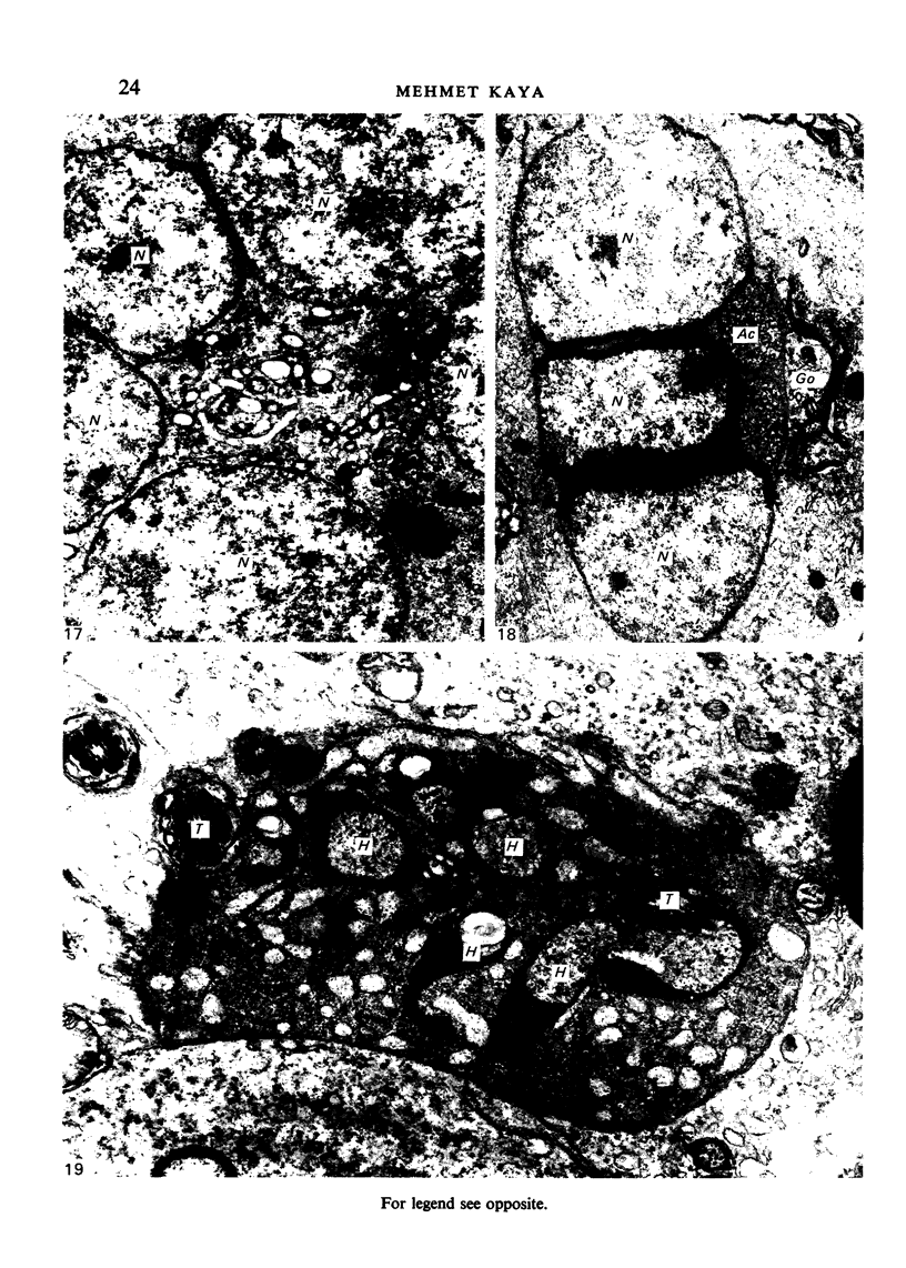
- Narbaitz R., Tolnai G., Jolly E. E., Barwin N., McKay D. E. Ultrastructural studies on testicular biopsies from eighteen cases of hypospermatogenesis. Fertil Steril. 1978 Dec;30(6):679–686. doi: 10.1016/s0015-0282(16)43696-4. [DOI] [PubMed] [Google Scholar]
- OETTLE A. G., HARRISON R. G. The histological changes produced in the rat testis by temporary and permanent occlusion of the testicular artery. J Pathol Bacteriol. 1952 Apr;64(2):273–297. doi: 10.1002/path.1700640204. [DOI] [PubMed] [Google Scholar]
- Rao A. R., Srivastava P. N. Giant cells in the gonads of the Indian desert gerbil, Meriones hurrianae Jerdon, on exposure to internal irradiation. Experientia. 1967 May 15;23(5):381–382. doi: 10.1007/BF02144530. [DOI] [PubMed] [Google Scholar]
- Sigg C., Hedinger C. Quantitative and ultrastructural study of germinal epithelium in testicular biopsies with "mixed atrophy". Andrologia. 1981 Sep-Oct;13(5):412–424. doi: 10.1111/j.1439-0272.1981.tb00074.x. [DOI] [PubMed] [Google Scholar]
- Sobhon P., Mitranond V., Tosukhowong P., Chindaduangrat W. Cytological changes in the testes of vitamin-A-deficient rats. II. Ultrastructural study of the seminiferous tubules. Acta Anat (Basel) 1979;103(2):169–183. doi: 10.1159/000145008. [DOI] [PubMed] [Google Scholar]
- Steinberger E., Tjioe D. Y. Spermatogenesis in rat testes after experimental ischemia. Fertil Steril. 1969 Jul-Aug;20(4):639–649. doi: 10.1016/s0015-0282(16)37091-1. [DOI] [PubMed] [Google Scholar]
- Tso E. C., Lacy D. An ultrastructural study of the testis and epididymis of the rat after treatment with prostaglandins E2 and F2 alpha (PGE2 and PGF2 alpha). J Anat. 1979 Jan;128(Pt 1):107–119. [PMC free article] [PubMed] [Google Scholar]
- Vydra G. Ultrastructure of testicular damage caused by varicocele. Acta Chir Acad Sci Hung. 1980;21(1):77–85. [PubMed] [Google Scholar]