Abstract
Objectives:
Volar locking plate (VLP) fixation is a very common procedure due to the high incidence of distal radius fractures (DRFs). Attritional flexor tendon rupture is a rare, but recognized complication after VLP fixation. There is no current consensus to prevent the condition. Our objective was to highlight the long-term risk for flexor tendon rupture after VLP fixation.
Methods:
We conducted a retrospective single-center review of patients with attritional flexor tendon rupture after VLP fixation for DRFs between 2016 and 2021. Patient demographics, DRF details including AO fracture classification, Soong grading and tendon reconstruction were collected. Thumb interphalangeal joint (IPJ) motion and Kapandji score were used as outcome measures for the tendon reconstruction.
Results:
We identified five patients with attritional flexor pollicis longus (FPL) ruptures. The median age of the patients at the time of DRF was 48 (34-56) years. All VLP fixations were Soong grade 2. Median time from VLP fixation to tendon rupture was 7 (3-14) years. Longest surgery-to-rupture interval was 14 years. One rupture was treated conservatively. Four were reconstructed using palmaris longus (PL) tendon graft. Thumb IPJ active range of motion median was 48 (20-55) degrees and Kapandji score 9/10 (7-9/10).
Conclusion:
Older generation VLP fixations with Soong grade 2 pose a long-term risk for attritional FPL rupture, which can be reconstructed with PL tendon graft with fair to good outcomes.
Key Words: Distal radius fracture, Flexor pollicis longus, Flexor tendon rupture, Tendon reconstruction, Volar locking plate fixation
Introduction
Nearly every fifth fracture is a distal radius fracture (DRF), making it the most common fracture in adults.1 The incidence of DRFs has been estimated to be 178.1 to 252.4 per 100,000 person-years among adults.2 Among elderly patients, distal radius fractures are four times more common in age-matched females than males due to the effect of postmenopausal osteoporosis.3,4
DRFs may either be treated operatively or non-operatively. When surgery is indicated, volar locking plate (VLP) fixation is currently the most commonly employed technique.5,6 AO type C fractures and DRFs among younger patients are more likely to be treated surgically. The existing evidence suggests that surgery leads to better clinical and radiologic outcomes and reduced complication rates compared to cast immobilization especially among patients with higher functional demands.7,8
VLP fixation has been considered a relatively safe procedure with low complication rates.9 Tendon irritation has been found to be a common indication for plate removal after DRF fixation and the flexor pollicis longus (FPL) tendon is the most commonly affected tendon due to its proximity to conventional volar implants.10,11 Based on an epidemiological meta-analysis, the tendon rupture rate has been estimated to be 1.5%, but the incidence rate rises, if symptoms of tendon irritation are present.12,13 Volar plate prominence has been implicated as a key cause of tendon rupture and the Soong classification system is widely used to estimate this plate prominence.14 To date, the longest-reported interval between internal fixation and tendon rupture is 10 years.15 There is no clear consensus on the prevention and early detection of post-operative flexor tendon rupture because of the low incidence of this complication and there is no manner to completely control the risk.
The aim of this study was to improve awareness of the long-term risk regarding attritional tendon ruptures after VLP fixation for DRFs. We present the details of a case series from our institution to outline our approach to manage and prevent such complications.
Materials and Methods
We performed a retrospective single-center case series of attritional flexor tendon ruptures after VLP fixation for DRFs over a 5-year period. Patients with diagnosed flexor tendon rupture after VLP fixation were included to the study. There were no exclusion criteria set. All available electronic medical records were reviewed and data on patient demographics, injury details, fracture type (based on the AO classification) and the index surgery were recorded. The plate position was graded according to Soong classification to determine plate prominence in relation to the watershed line of the distal radius.14 Data regarding the tendon rupture, such as affected tendon, interval between index surgery and rupture, and definitive management were also obtained. Patient outcomes included active thumb interphalangeal joint (IPJ) range of motion (ROM), Kapandji score, and patient satisfaction. The patient outcomes were based on medical chart review. The numerical data are presented as median and range unless otherwise stated. This study was approved by the hospital Institutional Review Board. The authors have no conflicting interests to declare regarding this study.
Results
We identified five patients (two males and three females) that met our inclusion criteria [Table 1]. The median age of the patients at the time of DRF was 48 years (range: 34 - 56). Four injuries were on the dominant (right) side and one occurred in the non-dominant (left) side. Three patients had comorbidities – asthma (one patient) and diabetes (two patients).
Table 1.
Patient demographicsa
| Patients (n) | 5 |
|---|---|
| Patient age at the time of DRF (median, range), years |
48 (34 - 56) |
| Gender, (%) | 3 female (60%) -2 male (40%) |
| Hand dominance (%) | 5 right (100%) |
| Effected side (%) | 4 right (80%)-1 left (20%) |
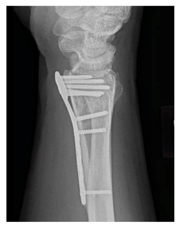
All 5 patients had been initially managed in other institutions. Two of the DRFs were AO Type C2 fractures. The initial injury radiographs were not available in the other three patients. Three VLP fixations involved older generation implants (DePuy Synthes, Indiana, USA), whereas two involved newer generation implants (Adaptive Trilock, Medartis, Basel, Switzerland; VA-plate, Synthes). All plate positions were graded as Soong 2 [Figure 1]. One patient had simultaneous dorsal plate fixation in addition to VLP fixation.
Figure 1.

All plate positions were categorized as Soong grade 2 as shown
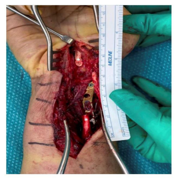
All five of these patients had closed attritional rupture of the FPL tendon. The median time from VLP fixation to FPL rupture was 7 years (range: 3-14). The medical records did not indicate that any of the patients had prodromal symptoms such as pain, discomfort, or a sensation of grating over the volar wrist. All the ruptures occurred suddenly during routine daily activities, except for one patient who sustained a blunt injury to the thumb from a heavy object. Notably, the patient with a 14-year interval between internal fixation and tendon rupture sustained the complication while attempting to open a packet of sauce [Figure 2]. All patients reported difficulty with tasks requiring key and lateral pinch. In addition, one of the patients reported occasional numbness and pain after the rupture. An ultrasound was used to confirm the diagnosis and locate the level of rupture in three patients.
Figure 2.

Case with 14-year interval from VLP fixation to FPL rupture. The fixed-angle volar locking plate was prominent (Soong 2). Attritional rupture of the FPL tendon was noted, with an approximately 5-centimetre gap between the retracted tendon ends (white asterisk). The FPL rupture was repaired within three months using an ipsilateral interpositional palmaris longus autograft. The patient also underwent removal of implants, flexor tenolysis, scar revision, and a carpal tunnel release. The final result was 45 degree of active motion at the thumb IPJ and the patient was satisfied
All patients were offered surgical treatment and one patient opted for non-operative treatment because she was satisfied with her level of function. All four patients underwent implant removal and FPL tendon reconstruction using a palmaris longus tendon graft. The previous volar incision was used to access and remove the plate. The flexor tendons were examined, a synovectomy was performed if necessary and the proximal end of the rupture FPL tendon was easily identified. The distal end of the ruptured FPL was identified by passively flexing the thumb interphalangeal joint and occasionally extending the volar incision. Three patients also had a concomitant open carpal tunnel release, which facilitated identification of the distal end of the FPL. If the distal end cannot be identified in the palm, a zig-zag incision should be made over the volar aspect of the thumb to locate the FPL tendon. We take great care to avoid incisions across the thenar region because of the potential for scarring and thumb adduction contracture. A palmaris longus (PL) tendon autograft was used in all cases. The tendon graft was harvested from the same incision, and wire / suture loops were used to pass the tendon graft from the distal forearm to the flexor sheath of the thumb. The segmental FPL tendon defect was reconstructed using a Pulvertaft weave technique. Adequate tendon tension and gliding was confirmed using passive joint motion and the observing the tenodesis effect. One patient, who underwent tendon reconstruction without carpal tunnel release, had occasional finger numbness, but no subsequent procedures were required. The median time from FPL rupture to reconstruction was 2 months (range: 1-5) and the median postoperative follow-up time was 6 months (range: 4-8).
?
Discussion
We identified five patients with attritional FPL tendon rupture following VLP fixation for DRFs. All cases corresponded with Soong grade 2 and the median interval between fixation and rupture was 7 years (range: 3-14). Four patients had implant removal and tendon reconstruction, and three had a simultaneous carpal tunnel release. The median active thumb IPJ ROM at final follow-up was 48 degrees (range: 20-55) and the median Kapandji score was 9 (range: 7-9).
Tendon irritation is the second most common indication for implant removal.12 Routine removal in asymptomatic patients has not been proven to correlate with better clinical outcomes and might even predispose the patient for possible complications related to the removal procedure itself, such as haematoma and re-fracture. Employing a strategy of routinely removing all implants may not prevent all tendon ruptures either, as attritional rupture might occur as early as 3 months after fixation.16,17 An alternative strategy is early hardware removal in patients who present with features of tendon irritation, such as crepitus, pain and/or swelling. The fundamental problem with this approach is that tendon rupture might be the first sign of tendon attrition related to VLP fixation. None of our patients had prodromal symptoms, although it is possible that patients may have failed to recognize mild symptoms that did not affect function. It has been estimated that 28% of attritional tendon rupture patients do not have any symptoms prior to tendon rupture, but existing symptoms might be underreported in general.13 We strongly advocate routine assessment for features of tendon irritation during post-operative outpatient visits and educate the patients regarding the possible clinical features that may indicate underlying flexor tendon irritation.
The FPL is the most frequently affected flexor tendon after VLP fixation. Most of the previously reported ruptures have been grade 1 on Soong´s criteria.13 It has been shown that a higher Soong grade is correlated with flexor tendon issues.18 Based on a systematic review, plate malposition has been reported to be the most common hardware problem related to flexor tendon ruptures.13 An ultra-distal fracture morphology demands distal plate positioning or use of highly contoured implants designed to fit the volar rim of the bone. In some of our cases, there was residual dorsal angulation which further accentuated volar plate prominence [Figure 1]. It is essential that dorsal tilt should be corrected as accurately as possible not just to maximize wrist function, but also to reduce the risk of flexor tendon irritation. The advent of variable-angle locking screws means that the plate may also be applied more proximally while still ensuring secure fixation of specific fragments. As with all operations, the surgeon should be familiar with the nuances of the implants being used and inexperienced surgeons must be aware of these technical points.
Three VLP fixations were done with older generation juxta-articular T-plates in our series. It has been suggested that older generation plates cause more tendon complications and the development of lower profile, anatomical plates may mitigate this risk.13 There are case reports of tendon ruptures with similar older generation devices, but the longest time interval between fixation and rupture was 40 months.17-20 Overall, the longest interval reported so far has been 10 years after fixation.15
In our own series, two patients with older-generation implants had tendon ruptures 10 and 14 years after internal fixation. This suggests that the risk for FPL rupture with these older generation plates do not subside over time. Floquet et al. reported a median fixation-to-rupture time interval of 8 months in their systematic review of 145 flexor rupture cases.13 our median time interval was significantly longer (7 years) and even the earliest rupture occurred 3 years after internal fixation. We now recommend early removal of implants (at 9 to 12 months after fixation) for implants that correspond to Soong grade 2. This seems to balance the risks of delayed tendon rupture and re-fracture from premature implant removal.21,22
For established tendon ruptures, the most common method of repair or reconstruction technique is tendon grafting23,24 Utilisation of palmaris longus tendon avoids sacrificing a flexor digitorum superficialis tendon and side-to-side tenorrhaphy with multiple weaves permits early active motion protocol. Concomitant carpal tunnel release should be considered to avoid subsequent median nerve compression after tendon grafting procedure.
There are several limitations of this study. First, the retrospective nature can introduce bias to patient selection. Second, it is a small size case series including only five patients. Third, some initial injury and immediate post-operative radiographs were not available for analysis from the databases. Fourth, the outcomes were based on chart review and no validated patient-reported outcomes were used.
Conclusion
Our findings indicate that VLP fixation with Soong grade 2 poses a long-term risk for FPL rupture that persists more than a decade after internal fixation. We now have a low threshold to removing VLP fixation implants with Soong grade 2, approximately 9 to 12 months after internal fixation. In the setting of an established FPL rupture, tendon reconstruction with PL tendon autograft provides acceptable outcomes without requiring sacrifice of a digital flexor tendon.
Acknowledgement
The results have been briefly presented at APFSSH meeting in Singapore June 2023.
Authors Contribution:
All authors contributed similarly to this article. All of them conceived and designed the analysis, collected the data, contributed to data analysis, and wrote the manuscript.
Declaration of Conflict of Interest:
The authors do NOT have any potential conflicts of interest with respect to this manuscript.
Declaration of Funding:
The authors received NO financial support for the preparation, research, authorship, and/or publication of this manuscript.
Declaration of Ethical Approval for Study:
This study was approved by the National University Hospital of Singapore´s Institutional Review Board and the Helsinki Declaration was followed with Ethical approval code: 2022/00296).
Declaration of Informed Consent:
There is no information in the submitted manuscript that can be used to identify patients.
References
- 1.Court-Brown CM, Caesar B. Epidemiology of adult fractures: a review. Injury. 2006;37(8):691–7. doi: 10.1016/j.injury.2006.04.130. [DOI] [PubMed] [Google Scholar]
- 2.Ponkilainen V, Kuitunen I, Liukkonen R, Vaajala M, Reito A, Uimonen M. The incidence of musculoskeletal injuries: a systematic review and meta-analysis. Bone Joint Res. 2022;11(11):814–825. doi: 10.1302/2046-3758.1111.BJR-2022-0181.R1. [DOI] [PMC free article] [PubMed] [Google Scholar]
- 3.Raudasoja L, Aspinen S, Vastamäki H, Ryhänen J, Hulkkonen S. Epidemiology and Treatment of Distal Radius Fractures in Finland-A Nationwide Register Study. J Clin Med. 2022;11(10):2851. doi: 10.3390/jcm11102851. [DOI] [PMC free article] [PubMed] [Google Scholar]
- 4.Ghafoori H, Kazemi M, Ghorbani S. Investigation of the History of Distal Radius Fractures in Patients Over 55 Years Old Suffering from Hip Fractures. Arch Bone Jt Surg. 2024;12(6):418–422. doi: 10.22038/ABJS.2023.75188.3477. [DOI] [PMC free article] [PubMed] [Google Scholar]
- 5.Kamal RN, Shapiro LM. American Academy of Orthopaedic Surgeons/American Society for Surgery of the Hand Clinical Practice Guideline Summary Management of Distal Radius Fractures. J Am Acad Orthop Surg. 2022 15;30(4):e480–e486. doi: 10.5435/JAAOS-D-21-00719. [DOI] [PMC free article] [PubMed] [Google Scholar]
- 6.Koo OT, Tan DM, Chong AK. Distal radius fractures: an epidemiological review. Orthop Surg. 2013;5(3):209–13. doi: 10.1111/os.12045. [DOI] [PMC free article] [PubMed] [Google Scholar]
- 7.Oldrini LM, Feltri P, Albanese J, Lucchina S, Filardo G, Candrian C. Volar locking plate vs cast immobilization for distal radius fractures: a systematic review and meta-analysis. EFORT Open Rev. 2022 19;7(9):644–652. doi: 10.1530/EOR-22-0022. [DOI] [PMC free article] [PubMed] [Google Scholar]
- 8.Ochen Y, Peek J, van der Velde D, et al. Operative vs Nonoperative Treatment of Distal Radius Fractures in Adults: A Systematic Review and Meta-analysis. JAMA Netw Open. doi: 10.1001/jamanetworkopen.2020.3497. [DOI] [PMC free article] [PubMed] [Google Scholar]
- 9.Lee JH, Lee JK, Park JS, et al. Complications associated with volar locking plate fixation for distal radius fractures in 1955 cases: A multicentre retrospective study. Int Orthop. 2020;44(10):2057–2067. doi: 10.1007/s00264-020-04673-z. [DOI] [PubMed] [Google Scholar]
- 10.Yamamoto M, Fujihara Y, Fujihara N, Hirata H. A systematic review of volar locking plate removal after distal radius fracture. Injury. 2017;48(12):2650–2656. doi: 10.1016/j.injury.2017.10.010. [DOI] [PubMed] [Google Scholar]
- 11.Cronin PK, Watkins IT, Riedel M, Kaiser PB, Kwon JY. Implant Removal Matrix for the upper Extremity Orthopedic Surgeon. Arch Bone Jt Surg. 2020;8(1):99–111. doi: 10.22038/abjs.2019.36525.1962. [DOI] [PMC free article] [PubMed] [Google Scholar]
- 12.Azzi AJ, Aldekhayel S, Boehm KS, Zadeh T. Tendon rupture and tenosynovitis following internal fixation of distal radius fractures: a systematic review. 2017;139(3):717e–724e. doi: 10.1097/PRS.0000000000003076. [DOI] [PubMed] [Google Scholar]
- 13.Floquet A, Druart T, Lavantes P, Vendeuvre T, Delaveau A. Flexor tendon rupture after volar plating of distal radius fracture: A systematic review of the literature. Hand Surg Rehabil. 2021;40(5):535–546. doi: 10.1016/j.hansur.2021.05.008. [DOI] [PubMed] [Google Scholar]
- 14.Soong M, Earp BE, Bishop G, Leung A, Blazar P. Volar locking plate implant prominence and flexor tendon rupture. J Bone Joint Surg Am. 2011;93(4):328–35. doi: 10.2106/JBJS.J.00193. [DOI] [PubMed] [Google Scholar]
- 15.Monda MK, Ellis A, Karmani S. Late rupture of flexor pollicis longus tendon 10 years after volar buttress plate fixation of a distal radius fracture: a case report. Acta Orthop Belg. 2010;76(4):549–51. [PubMed] [Google Scholar]
- 16.Marsh JL, Slongo TF, Agel J, et al. Fracture and dislocation classification compendium - 2007: Orthopaedic trauma association classification, database and outcomes committee. J Orthop Trauma. 2007;21(10 Suppl):S1–133 . doi: 10.1097/00005131-200711101-00001. [DOI] [PubMed] [Google Scholar]
- 17.Drobetz H, Kutscha-Lissberg E. Osteosynthesis of distal radial fractures with a volar locking screw plate system. Int Orthop. 2003;27(1):1–6. doi: 10.1007/s00264-002-0393-x. [DOI] [PMC free article] [PubMed] [Google Scholar]
- 18.Vasara H, Tarkiainen P, Stenroos A, et al. Higher Soong grade predicts flexor tendon issues after volar plating of distal radius fractures - a retrospective cohort study. BMC Musculoskelet Disord. 2023 10;24(1):271 . doi: 10.1186/s12891-023-06313-0. [DOI] [PMC free article] [PubMed] [Google Scholar]
- 19.Marimuthu R, Jaiswal P, Mani GV. Volar plates on distal radius may cause late rupture of flexor pollicis longus. Injury Extra. 2006;37(1):1–3. [Google Scholar]
- 20.Adham MN, Porembski M, Adham C. Flexor tendon problems after volar plate fixation of distal radius fractures. Hand (N Y) 2009;4(4):406–9. doi: 10.1007/s11552-009-9180-0. [DOI] [PMC free article] [PubMed] [Google Scholar]
- 21.Ateschrang A, Stuby F, Werdin F, Schaller HE, Weise K, Albrecht D. Flexor tendon irritations after locked plate fixation of the distal radius with the 3 5 mm T-plate: identification of risk factors. Zeitschrift fur Orthopadie und Unfallchirurgie. 2010;148(3):319–25. doi: 10.1055/s-0029-1241027. [DOI] [PubMed] [Google Scholar]
- 22.Cho CH, Lee KJ, Song KS, Bae KC. Delayed rupture of flexor pollicis longus after volar plating for a distal radius fracture. Clin Orthop Surg. 2012;4(4):325–8. doi: 10.4055/cios.2012.4.4.325. [DOI] [PMC free article] [PubMed] [Google Scholar]
- 23.Koo SC, Ho ST. Delayed rupture of flexor pollicis longus tendon after volar plating of the distal radius. Hand Surg. 2006;11(1-2):67–70. doi: 10.1142/S0218810406003048. [DOI] [PubMed] [Google Scholar]
- 24.Honoki K, Nishida K, Che YH, et al. The sole rupture of flexor digitorum tendons to the index finger following volar locking plate fixation of distal radius fracture: a case report. Trauma Case Rep. 2020;30:100369. doi: 10.1016/j.tcr.2020.100369. [DOI] [PMC free article] [PubMed] [Google Scholar]


