



Abstract
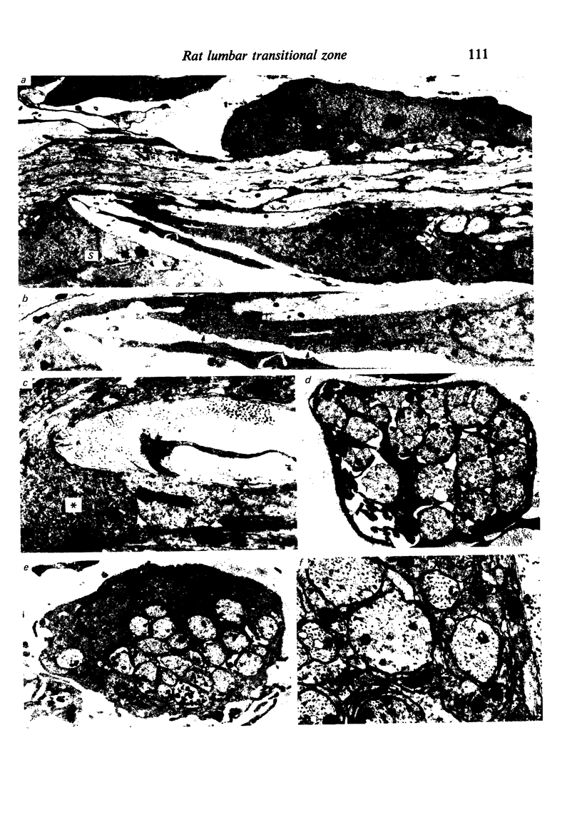
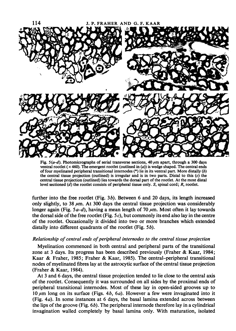
In the rat lumbar ventral spinal nerve rootlets, the central-peripheral transition occurs at the surface of a distal projection of central tissue into the rootlet. This changes considerably in appearance during development. In the first week after birth, it grows distally into the rootlet to form an irregular, tapering projection. In the second week after birth, it is retracted and becomes splayed out; its distal surface is irregular and lies just above the surrounding spinal cord. After this, it again grows distally into the rootlet. It forms a tapering projection which generally lies eccentrically in the rootlet, most often towards its dorsal surface. The central ends of the proximal transitional myelinated peripheral internodes generally lie in grooves on the surface of the central tissue projection. However, for a time during the second week after birth, many lie in invaginations into it. Occasional invaginations of the central tissue projection contain large numbers of collagen fibres rather than axons. A ring of collagen fibres surrounds the rootlet immediately distal to its attachment to the cord surface. Though the central tissue projection contains occasional astrocytic perikarya, it consists mostly of closely interwoven astrocyte processes derived from cell bodies situated at the cord surface surrounding the rootlet attachment. Changes in the form and size of the central tissue projection probably result largely from active growth and reorganisation of astrocyte processes. The barrier which these processes constitute probably prevents invasion of the cord by transitional Schwann cells. Before the central tissue projection develops, such invasion is probably prevented by the arrangement of transitional Schwann cells as a close-knit epithelium on the surface of the rootlet. The central tissue projection of ventral rootlets is smaller, more irregular in shape and less highly organised than that of dorsal rootlets. Central-peripheral transitional nodes lie close to the surface of the central tissue projection. They are therefore offset relative to one another and so are less likely to discharge one another. This arrangement may also protect the rootlet against mechanical damage due to traction.
Full text
PDF





Images in this article


Selected References
These references are in PubMed. This may not be the complete list of references from this article.
- Berthold C. H., Carlstedt T. Observations on the morphology at the transition between the peripheral and the central nervous system in the cat. II. General organization of the transitional region in S1 dorsal rootlets. Acta Physiol Scand Suppl. 1977;446:23–42. [PubMed] [Google Scholar]
- Blakemore W. F. Limited remyelination of CNS axons by Schwann cells transplanted into the sub-arachnoid space. J Neurol Sci. 1984 Jun;64(3):265–276. doi: 10.1016/0022-510x(84)90175-8. [DOI] [PubMed] [Google Scholar]
- Blakemore W. F., Patterson R. C. Observations on the interactions of Schwann cells and astrocytes following X-irradiation of neonatal rat spinal cord. J Neurocytol. 1975 Oct;4(5):573–585. doi: 10.1007/BF01351538. [DOI] [PubMed] [Google Scholar]
- Carlsen F., Behse F. Three dimensional analysis of Schwann cells associated with unmyelinated nerve fibres in human sural nerve. J Anat. 1980 May;130(Pt 3):545–557. [PMC free article] [PubMed] [Google Scholar]
- Carlstedt T. An electron-microscopical study of the developing transitional region in feline S1 dorsal rootlets. J Neurol Sci. 1981 Jun;50(3):357–372. doi: 10.1016/0022-510x(81)90148-9. [DOI] [PubMed] [Google Scholar]
- Cravioto H. The role of Schwann cells in the development of human peripheral nerves. An electron microscopic study. J Ultrastruct Res. 1965 Jun;12(5):634–651. doi: 10.1016/s0022-5320(65)80053-3. [DOI] [PubMed] [Google Scholar]
- Fraher J. P., Kaar G. F. The development of alpha and gamma motoneuron fibres in the rat. II. A comparative ultrastructural study of their central and peripheral myelination. J Anat. 1985 Aug;141:89–103. [PMC free article] [PubMed] [Google Scholar]
- Fraher J. P., Kaar G. F. The maturation of the ventral root-spinal cord transitional zone. Part 2. A quantitative ultrastructural study of the dynamics of its early development. J Neurol Sci. 1982 Jan;53(1):63–75. doi: 10.1016/0022-510x(82)90080-6. [DOI] [PubMed] [Google Scholar]
- Fraher J. P., Kaar G. F. The transitional node of Ranvier at the junction of the central and peripheral nervous systems: an ultrastructural study of its development and mature form. J Anat. 1984 Sep;139(Pt 2):215–238. [PMC free article] [PubMed] [Google Scholar]
- Fraher J. P. Probable glial islands in a rat spinal nerve root--a longitudinal study. J Neuropathol Exp Neurol. 1974 Aug;33(4):552–560. doi: 10.1097/00005072-197408000-00006. [DOI] [PubMed] [Google Scholar]
- Fraher J. P., Rossiter J. P. Cell clusters on fetal rat ventral roots: prenatal development. J Anat. 1983 Jan;136(Pt 1):111–128. [PMC free article] [PubMed] [Google Scholar]
- Fraher J. P., Rossiter J. P. Cell clusters on rat ventral roots: postnatal development. J Anat. 1983 Oct;137(Pt 3):555–571. [PMC free article] [PubMed] [Google Scholar]
- Fraher J. P. The maturation of the ventral root-spinal cord transitional zone. An ultrastructural study. J Neurol Sci. 1978 May;36(3):427–449. doi: 10.1016/0022-510x(78)90049-7. [DOI] [PubMed] [Google Scholar]
- GAMBLE H. J. COMPARATIVE ELECTRON-MICROSCOPIC OBSERVATIONS ON THE CONNECTIVE TISSUES OF A PERIPHERAL NERVE AND A SPINAL NERVE ROOT IN THE RAT. J Anat. 1964 Jan;98:17–26. [PMC free article] [PubMed] [Google Scholar]
- Gamble H. J., Eames R. A. Electron microscopy of human spinal-nerve roots. Arch Neurol. 1966 Jan;14(1):50–53. doi: 10.1001/archneur.1966.00470070054006. [DOI] [PubMed] [Google Scholar]
- Gamble H. J. Further electron microscope studies of human foetal peripheral nerves. J Anat. 1966 Jul;100(Pt 3):487–502. [PMC free article] [PubMed] [Google Scholar]
- Gilmore S. A., Sims T. J., Heard J. K. Autoradiographic and ultrastructural studies of areas of spinal cord occupied by Schwann cells and Schwann cell myelin. Brain Res. 1982 May 13;239(2):365–375. doi: 10.1016/0006-8993(82)90515-7. [DOI] [PubMed] [Google Scholar]
- Kaar G. F., Fraher J. P. The development of alpha and gamma motoneuron fibres in the rat. I. A comparative ultrastructural study of their central and peripheral axon growth. J Anat. 1985 Aug;141:77–88. [PMC free article] [PubMed] [Google Scholar]
- Maxwell D. S., Kruger L., Pineda A. The trigeminal nerve root with special reference to the central-peripheral transition zone: an electron microscopic study in the macaque. Anat Rec. 1969 May;164(1):113–125. doi: 10.1002/ar.1091640108. [DOI] [PubMed] [Google Scholar]
- Moll C., Meier C. The central-peripheral transition zone of cervical spinal nerve roots in Jimpy mutant and normal mice. Light- and electron-microscopic study. Acta Neuropathol. 1983;60(3-4):241–251. doi: 10.1007/BF00691872. [DOI] [PubMed] [Google Scholar]
- Nemecek S., Parízek J., Spacek J., Nemecková J. Histological, histochemical and ultrastructural appearance of the transitional zone of the cranial and spinal nerve roots. Folia Morphol (Praha) 1969;17(2):171–181. [PubMed] [Google Scholar]
- Ross M. D., Burkel W. Electron microscopic observations of the nucleus, glial dome, and meninges of the rat acoustic nerve. Am J Anat. 1971 Jan;130(1):73–91. doi: 10.1002/aja.1001300106. [DOI] [PubMed] [Google Scholar]
- Steer J. M. Some observations on the fine structure of rat dorsal spinal nerve roots. J Anat. 1971 Sep;109(Pt 3):467–485. [PMC free article] [PubMed] [Google Scholar]