

Abstract
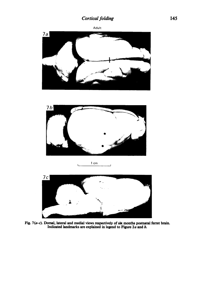
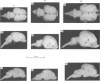
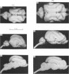
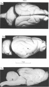
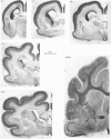
The external features of gyrus formation in the postnatal ferret cerebral cortex are described and correlated with certain internal changes. The observations indicate that gyri are formed by longitudinal and radial expansion of the cortical compartment occurring between relatively fixed areas which form the sulcal floors. The gyri were initially rounded with open sulci and the cerebrum had a rectangular outline when seen in lateral and dorsal view. By adult life the hemisphere had been subjected to considerable moulding by the growing skull, so that the frontal pole of the cerebrum became pointed while the sulcal walls became closely opposed and the gyral crowns flattened.
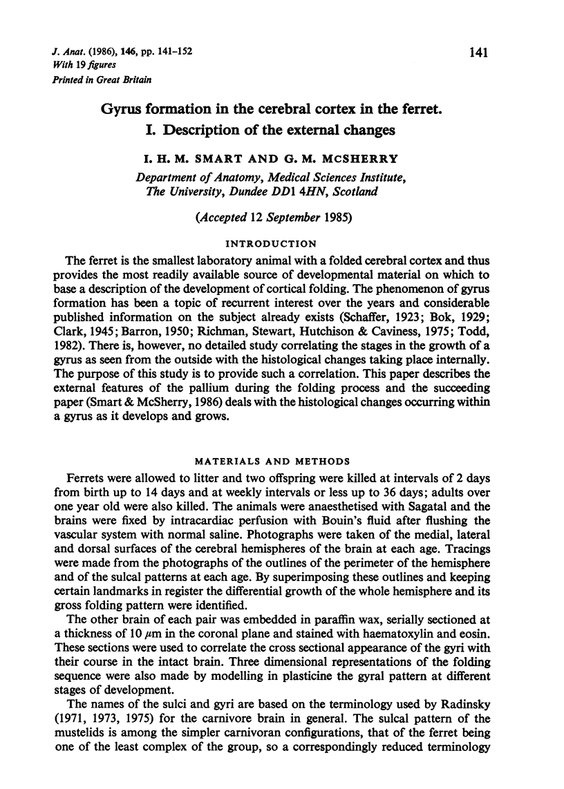
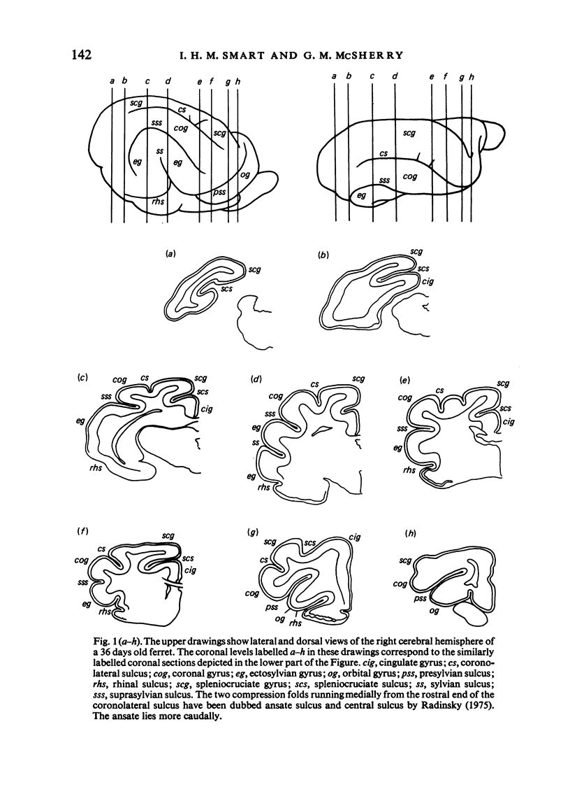
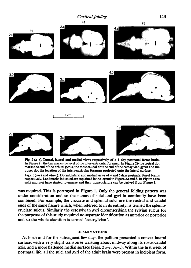
Full text
PDF
Images in this article
Selected References
These references are in PubMed. This may not be the complete list of references from this article.
- Marin-Padilla M. Dual origin of the mammalian neocortex and evolution of the cortical plate. Anat Embryol (Berl) 1978 Feb 20;152(2):109–126. doi: 10.1007/BF00315920. [DOI] [PubMed] [Google Scholar]
- McSherry G. M. Mapping of cortical histogenesis in the ferret. J Embryol Exp Morphol. 1984 Jun;81:239–252. [PubMed] [Google Scholar]
- Radinsky L. Evolution of the canid brain. Brain Behav Evol. 1973;7(3):169–202. doi: 10.1159/000124409. [DOI] [PubMed] [Google Scholar]
- Radinsky L. Viverrid neuroanatomy: Phylogenetic and behavioral implications. J Mammal. 1975 Feb;56(1):130–150. [PubMed] [Google Scholar]
- Richman D. P., Stewart R. M., Hutchinson J. W., Caviness V. S., Jr Mechanical model of brain convolutional development. Science. 1975 Jul 4;189(4196):18–21. doi: 10.1126/science.1135626. [DOI] [PubMed] [Google Scholar]
- Smart I. H. Proliferative characteristics of the ependymal layer during the early development of the mouse neocortex: a pilot study based on recording the number, location and plane of cleavage of mitotic figures. J Anat. 1973 Oct;116(Pt 1):67–91. [PMC free article] [PubMed] [Google Scholar]
- Smart I. H. Radial unit analysis of hippocampal histogenesis in the mouse. J Anat. 1982 Dec;135(Pt 4):763–793. [PMC free article] [PubMed] [Google Scholar]
- Smart I. H. Three dimensional growth of the mouse isocortex. J Anat. 1983 Dec;137(Pt 4):683–694. [PMC free article] [PubMed] [Google Scholar]
- Todd P. H. A geometric model for the cortical folding pattern of simple folded brains. J Theor Biol. 1982 Aug 7;97(3):529–538. doi: 10.1016/0022-5193(82)90380-0. [DOI] [PubMed] [Google Scholar]