Abstract
As the evolutionary ancestor of Cas9 nuclease, IscB proteins serve as compact RNA-guided DNA endonucleases and nickases, making them strong candidates for base editing. Nevertheless, the narrow targeting scope limits the application of IscB systems; thus, it is necessary to find more IscBs that recognize different target-adjacent motifs (TAMs). Here, we identified 10 of 19 uncharacterized IscB proteins from uncultured microbes with activity in mammalian cells. Through protein and ωRNA engineering, we further enhanced the activity of IscB ortholog IscB.m16 and expanded its TAM scope from MRNRAA to NNNGNA, resulting in a variant named IscB.m16*. By fusing the deaminase domains with IscB.m16* nickase, we generated IscB.m16*-derived base editors that exhibited robust base-editing efficiency in mammalian cells and effectively restored Duchenne muscular dystrophy proteins in diseased mice through single adeno-associated virus delivery. Thus, this study establishes a set of compact base-editing tools for basic research and therapeutic applications.
Subject terms: Biological techniques, Bacteria
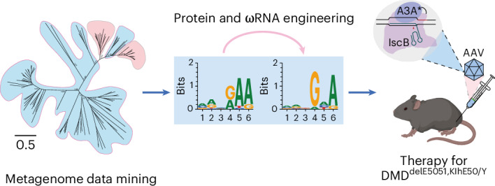
Xiao et al. characterize 19 previously unidentified IscB proteins, 10 of which show activity in mammalian cells. They engineer one of the proteins into a robust in vivo genome-editing tool with a broad target-adjacent motif scope and further develop it into a base editor.
Main
Clustered regularly interspaced short palindromic repeat (CRIPSR) Cas systems, such as type II Cas9 and type V Cas12 systems, serving as the prokaryotic adaptive immunity system against viruses, have been developed into genome-editing tools in basic research and gene therapy1–3. The engineered Cas9 nickase (nCas9) or deactivated Cas9 (dCas9) versions fused with various domains have been established as base-editing, prime-editing and epigenome-editing technologies4–6. However, the large size of Cas9 and Cas12, particularly nCas9-based gene-editing tools, hinders the application of gene editing based on adeno-associated virus (AAV) vectors. Recently, compact Cas9 (refs. 7–9), Cas12f homologs10–14 (400–700 aa) and TnpB15,16 (~400 aa), the ancestral branch of Cas12, have been reported. However, because of their poor editing activity or lack of an HNH domain, these proteins have limited base-editing activity.
IscB proteins are encoded in a distinct family of IS200/IS605 transposons possessing HNH and RuvC domains, such as Cas9, and are thought to be the ancestor of Cas9 (refs. 17,18). However, the size of IscB proteins is only two fifths of that of Cas9 (~400 aa). Recent studies have shown that the IscB system (IscB–ωRNA) is a programmable long noncoding RNA (referred to as ωRNA)-guided DNA endonuclease and engineered OgeuIscB-based base editors (enOgeuIscB-BEs) exhibit high base-editing efficiency in mammalian cells19. The IscB system requires a 3′ terminal target-adjacent motif (TAM) to recognize the target DNA (usually 6 nt) and the recently reported enOgeuIscB requires 4 nt (NWRRNA). Complex TAM sequences greatly reduce the number of sites that can be edited. The narrow TAM range of OgeuIscB in mammalian cells has become an obvious limitation. Therefore, it is necessary to develop higher-efficiency miniature base editors with a broader TAM range. Here, we identified 19 natural IscB–ωRNA systems with various TAM scopes from metagenome datasets. By engineering both the ωRNA and the IscB.m16 protein, we generated the IscB.m16* system (IscB.m16 containing E326R;T459E;P460S;T462H substitutions (IscB.m16RESH) and enωRNA) with robust editing activity and expanded the TAM range to NNNGNA in mammalian cells. We further developed IscB.m16*-based adenine and cytosine base editors demonstrating robust base-editing efficiency and broad target recognition in mammalian cells and mouse models. Moreover, we provide a comprehensive dataset of IscB–ωRNA systems with diverse TAM scopes and a strategy to widen the TAM range.
Results
Functional identification of uncharacterized IscB orthologs
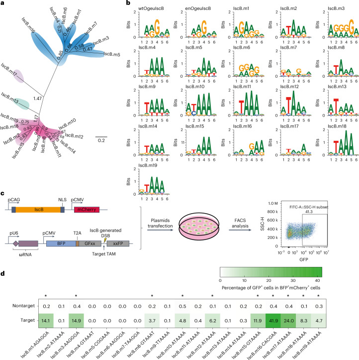
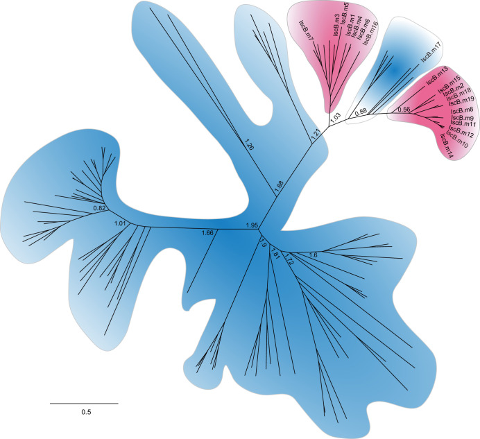
To identify additional IscB–ωRNA systems with diverse TAMs, we downloaded 200 Gb of rumen metagenome-assembled genomes20. We used a computational pipeline to annotate IscB orthologs and their corresponding ωRNAs, which led to the discovery of 19 uncharacterized IscB systems. These systems were phylogenetically clustered into three subgroups on the basis of a sequence alignment of IscB effector proteins (Fig. 1a and Extended Data Fig. 1). Through the protein sequence alignment encompassing 500 aa, we identified the conserved residues within the RuvC domain, HNH domain, P1D (P1 interaction domain) and TID (TAM interaction domain), suggesting the possibility of nuclease and nickase activity (Supplementary Fig. 1).
Fig. 1. Identification and characterization of functional IscB orthologs.
a, Phylogenetic tree of 19 uncharacterized IscB orthologs. b, TAMs of 19 active IscB proteins and two OgeuIscB variants19 determined by bacterial depletion assay. c, Schematics describing the detection of editing activity based on the fluorescence signal of GFxxFP reporter activation in HEK293T cells. d, Fluorescence signal of EGFP activated by IscB-mediated DSBs quantified by flow cytometry. Nontarget denotes a spacer with a random sequence. Asterisks denote a > 9-fold ratio of target and nontarget, with target recognition > 3.0%, representing variants with activity in HEK293T cells. Values represent the mean of three independent biological replicates.
Extended Data Fig. 1. Maximum-likelihood tree of identified IscB orthologs and previously reported IscBs.
The name of 19 identified IscB orthologs are shown. The evolutionary distance scale of 0.5 is shown.
To detect whether these IscB proteins and predicted corresponding ωRNAs were capable of cleaving DNA and characterized by TAM recognition, we performed a bacterial depletion assay. We cotransformed Escherichia coli cells with plasmids carrying IscB and its cognate ωRNA with a spacer, as well as a TAM library plasmid carrying target sequences complementary to the spacer and 8-bp randomized sequences (Supplementary Fig. 2a). Through this assay, a series of specific depleted TAM sequences were enriched associated with each IscB system, indicating that these natural IscB orthologs have RNA-guided endonuclease activity in prokaryotes (Fig. 1b and Supplementary Fig. 2b). Subsequently, we analyzed the relationship between the divergence of IscB proteins and differences in TAMs and observed that most IscB proteins have notable distinctions in both their amino acid sequences and their respective optimal TAMs (Supplementary Fig. 2c).
To further assess the nuclease activity of these IscB orthologs in human cells, we used a fluorescence reporter system. This system involved cotransfecting a plasmid expressing the IscB protein and its corresponding target ωRNA, along with a reporter plasmid encoding GFxxFP, into cultured HEK293T cells. Green fluorescent protein (GFP) activation based on the GFxxFP reporter (GFxx–target site–xxFP) was carried out by endonuclease-mediated double-strand breaks (DSBs) to the target site and single-strand annealing (SSA)-mediated repair. We then measured the enhanced GFP (EGFP) signal intensity of the inactivated GFxxFP reporter, which was activated by IscB-mediated DSBs21 (Fig. 1c). Using the GFxxFP reporter with the experimentally determined TAM for each IscB, 10 of 19 IscBs exhibited a significant increase (>9-fold ratio of target and nontarget, with target recognition >3.0%) in EGFP signal intensity relative to nontarget sequences. Notably, IscB.m16 exhibited the highest signal intensity (Fig. 1d).
Engineering ωRNA to improve editing efficiency
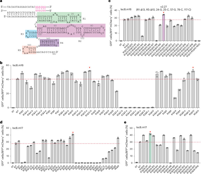
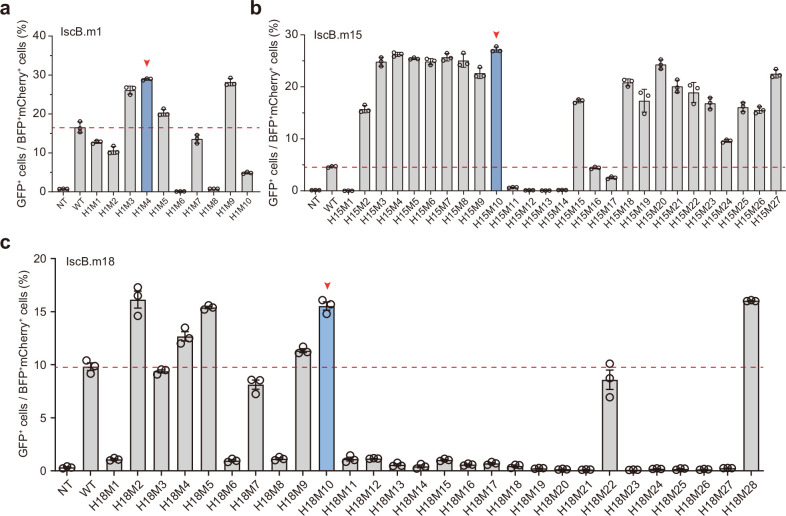
Guide RNA (gRNA) engineering strategies have been widely applied to enhance the cleavage activity of RNA-guided nucleases11,12,19. To enhance the activity of the natural IscB.m16 system, we engineered its ωRNA by truncation or mutagenesis, generating ωRNA variants in five stem-loop regions: R1, R2, R3, R4 and R5 (Fig. 2a and Supplementary Fig. 3a). We performed the truncations by shrinking loops and truncating long stems. We screened the editing efficiency using the GFxxFP reporter and observed increased editing activity in the case of R1 (R1-Δ13) and R5 (R5-Δ10) truncated ωRNA (Fig. 2b). To increase the stability of the ωRNA, we replaced the A•U or mismatched base pairs in stem regions with thermodynamically stable G•C base pairs. We replaced the mismatched G•U and partial A•U base pairs in stem regions with thermodynamically stable G•C base pairs and five variants exhibited increased activity (Supplementary Fig. 4). For the R1-Δ13 and R5-Δ10 truncated ωRNA, we further combined five mutations and found that v2.27 (del15–20, del29–35, del171–180, 24-G, 25-C, 57-G, 79-C and 117-C) showed enhanced editing activity at the AAAGCA TAM reporter (Fig. 2c). Similarly, to improve the activity of the IscB.m17 system, we truncated six stem loops of the ωRNA on the basis of their secondary structure and found that R1 (R1-Δ59) and R6 (R6-Δ9) truncated ωRNA showed increased editing activity (Fig. 2d and Supplementary Fig. 3b). We then generated a variant with slightly improved editing activity by replacing A•U with C•G base pairs in the R1 stem loop of the truncated ωRNA (R1-Δ59) (Fig. 2e). Notably, a truncation of the first (R1) or last (R5 or R6) stem loop of the ωRNA improved the IscB activity, while truncation of the intermediate (R2, R3, R4 or R5) stem loops markedly reduced activity. To further test this hypothesis and obtain more IscB systems with high activity, we trimmed the R1 and/or R5 stem loops of the ωRNA from four other IscB systems with different TAM ranges and activity in mammalian cells. We found that the truncation of R1 and/or R5 from IscB.m1, IscB.m15 and IscB.m18 markedly improved the editing activity (Supplementary Fig. 3c–e and Extended Data Fig. 2). Taken together, our extensive engineering of ωRNA resulted in numerous active IscB systems, particularly IscB.m16 and IscB.m17.
Fig. 2. Engineering of various IscB ωRNAs to improve editing efficiency in mammalian cells.
a, Secondary structure of IscB.m16 ωRNA predicted by RNAfold. Five regions are indicated as R1, R2, R3, R4 and R5. b, Increased EGFP signal induced by IscB.m16 caused by truncation of the stem loops in R1 (R1-Δ13b) and R5 (R5-Δ10) of the ωRNA. c, Substitutions of A•U to G•C based on the trimmed ωRNA (R1-Δ13b and R5-Δ10) enhanced the EGFP fluorescence signal mediated by IscB.m16. Here, v2.27 represents the IscB.m16–ωRNA variant with R1-Δ13, R5-Δ10, 24-G, 25-C, 57-G, 79-C and 117-C. d,e, ωRNA engineering for IscB.m17. Truncation of the stem loop and/or substitutions of A•U to G•C in the ωRNA improved the editing efficiency of IscB.m17. Nontarget (NT) denotes a spacer with a random sequence. Values represent the mean of three independent biological replicates. The red dashed lines represent the value of the WT. The red arrows represent the current optimal variants for each IscB ωRNA. Values and error bars represent the mean ± s.d. (n = 3 independent biological replicates).
Extended Data Fig. 2. Engineering various IscB ωRNAs to improve their editing efficiency in mammalian cells.
a–c, IscB.m1 (a) improved EGFP signal by truncation of stem loop in R1 of ωRNA, and IscB.m15 (b) and IscB.m18 (c) improved EGFP signal by truncation of stem loop in R1 and R5 of ωRNA. The red dashed lines represent the value of WT. The red arrows represent the current optimal variants for each IscB ωRNA. Data are shown as mean ± s.d., n = 3 independent biological replicates.
Engineering IscB to expand recognition and enhance activity
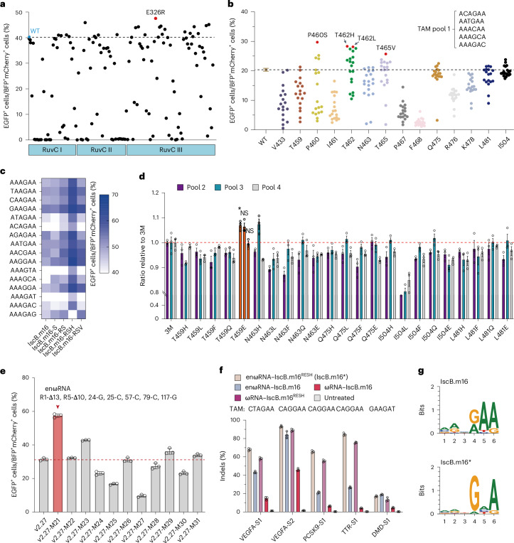
Substitutions of amino acid residues in the DNA-binding pocket or cleavage domains with positively charged arginine have been shown to enhance the editing activity of RNA-guided nucleases in eukaryotic cells21–23. We performed a sequence alignment analysis of IscB.m16 and OgeuIscB. According to conserved sequences, we divided the different structure domains of IscB.m16 and further performed an arginine scanning mutagenesis in the P1D, TID and RuvC domain. According to the activated EGFP fluorescence intensity of cells with an AAAGAA TAM reporter, over 20 of 138 variants in the RuvC domain exhibited improved editing activity compared to wild-type (WT) IscB, with one variant (E326R) showing the highest editing activity (Fig. 3a).
Fig. 3. Protein engineering of IscB.m16 to improve editing efficiency and expand TAM range in mammalian cells.
a, Screening for highly efficient variants by substitutions of amino acid residues in the RuvC domain of IscB.m16 protein with arginine. Each dot represents the editing activity for a single variant. The dashed line indicates the editing activity of the WT. b, Screening for highly efficient variants with saturation mutagenesis at selected sites using GFxxFP reporters containing a pooled TAM target. Each dot represents the editing activity for a single variant. The dashed line indicates the editing activity of the WT. c, Comparison of editing activity among WT IscB.m16 and its variants at 16 GFxxFP reporters with different TAMs. IscB.m16-S represents the variant P460S, IscB.m16-RS represents the variant with a combination of E326R and P460S, IscB.m16-RSH represents the variant with a combination of E326R, P460S and T462H and IscB.m16-RSV represents the variant with a combination of E326R, P460S and T465V. Colored dots reflect the mean of three independent biological replicates. d, Screening for variants with improved editing frequency based on GFxxFP reporters containing three different TAM pools along with the same ωRNA-v2.27. The orange bar represents the IscB.m16RESH variant with a combination of IscB.m16-RSH and T495E. P values were determined by Tukey’s multiple comparisons test following ordinary one-way ANOVA. *P < 0.05. NS, not significant. e, The second round of ωRNA engineering by substituting C•G base pairs on ωRNA-v2.27 (R1-Δ13, R5-Δ10, 24-G, 25-C, 57-G, 79-C and 117-C) based on IscB.m16RESH. f, Comparison of indel frequency of WT IscB.m16 and its variants at five endogenous sites in HEK293T cells. g, TAM logos of IscB.m16 and IscB.m16* systems. Values and error bars represent the mean ± s.d. (n = 3 independent biological replicates).
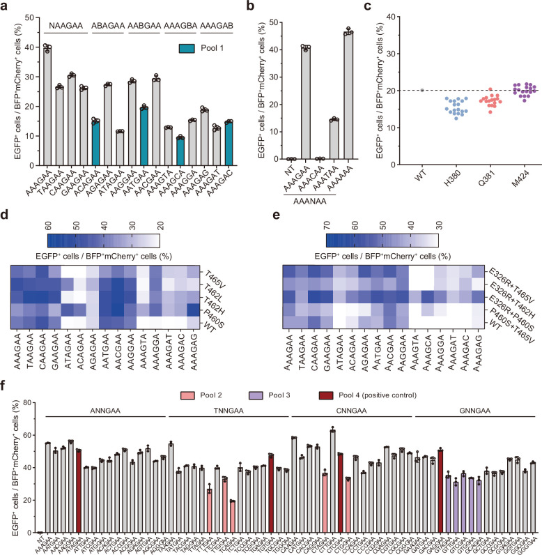
Considering that the P1D and TID domains are related to TAM recognition, we next screened 124 variants in these two domains using six GFxxFP reporters with different TAMs to broaden the TAM range. These reporters had the same target sequences but different 6-nt TAMs (AAAGAA, CAAGAA, ACAGAA, AACGAA, AAAGCA and AAAGAC). Compared to WT IscB.m16, seven variants (M424R, T462R, N463R, T465R, Q475R, K478R and I504R) showed improved activity and TAM recognition, as evidenced by the increase (>1.05-fold) in EGFP fluorescence intensity for all six reporters relative to the WT (Supplementary Fig. 5). Meanwhile, through predicted structure analysis of IscB.m16, we identified 11 potential sites associated with TAM recognition: H380, Q381, V433, T459, P460, I461, F467, Y468, R476, K478 and L481. In order to broaden the TAM range with improved editing efficiency, we conducted saturation mutagenesis at these 18 sites—the 7 sites from P1D and TID screening and the 11 predicted sites. We then screened these mutants using a TAM pool characterized by low activity. TAM pool 1 consisted of ACAGAA, AATGAA, AAACAA, AAAGCA and AAAGAC, which had relatively low editing efficiency recognized by the IscB.m16 WT (Extended Data Fig. 3a,b). Using a similar fluorescence reporter system, we found that some variants, especially P460S, T462H, T462L and T465V, greatly enhanced the editing activity of TAM pool 1 (Fig. 3b and Extended Data Fig. 3c). To validate the enhanced activity of these four variants, we performed TAM recognition with 16 reporters including NAAGAA, ANAGAA, AANGAA, AAAGNA and AAAGAN. The results demonstrated that four variants exhibited higher EGFP fluorescence intensity relative to the WT, suggesting their superior editing activity (Extended Data Fig. 3d). On the basis of the results of the 16 reporters described above, we combined the E326R, P460S, T462H, T462L and T465V substitutions and obtained the best-performing combination variant with E326R, P460S and T462H, named IscB.m16-RSH (Fig. 3c and Extended Data Fig. 3e). To test the TAM preference of IscB.m16-RSH, we detected EGFP activation using 64 TAM reporters with the 5′-NNNGAA-3′ TAM and IscB.m16-RSH showed high editing activity for most TAMs but remained low for others (Extended Data Fig. 3f). Considering the characteristics of TAM recognition, we designed three additional TAM pools, pool 2 (TTTGAA, TTGGAA, TCAGAA, CTAGAA and CTGGAA), pool 3 (GTAGAA, GTTGAA, GTCGAA, GTGGAA and GCAGAA) and pool 4 (ATAGAA, TGTGAA, CTCGAA and GAGGAA) as positive pools (Extended Data Fig. 3f). To further improve the activity at further TAMs, we selected sites that showed improved activity in TAM pool 1. We then separately combined the mutants at sites T459, N643, Q475, L481 or I504 with IscB.m16-RSH and evaluated the variants using reporters from pools 2 to 4 (Fig. 3d). We found that the combination of T459E with IscB.m16-RSH, named IscB.m16RESH, exhibited increased editing efficiency of reporters from pool 2 relative to IscB.m16-RSH, with comparable editing efficiency of reporters from pools 3 and 4 (Fig. 3d). To assess the target range and editing activity of IscB.m16RESH, we used 64 NNNGAA and 16 AAAGNN TAM reporters and found that IscB.m16RESH exhibited significantly improved editing efficiency of these reporters compared to WT IscB.m16 (Supplementary Fig. 6).
Extended Data Fig. 3. TAM profiling of IscB.m16 WT and its variants using GFxxFP fluorescence reporter system in HEK293T cells.
a, b, TAM profiling of IscB.m16 at 19 reporters containing different TAMs. c, Screening for highly active variants from saturated mutants at H380, Q381 and M424 of IscB.m16 proteins based on GFxxFP reporters containing a TAM pool1 target. Each dot represents the editing activity for a single variant. The dashed line indicates the editing activity of WT. Colored dots reflect the mean of three independent biological replicates. d, e, Comparison of editing activity among IscB.m16 WT and its variants at 19 GFxxFP reporters, which contain different TAMs. Data are shown as mean of 3 independent biological replicates. f, TAM profiling of IscB.m16RSH at 64 reporters containing the same spacer and 5’-NNNGAA-3’ TAM sequences. Values and error bars represent mean and s.d., n = 3 independent biological replicates.
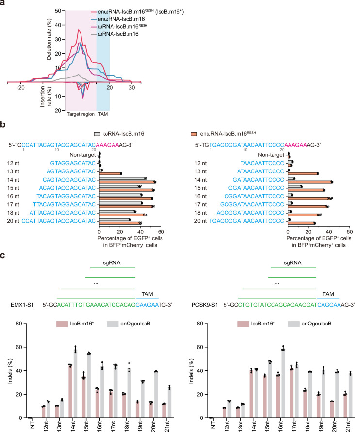
To further optimize ωRNA-v2.27 (del15–20, del29–35, del171–180, 24-G, 25-C, 57-G, 79-C and 117-C) based on IscB.m16RESH, we flipped the G•C base pairs of existing mutations (for example, converting 57G•117C to 57C•117G) or replaced the remaining mismatched G•U base pairs with G•C or C•G, guided by the ωRNA secondary structure. We found that v2.27-M21 (R1-Δ13, R5-Δ10, 24-G, 25-C, 57-C, 79-C, 117-C and 189-G) showed significantly enhanced editing efficiency, hereafter named enωRNA (Fig. 3e). Then, we examined the indel efficiency of IscB.m16RESH with enωRNA at five endogenous loci in cultured HEK293T cells and found that enωRNA–IscB.m16RESH (named IscB.m16*) showed the highest activity and the broadest range of deletion (Fig. 3f and Extended Data Fig. 4a). We also explored a range of spacer lengths for IscB.m16* using fluorescence reporters at two different targets and two endogenous loci in the human genome and found that IscB.m16* exhibited the highest activation with a guide spacer length of 14–21 nt (Extended Data Fig. 4b,c). Furthermore, TAM identification of IscB.m16* using bacterial depletion indicated that IscB.m16* recognized a 5′-NNNGNA-3′ TAM, while IscB.m16 WT recognized a 5′-MRNRAA-3′ TAM (Fig. 3g). To investigate off-target activity, we performed primer-extension-mediated sequencing (PEM-seq) experiments on IscB.m16*, enOgeuIscB and SpG Cas9. IscB.m16* showed similar translocation events to enOgeuIscB and SpG at the vascular endothelial growth factor A (VEGFA)-S6 site (Supplementary Fig. 7). Together, these results demonstrate that IscB.m16* exhibits high editing efficiency with highly flexible 5′-NNNGNA-3′ TAM recognition.
Extended Data Fig. 4. Characterization of IscB.m16 and IscB.m16* editing activities in HEK293T cells.
a, The indels patterns generated by wild-type IscB.m16 and its variants at VEGFA-S1 site. b, Cleavage activity of wild-type IscB.m16 and its variants are affected by spacer length at two target sites on GFxxFP reporters. c, Spacer length-dependent effects on cleavage activity in IscB.m16* and enOgeuIscB systems targeting two sites in human genome. Values and error bars were shown as mean and s.d., n = 3 independent biological replicates.
IscB.m16*-mediated base editing in mammalian cells
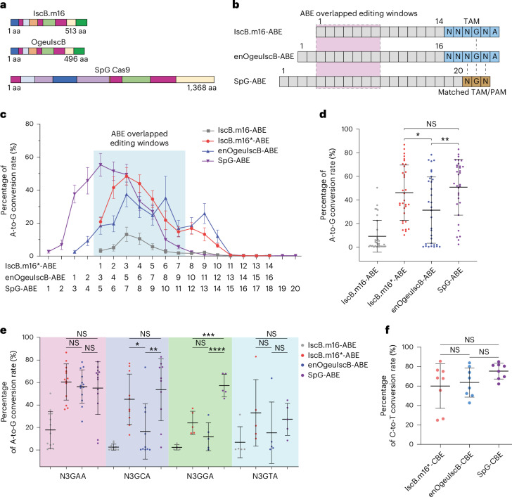
Using prior information of the catalytic residues for IscB19 or SpCas9 (refs. 5,6), we constructed the inactive mutant D61A in the RuvC-I domain, H248A in the HNH domain and D61A;H248A on the basis of IscB.m16 and IscB.m16*. We tested nickase activity using the dual target reporter according to a previous study19. Consistently, IscB.m16*D61A showed the highest nickase activity and IscB.m16*-D61A;H248A showed no activity (Supplementary Fig. 8). In view of the compact size of IscB (Fig. 4a), we next fused IscB.m16*D61A with TadA8e-V106W to generate IscB.m16*-ABE (adenosine base editor) or with human APOBEC3A-W104A to generate IscB.m16*-CBE (cytosine base editor)24,25.
Fig. 4. Characterization of IscB-derived and SpG-derived base editors in mammalian cells.
a, Schematic of IscB, OgeuIscB and SpG with different sizes. b, Overview of TAM-matched and PAM-matched sites used to compare IscB.m16-derived ABE to enOgeuIscB-ABE and SpG-ABE. c, Editing window and base-editing activity of IscB.m16-ABE, IscB.m16*-ABE, enOgeuIscB-ABE and SpG-ABE at all protospacer positions. Data are presented as the mean ± s.e.m. Values are average editing efficiencies at each position of A within the target of three independent biological replicates from 33 endogenous sites. d, Comparison of the A-to-G conversion efficiency of IscB.m16-ABE, IscB.m16*-ABE, enOgeuIscB-ABE and SpG-ABE at 33 endogenous loci. Data were collected from 33 endogenous sites and are presented as the mean ± s.d. Each dot represents the average highest base-editing activity at each endogenous target site of three independent biological replicates. Adjusted P (Padj) values are 0.8453, 0.046 and 0.0041, respectively. e, Comparison of the A-to-G conversion efficiency of IscB.m16-ABE, IscB.m16*-ABE, enOgeuIscB-ABE and SpG-ABE grouped by TAM at 33 target sites. Data are presented as the mean ± s.d. The number of values from left to right is 13, 10, 5 and 5, respectively, and the values represent optimal editing efficiencies within the target as the mean of three independent biological replicates from endogenous sites. The Padj values of N3GAA sites are 0.8698, 0.9404 and 0.9974, respectively. The Padj values of N3GCA sites are 0.8154, 0.0271 and 0.0027, respectively. The Padj values of N3GGA sites are 0.0003, 0.2172 and 0.000007, respectively. The Padj values of N3GTA sites are 0.9744, 0.6102 and 0.8342, respectively. f, Comparison of the C-to-T conversion efficiency of IscB.m16*-CBE, enOgeuIscB-CBE and SpG-CBE at eight target sites. Data were collected from eight endogenous sites and are exhibited as the mean ± s.d. Each dot represents the average highest base-editing activity within the target at each endogenous target site of three independent biological replicates. All P values were determined by Tukey’s multiple comparisons test following ordinary ANOVA. *P < 0.05, **P < 0.01, ***P < 0.001 and ****P < 0.0001. NS, not significant.
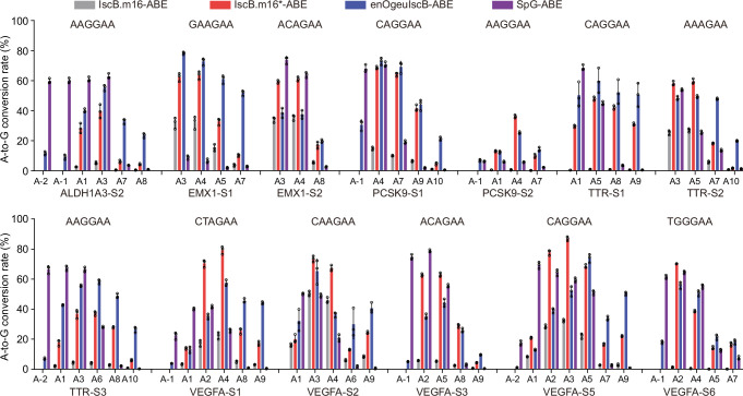
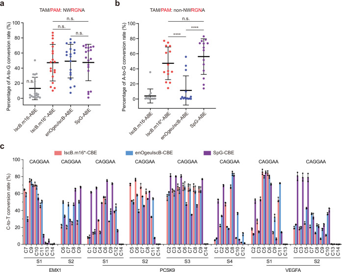
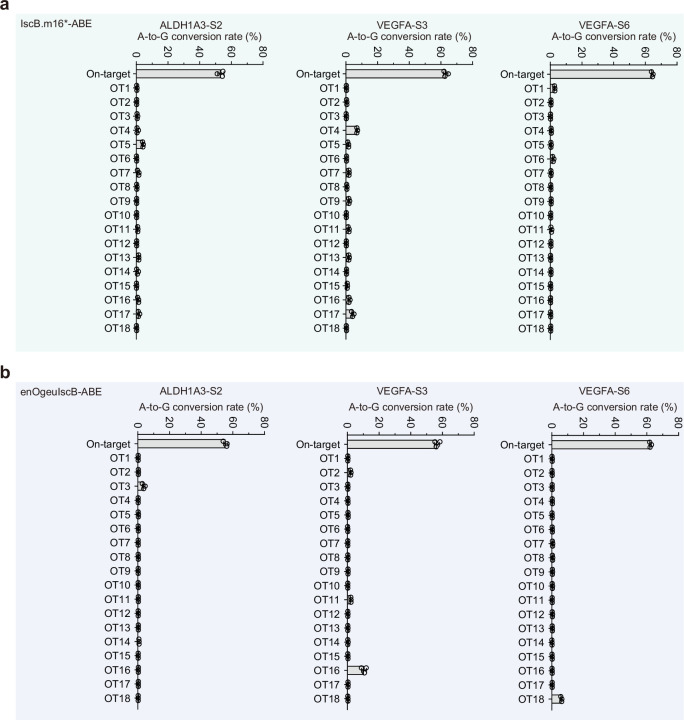
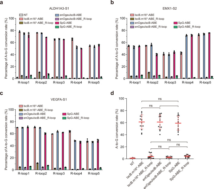
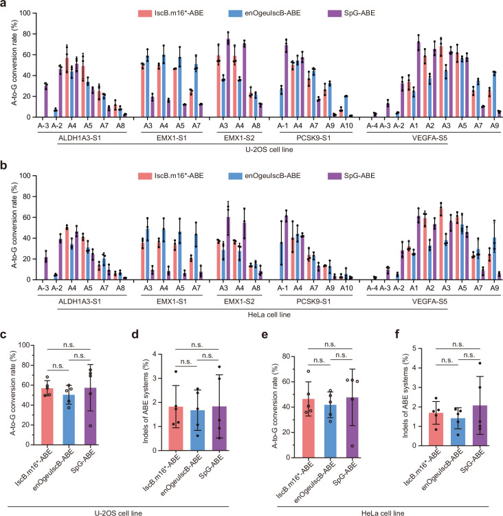
To comprehensively evaluate the editing performance of IscB.m16*-ABE, we designed dozens of TAM-matched and protospacer-adjacent motif (PAM)-matched endogenous loci for IscB.m16-ABE, IscB.m16*-ABE, enOgeuIscB-ABE19 and SpG-ABE22 (Fig. 4b). We found that the editing window of IscB.m16*-ABE ranged from positions 1 to 10 (counting the TAM as positions 15–20), while the optimal editing window occurred within positions 2–5 (Fig. 4c). At these matched G-containing TAM and PAM sites in HEK293T cells, IscB.m16*-ABE showed significantly higher A-to-G base-editing efficiency (46.15% ± 4.08%) than IscB.m16-ABE (9.19% ± 2.34%) and enOgeuIscB-ABE (31.34% ± 4.90%) and comparable base-editing efficiency to SpG-ABE (50.77% ± 4.13%) (Fig. 4d, Extended Data Fig. 5 and Supplementary Fig. 9). In addition, the indel activity of IscB.m16*-ABE was similar to that of enOgeuIscB-ABE but lower than that of SpG-ABE (Supplementary Fig. 10a,b). To characterize the TAM compatibility of IscB.m16*-ABE, we further analyzed the base-editing results and found that it showed A-to-G base editing at all TAM sites, while enOgeuIscB-ABE showed no activity at some TAM sites such as N3GCA, N3GGA and N3GTA (Fig. 4e and Supplementary Fig. 9). Among the 33 designed TAM sequences, 19 TAM sequences were NWRGNA, which conformed to the canonical TAM and PAM sequences for each nuclease (IscB.m16*, NNNGNA; enOgeuIscB, NWRRNA; SpG, NGN). For the 19 NWRGNA TAM sites, IscB.m16*-ABE, enOgeuIscB-ABE and SpG-ABE exhibited comparable A-to-G efficiency (Extended Data Fig. 6a). For the 14 non-NWRGNA TAM sequences, IscB.m16*-ABE showed significantly higher A-to-G base editing than enOgeuIscB-ABE and comparable base editing to SpG-ABE (Extended Data Fig. 6b). At some sites such as EMX1-S1 (GAAGAA) and VEGFA-S4 (AAAGCA), enOgeuIscB-ABE exhibited higher editing efficiency than IscB.m16*-ABE and SpG-ABE (Extended Data Fig. 5 and Supplementary Fig. 9). This result also indicated the different preferred recognition TAM of each nuclease. To further evaluate the specificity of IscB.m16*-ABE in HEK293T cells, we conducted gRNA-dependent off-target DNA editing at predictive sites using Cas-OFFinder26 and gRNA-independent off-target DNA editing using the orthogonal R-loop assay27 at the ALDH1A3-S1, VEGFA-S1 and EMX1-S2 target sites. Targeted deep sequencing analysis revealed that IscB.m16*-ABE exhibited similar gRNA-dependent off-target effects to enOgeuIscB-ABE and SpG-ABE at predicted off-target sites (Extended Data Fig. 7 and Supplementary Fig. 11). Using five previously reported SaCas9 target sites, we observed that IscB.m16*-ABE showed comparable low gRNA-independent off-target events to enOgeuIscB-ABE and SpG-ABE (Extended Data Fig. 8). In addition, IscB.m16*-CBE exhibited comparable base-editing activity and indels to enOgeuIscB-CBE and SpG-CBE, with base-editing efficiencies of 60.01% ± 8.08%, 63.72% ± 5.33% and 75.42% ± 8.12%, respectively (Fig. 4f, Extended Data Fig. 6c and Supplementary Fig. 10c,d). In addition, we detected IscB.m16*-ABE, enOgeuIscB-ABE and SpG-ABE at five endogenous sites (ALDH1A3-S1, EMX1-S1, EMX1-S2, PCSK9-S1 and VEGFA-S5) in the U-2OS and HeLa cell lines. Consistent with its editing efficiency in the HEK293T cell line, IscB.m16*-ABE exhibited high editing efficiency in the U-2OS and HeLa cell lines (Extended Data Fig. 9). Collectively, these results indicate that IscB*-based base editors exhibit highly active editing, a broad target range and low off-target effects in mammalian cells.
Extended Data Fig. 5. A-to-G conversion efficiency of IscB- and SpG-derived adenine base editors at endogenous loci in HEK293T cells.
Comparisons of A-to-G conversion efficiency of IscB.m16-ABE, IscB.m16*-ABE, enOgeuIscB-ABE and SpG-ABE at 13 target sites containing various TAMs in HEK293T cells. Value and error bars are presented as means ± s.d., n = 3 independent biological replicates.
Extended Data Fig. 6. Base editing efficiency of IscB- and SpG-based adenine and cytosine base editors at endogenous loci in HEK293T cells.
a, b, Comparison of the A-to-G conversion efficiency of IscB.m16-ABE, IscB.m16*-ABE, enOgeuIscB-ABE, and SpG-ABE at 19 TAM sites of NWRGNA (a) and 14 TAM sites of NNNGNA, but non-NWRGNA (b). Data are collected from 19 and 14 endogenous sites, respectively, and presented as means ± s.d. Each dot represents the average highest base editing activity at each endogenous target site of three independent biological replicates. P values determined by Tukey’s multiple comparisons test following ordinary one-way analysis of variance. The Padj. are 0.613, 0.00005 and 0.0000007, respectively. c, Comparisons of C-to-T conversion efficiency of IscB.m16-CBE, IscB.m16*-CBE, enOgeuIscB-CBE, SpG-CBE at 8 target sites containing various TAMs in HEK293T cells. Value and error bars are presented as means ± s.d., n = 3 independent biological replicates.
Extended Data Fig. 7. The gRNA-dependent off-target levels of ABEs at in-silico predicted off-target sites, determined by targeted deep sequencing.
a, b A-to-G conversion rate of IscB.m16*-ABE (a) and enOgeuIscB-ABE (b) targeting ALDH1A3-S2, VEGFA-S3 and VEGFA-S6 sites respectively. Value and error bars are presented as means ± s.d., n = 3 independent biological replicates.
Extended Data Fig. 8. The gRNA-independent off-target levels of IscB- and SpG-derived adenine base editors at five R-loops formed by dSaCas9 in HEK293T cells.
a–c, The gRNA-independent off-target levels of base editors at ALDH1A3-S1 (a), EMX1-S2 (b), and VEGFA-S1 (c). d, Statistical analysis of gRNA-independent off-target levels at 3 endogenous sites. Data collected from 3 endogenous sites and each site with 5 R-loops of each editor respectively. The single dot presents mean of the highest edits at one site and one of the R-loop. Value and error bars are presented as means ± s.d., P values determined by Tukey’s multiple comparisons test following ordinary one-way analysis of variance. NS, not significant.
Extended Data Fig. 9. A-to-G conversion efficiency of IscB- and SpG-based adenine base editors at endogenous loci in U-2OS and HeLa cells.
a, b, Comparisons of A-to-G conversion efficiency of IscB.m16*-ABE, enOgeuIscB-ABE, SpG-ABE at five target sites containing various TAMs in U-2OS (a) and HeLa (b) cells. Value and error bars are presented as means ± s.d., n = 3 independent biological replicates. c–f, Statistical of A-to-G editing efficiency (c, e) and indels (d, f) at five endogenous sites in U-2OS (c, d) and HeLa (e, f) cells, respectively. Value and error bars are presented as means ± s.d., n = 3 independent biological replicates. P values determined by Tukey’s multiple comparisons test following ordinary one-way analysis of variance. NS, not significant.
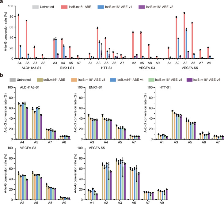
Considering the bystander editing of base editors, we used high-fidelity TadA8e variants and different linkers combined with IscB to narrow the editing window (Supplementary Fig. 12). To address this concept, we tested high-fidelity deaminases and different linkers. We replaced TadA8e-V106W of IscB.m16*-ABE with TadA8e-N108Q or TadA8e-N108Q;L145T (ref. 28) and found a narrower editing window but lower editing efficiency compared to IscB.m16*-ABE (Extended Data Fig. 10a). The replacement of linkers between IscB and TadA8e-V106W showed no significant improvement with respect to narrowing the editing window (Extended Data Fig. 10b).
Extended Data Fig. 10. A-to-G conversion efficiency of IscB.m16*-ABE with different TadA8e variants and different linkers.
a, Comparison of editing window and base editing activity of IscB.m16*-ABEs with different TadA8e variants at 5 endogenous loci. b, Comparison of editing window and base editing activity of IscB.m16*-ABEs with different linkers at 5 endogenous sites. Value and error bars are presented as means ± s.d., n = 3 independent biological replicates.
IscB-derived CBE restores dystrophin expression in mice
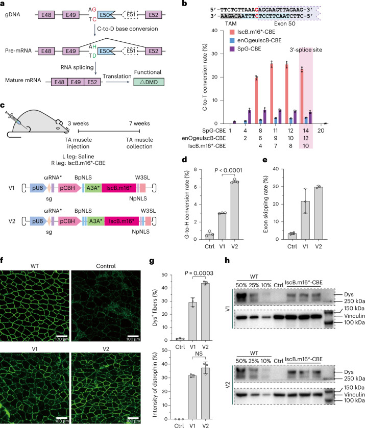
Taking advantage of its small size, the IscB*-derived base editor can be packaged with its ωRNA into a single rAAV vector, making it a greatly promising candidate for the treatment of certain genetic diseases, such as Duchenne muscular dystrophy (DMD)29,30. Previous studies have shown that exon 50 skipping of the dystrophin gene can restore dystrophin expression in a mouse model with an exon 51 deletion, a mutation occurring in nearly 8% of patients with DMD31,32. To access IscB.m16*-based base editing in DMD therapy, we devised a strategy whereby IscB.m16*-CBE disrupted the splicing signal by converting the G (in the paired chain of C) within the splicing acceptor site (‘AG’) to other bases (A, C or T), resulting in exon skipping (Fig. 5a). We first tested IscB.m16*-CBE with the ωRNA targeting the AG site adjacent to exon 50 in HEK293T cells. We observed that IscB.m16*-CBE displayed approximately 25% activity at position 10, which is the splicing acceptor site, while enOgeuIscB-CBE and SpG-CBE showed almost no base-editing activity (Fig. 5b). To conveniently package tools into a single AAV, we chose IscB.m16*-CBE (4.0 kb) without the uracil DNA glycosylase inhibitor (UGI) domain (two UGI domains and a linker, 196 aa) to be packaged into AAV9 and detected the base-editing activity in mice. Two versions of IscB.m16*-CBE carrying different nuclear localization signals (NLSs) were designed and delivered to the muscle of mice with humanized exon 50 knock-in and exon 51 deletion (Fig. 5c). Then, 4 weeks after injection, we performed an editing efficiency evaluation, western blot analysis and histological staining for dystrophin expression. Targeted deep sequencing analysis showed that IscB.m16*-CBE-v2 achieved an approximate 7% conversion of G-to-H (G-to-A, G-to-T and G-to-C) and up to 30% level of exon 51 skipping (Fig. 5d,e). Western blotting and histological staining quantitative analysis of the tibialis anterior (TA) muscle and immunostaining results indicated that IscB.m16*-CBE-v2 restored the dystrophin protein levels in myofibers to 40% of the WT control (Fig. 5f–h). Together, these results indicate that the IscB.m16*-derived base editor, as a highly effective and broad-TAM miniature base-editing tool, provides a promising approach for basic research and therapeutic applications.
Fig. 5. IscB.m16*-based cytosine base editor mediates effective base editing and restores dystrophin expression in humanized DMDE51del mice.
a, Schematic of the strategy of IscB.m16*-CBE DMD treatment. IscB.m16*-CBE disrupts the conserved guanine within the splice acceptor site for programmable exon 50 skipping, leading to the restoration of dystrophin expression. b, The C-to-T conversion efficiency of IscB.m16*-CBE, enOgeuIscB-CBE and SpG-CBE at the splice acceptor site of the DMD intron between exon 49 and exon 50 in HEK293T cells. Data are shown as the mean ± s.d. (n = 3 independent biological replicates). c, Schematics of single AAV9 carrying two versions of IscB.m16*-CBE delivered to the muscles in mice by TA muscle injection. Saline was injected in the left leg, while AAV9 cargo IscB.m16*-CBE was injected in the right. d,e, The in vivo G-to-H (C-to-D, including C-to-T, C-to-A and C-to-G) editing efficiencies (d) and RNA level of exon 50 skipping (e) of AAV9-IscB.m16*-CBE were detected by targeted deep sequencing. Data are presented as the mean ± s.d. (n = 3 independent biological replicates; n = 4 for the control in e). The Padj value is 0.000002 in d. f, Dystrophin immunohistochemistry showing the restoration of dystrophin expression 4 weeks after TA injection of IscB.m16*-CBE. Dystrophin is shown in green. Scale bars, 100 µm. g, Quantification of Dys+ fibers and dystrophin in cross sections of TA muscles from f. Data are presented as the mean ± s.d. (n = 3 independent biological replicates). h. Western blot analysis of dystrophin and vinculin expression in TA muscles 4 weeks after injection with AAV9-IscB.m16*-CBE or saline.
Discussion
In summary, through computational mining of metagenomic sequence datasets, we identified 19 natural IscB orthologs with various TAM recognition sites and 10 of the IscBs showed activity in mammalian cells, highlighting the diversity of the IscB family. By examining the results of six engineered ωRNAs, we found that the truncation of the first (R1) and last (R5 or R6) stem loops of the ωRNA usually enhanced the editing activity of IscBs. Through structure-guided design and protein engineering of the P1D, TID and RuvC domain of IscB, we developed the IscB.m16* system that exhibited improved editing activity and extended the TAM scope to 5′-NNNGNA-3′. This is a notably broader recognition range than the previously reported enOgeuIscB with a 5′-NWRRNA-3′ TAM19, although we found that enOgeuIscB showed efficient activity with a broader TAM (not only NWRRNA) in mammalian cells. Furthermore, IscB.m16*-derived base editors showed editing activity comparable to SpG-BE and even higher editing activity than SpG-BE and enOgeuIscB-BE at some disease-related loci, such as DMD. Therefore, considering their compact size and extended editing scope, IscBm16*-derived base editors have the potential to be alternatives to enOgeuIscB-derived and Cas9-derived base editors for AAV-based therapeutic applications.
Additionally, we found that both IscB.m16* and enOgeuIscB showed indel activity with a guide containing a spacer length of 14–21 nt (Extended Data Fig. 4c). This is similar to a previous study showing that OgeuIscB exhibited indel activity with a guide containing a spacer length of 14–26 nt17. Thus, most IscB–ωRNA systems have less stringent requirements with regard to spacer length. Given the potential for off-target effects with short spacer lengths, screening variants to specifically bind long spacers (for example, 20 nt), increasing the mismatch of spacers to extend the spacer length or designing a specific stable lock-and-key structure33 may minimize the off-target effects. Considering the 14-nt spacer length, a 2-nt TAM may achieve the best balance between targeting range and specificity. Therefore, developing more highly active IscB orthologs with different TAM recognition sites may be more important for future applications than developing a near-TAMless IscB. Given the experience gained in engineering the IscB nuclease and ωRNA, the activity and TAM editing range of other IscB orthologs such as OgeuIscB could be improved and expanded using similar strategies. Together with IscB.m16*, a set of engineered IscB orthologs may constitute a miniature genome base-editing toolbox.
Overall, the engineered compact IscB-derived base editors were proven to be a platform with highly efficient, specific and broad-TAM-scope DNA base editing in mammalian cells and in mouse models of diseases, highlighting their potential in gene therapy.
Methods
Computational analysis of IscB systems
More than 200 Gb of metagenome assemblies were downloaded from the European Nucleotid Archive database (accession number PRJEB31266). Firstly, we used TBLASTN and the OgeuIscB protein to identify IscB-containing sequences of metagenomes with an E value < 1 × 10−50 (ref. 20). Then, Prodigal was used to annotate the proteins of the IscB-containing sequences34. We further searched for previously trained ωRNA models to annotate the ωRNA sequences with an E value < 1 × 10⁻¹⁰. RNAfold was used to predict the secondary structure of the ωRNA35,36. MEGAX was used to construct the phylogenetic tree37. All newly IscB protein sequences and ωRNA scaffolds (DNA sequences) are provided in Supplementary Table 1.
Plasmid construction
All E. coli codon-optimized IscB-encoding genes and their associated ωRNA scaffolds were synthesized by Shanghai Huagene Biotechnology and assembled into a pUC19-derived vector (EcoNI + XbaI) under the lac and J23119 promoters using a 2× pEASY basic seamless cloning and assembly kit (TransGen Biotech). All human codon-optimized IscB-encoding sequences were synthesized by GenScript and incorporated into a mammalian expression vector under the CBh promoter. For endogenous genome-editing experiments in HEK293T cells, the gRNA oligos were synthesized and cloned into a BpiI-digested backbone of the U6 promoter using T4 ligase (Thermo Fisher Scientific). All colonies were sequence-verified from promoter to poly(A) using Sanger sequencing (Genewiz). Information on all IscB expression sequences is provided in Supplementary Tables 2–4.
Generation of the TAM library and TAM depletion assay
A randomized TAM library containing a target sequence followed by eight randomized bases downstream was constructed. The synthesized single-stranded DNA (ssDNA) (HuaGene) was converted into double-stranded DNA (dsDNA) by annealing with a short ssDNA and second-strand synthesis using the Large (Klenow) fragment (New England Biolabs). The resulting dsDNA was then assembled into pACYC184 vectors using Gibson assembly (New England Biolabs). The products were purified using isopropanol, electroporated into TransforMax EC100 electrocompetent E. coli according to the manufacturer’s instructions and plated on chloramphenicol plates. After 13 h of growth at 37 °C, E. coli cells were scraped from the plates and extracted using a NucleoBond Xtra Midiprep kit (Machery Nagel).
For the bacterial TAM depletion assay, we cotransformed 200 ng of TAM library plasmids and 300 ng of plasmids expressing E. coli codon-optimized IscB and ωRNA into TransforMax EC100 electrocompetent E. coli cells by electroporation. Then, the transformed cells were recovered for 1 h at 37 °C with antibiotic-free medium and plated on 250 mm × 250 mm carbenicillin and chloramphenicol plates. After 13 h of growth, cells were harvested and plasmid DNA was extracted using a NucleoBond Xtra Midiprep kit (Machery Nagel). The TAM-containing region was amplified by Phanta Max super-fidelity DNA polymerase (Vazyme Biotech) for 12 cycles and Illumina adaptors and unique barcodes were added by a second round of PCR for 18 cycles. The resulting PCR products were purified with a gel extraction kit (Omega) and sequenced by a Illumina NovaSeq 6000 platform with 150-bp paired-end reads (Genewiz).
TAM regions were extracted, counted and then normalized to the total TAM counts for each sample. For each specific TAM, TAMs that appeared more than once were filtered and the log fold change (logFC) of its frequency was measured as the log ratio compared to nontarget control. Depletions with a logFC < −3σ (s.d.) were considered statistically significant. A position weight matrix (PWM) was built from all significantly depleted sequences, with −logFC values serving as the corresponding weight. A sequence logo was generated on the basis of this PWM using WebLogo (version 3.7.12)17.
Cell culture and transfection
HEK293T cells were cultivated in DMEM (Sigma) supplemented with 10% FBS (Gibco), 1% penicillin–streptomycin–glutamine (Gibco) and 1% minimum essential medium nonessential amino acids (Gibco) in a humidified incubator at 37 °C with 5% CO2. For the detection of IscB nuclease activities and screening of its variants, HEK293T cells were seeded in 24-well plates with 70–80% confluence. After a 12-h incubation, 1.6 μg of plasmids were cotransfected into HEK293T cells using polyethylenimine (PEI) following the manufacturer’s manual. The plasmids included those encoding the BFP–T2A–GFxxFP and IscB systems, with a molar ratio of 1:1. For genome or base editing at endogenous loci, 1.6 μg of all-in-one plasmids were transfected to express gRNA and the nuclease-editing or base-editing system. After 48 h, cells were sorted by fluorescence-activated cell sorting (FACS) analysis.
FACS analysis
Before FACS analysis, cells were subjected to treatment with 0.25% trypsin-EDTA (Gibco) for dissociation and suspended in FBS-containing DMEM. For the assessment of IscB nuclease activity and screening of variants using the fluorescence reporter system, cells were analyzed for EGFP, mCherry and BFP fluorescence. A total of 25,000 single cells were recorded to analyze efficiency using a Beckman CytoFlex flow cytometer 48 h after transfection. Data analysis was performed by FlowJo X (version 10.0.7). For genome-editing analysis, approximately 15,000 transfection-positive cells (defined as those with a fluorescence intensity ≥ 103 among fluorescence-positive cells) were sorted 48 h after transfection using a BD FACS Aria III flow cytometer. Following FACS sorting, genomic DNA from the collected cells was extracted by cell lysis with 25 μl of proteinase K-added lysis buffer (Vazyme Biotech) per sample, as described previously. The cell lysates were stored at −20 °C until further use.
Targeted deep sequencing and analysis
To detect the editing efficiency at endogenous loci, the target genome regions of interest were amplified from cell lysates by PCR using Phanta Max super-fidelity DNA polymerase (Vazyme Biotech). For targeted deep sequencing analysis, PCR reactions were performed using primers with unique barcodes. The amplified products were purified using a gel extraction kit (Omega) and sequenced by an Illumina NovaSeq 6000 platform with 150-bp paired-end reads (Genewiz). The deep sequencing data were first demultiplexed by a custom script based on sample barcodes. The demultiplexed reads were then analyzed by CRISPResso2 (version 2.0.20b)38 for the quantification of the editing efficiency, including indels and base conversions at each target locus. All targeting sites and primers used are provided in Supplementary Table 5.
PEM-seq assay
PEM-seq in HEK293T cells was performed as previously described39,40. Specifically, all-in-one plasmids containing IscB.m16*, enOgeuIscB and SpCas9-SpG with targeting VEGFA-S6 ωRNA were transfected into HEK293T cells using PEI; after 48 h, positive cells were harvested for DNA extraction. A total of 10 μg of genomic DNA was fragmented with a peak length of 300–700 bp by Covaris sonication. Those DNA fragments were firstly tagged with biotin through a one-round biotinylated primer extension at the 5′ end and then primers were removed by AMPure XP beads and purified by streptavidin beads. Then, the ssDNA attached to the streptavidin beads was ligated with a 14-bp random molecular barcode bridge adapter and a nested PCR was performed for enriching the DNA fragment containing the bait DSB and tagging the DNA fragment with Illumina adaptor sequences. The prepared sequencing library was subjected to high-throughput sequencing on a Hi-seq 2500 with 2 × 150 bp reads.
gRNA-dependent off-target analysis
To examine the gRNA-dependent off-target effects of IscB.m16*-ABE, enOgeuIscB-ABE and SpG-ABE, CRISPR RGEN Tools (Cas-OFFinder, http://www.rgenome.net/cas-offinder/) was used to predict potential off-target sites as described previously26. For the ABE based on IscB.m16*, the search queries covered both the 14-nt target spacer sequences and ‘NNNGNA’. The PAM of the search was set as ‘NNN’ and the number of mismatches was set to three. The search queries of enOgeuIscB-ABE were set similarly but with a 16-nt spacer sequence and a 6-nt TAM sequence containing ‘NWRRNA’. For the ABE based on SpG, search queries covered 20-nt target spacer sequences, the PAM type was set to ‘NG’ and the number of mismatches was set to four. All other parameters were default. Off-target sites for each gRNA in each group were manually selected in order of the number of mismatches from low to high. Sites with a 5′-NNNGNA-3′ TAM were retained for IscB.m16*-ABE and sites with a 5′-NWRRNA-3′ TAM were retained for enOgeuIscB-ABE. All potential sites and primers are provided in Supplementary Tables 6–15.
Orthogonal R-loop assay
An orthogonal R-loop assay was performed to detect the gRNA-independent off-target editing as described previously27. First, 0.8 μg of plasmids that encode IscB.m16*-ABE, enOgeuIscB-ABE or SpG-ABE with their respective ωRNA or single-guide RNA (sgRNA) and 0.8 μg of dSaCas9 plasmids with their corresponding sgRNA targeting five previously reported R-loop sites were cotransfected into HEK293T cells using PEI. After a 48-h cultivation, transfected cells were analyzed by FACS followed by genomic DNA extraction with 25 μl of freshly prepared lysis buffer (Vazyme) containing proteinase K. Amplification and targeted deep sequencing were performed at the ABE target sites and dSaCas9 R-loop off-target sites. All targeting sequences and primers are provided in Supplementary Table 16.
Animals
All animal experiments in this study were performed following approved protocols and guidelines set by the Animal Care and Use Committee of Huidagene Therapeutics. Mice were housed in a controlled barrier facility with a 12-h light–dark cycle at 18–23 °C with 40–60% humidity. Diet and water were accessible at all times. DMDΔmE5051, KIhE50/Y mice were generated in the C57BL/6J background using the CRISPR–Cas9 system. DMD is the most common sex-linked lethal disease in humans; thus, male mice were selected for this study.
Production and delivery of AAV9 to DMDΔmE5051, KIhE50/Y mice
AAVs were manufactured by HuidaGene Therapeutics. Briefly, cells were grown in culture until they reached a confluency of 70–90%. Before transfection, the growth medium was replaced with prewarmed growth medium. For each 15-cm dish, a mixture of 20 μg of pHelper, 10 μg of pRepCap and 10 μg of the gene-of-interest plasmid was prepared and added dropwise to the cell medium. After a 3-day incubation period, AAVs were harvested and purified using iodixanol density gradient centrifugation. For intramuscular injection, 3-week-old DMDΔmE5051, KIhE50/Y mice were anesthetized and their TA muscle was injected with either 30 μl of AAV9 (2.5 × 10¹¹ vg) preparations or an equivalent volume of saline solution. Tissue samples were collected for genomic DNA, RNA, immunoblotting and immunofluorescence analyses 4 weeks after treatment.
Western blot analysis
Tissue samples were homogenized using radioimmunoprecipitation assay buffer supplemented with a protease inhibitor cocktail. The supernatants of the lysates were quantified using a Pierce BCA protein assay kit (Thermo Fisher Scientific, 23225) and adjusted to a uniform concentration using H2O. Equal volumes of the samples were mixed with NuPAGE LDS sample buffer (Invitrogen, NP0007) and 10% β-mercaptoethanol and then subjected to boiling at 70 °C for 10 min. A total of 10 µg of protein per lane was loaded into 3–8% Tris-acetate gels (Invitrogen, EA03752BOX) and underwent electrophoresis for 1 h at 200 V. Proteins were then transferred onto a PVDF membrane under wet conditions at 350 mA for 3.5 h. The membrane was then blocked in 5% nonfat milk in TBST buffer and incubated with the primary antibody to mark the target protein. After three washes with TBST, the membrane was incubated with a horseradish peroxidase-conjugated secondary antibody specific to the IgG of the species from which the primary antibody against dystrophin (Sigma, D8168) or vinculin (CST, 13901S) was derived. The target proteins were visualized using chemiluminescent substrates (Invitrogen, WP20005).
Immunofluorescence
Tissues were encased in optimal cutting temperature compound and rapidly frozen in liquid nitrogen. Serial frozen cryosections, each measuring 10 µm in thickness, were fixed for 2 h at 37 °C, followed by permeabilization with PBS containing 0.4% Triton-X for 30 min. After washing with PBS, samples were blocked with 10% goat serum for 1 h at room temperature. Following this, the slides were incubated overnight at 4 °C with primary antibodies against dystrophin (Abcam, ab15277) and spectrin (Millipore, MAB1622). The following day, samples were thoroughly washed with PBS and incubated with compatible secondary antibodies (Alexa Fluor 488 AffiniPure donkey anti-rabbit IgG (Jackson ImmunoResearch labs, 711-545-152) or Alexa Fluor 647 AffiniPure donkey anti-mouse IgG (Jackson ImmunoResearch labs, 715-605-151)) and DAPI for 3 h at room temperature. After a 15-min PBS wash, slides were sealed with fluoromount-G mounting medium. All images were captured using a Nikon C2 camera. The number of Dys+ muscle fibers was represented as a percentage of the total spectrin-positive muscle fibers.
Statistical analysis
All values are shown as the mean ± s.d. except for values of the editing window from base editors, which were shown as the mean ± s.e.m. A one-way analysis of variance (ANOVA) was used for statistical comparisons and a P value < 0.05 was considered statistically significant. Details of statistical values are provided in corresponding figure legends. The experiments were not randomized and the investigators were not blinded to allocation during experiments and outcome assessment. GraphPad Prism (version 8.2.1) was used for statistical analysis (www.graphpad.com/).
Reporting summary
Further information on research design is available in the Nature Portfolio Reporting Summary linked to this article.
Online content
Any methods, additional references, Nature Portfolio reporting summaries, source data, extended data, supplementary information, acknowledgements, peer review information; details of author contributions and competing interests; and statements of data and code availability are available at 10.1038/s41589-024-01706-1.
Supplementary information
Supplementary Figs. 1–13.
Supplementary Tables 1–16: IscB ortholog sequences, targeting sequences and primers.
Source data for supplementary figures.
Source data
Statistical source data.
Statistical source data.
Statistical source data.
Statistical source data.
Statistical source data.
Unprocessed western blots.
Statistical source data.
Statistical source data.
Statistical source data.
Statistical source data.
Statistical source data.
Statistical source data.
Statistical source data.
Statistical source data.
Statistical source data.
Acknowledgements
This work was supported by HUIDAGENE Therapeutics Inc. We thank Y. Yuan for assistance with intellectual property. This work was supported by the National Natural Science Foundation of China (31925016, 82021001 and 82271048), National Science and Technology Innovation 2030 Major Program (2021ZD0200900), Basic Frontier Scientific Research Program of the Chinese Academy of Sciences From 0 to 1 Original Innovation Project (ZDBS-LY-SM001), Project of Shanghai Municipal Science and Technology Commission (23HC1401100), Science and Technology Commission of Shanghai Municipality (22S11900200 and 23XD1420500), Project of Shanghai Xuhui District Science and Technology (2020-015), Program for Professor of Special Appointment (Eastern Scholar, TP2022046) at the Shanghai Institutions of Higher Learning and Shanghai Sailing Program (21YF1453100).
Extended data
Author contributions
Q.X. and H.Z. jointly conceptualized the project and designed the experiments. D.H. conducted the bacterial depletion assay and performed the orthogonal R-loop assay. Q.X., D.H., H.W. and G.L. performed the ωRNA and protein engineering and reporter assay. Q.X., M.Y. and T.M. conducted the experiments related to genome editing and base editing and the off-target assays. G.L. performed the animal experiments. Q.X. performed the bioinformatics analysis. M.Y. and T.M. constructed plasmids and performed the FACS analysis. J.Z., Y.Z., X.Z., B.H., Y.Y., J.H. and L.S. assisted with experiments. J.H., H.Y. and H.Z. supervised the project. T.L., J.H., H.Y. and H.Z. wrote and revised the manuscript with data contributed by all authors who participated in the project.
Peer review
Peer review information
Nature Chemical Biology thanks the anonymous reviewers for their contribution to the peer review of this work.
Data availability
All sequencing data were deposited to the National Center for Biotechnology Information Sequence Read Archive under accession number PRJNA1043847. All materials are available upon reasonable request. Source data are provided with this paper.
Competing interests
H.Z. and Q.X. disclose a patent application (PCT/CN2024/071744) related to the proteins described in this manuscript. H.Y. and L.S. are cofounders of HUIDAGENE Therapeutics. The other authors declare no competing interests.
Footnotes
Publisher’s note Springer Nature remains neutral with regard to jurisdictional claims in published maps and institutional affiliations.
These authors contributed equally: Qingquan Xiao, Guoling Li, Dingyi Han, Haoqiang Wang, Mingyu Yao.
Contributor Information
Tong Li, Email: tongli@huidagene.com.
Hui Yang, Email: huiyang@huidagene.com.
Jinhai Huang, Email: jinhaihuang@fudan.edu.cn.
Hainan Zhang, Email: zhanghainan@simm.ac.cn.
Extended data
is available for this paper at 10.1038/s41589-024-01706-1.
Supplementary information
The online version contains supplementary material available at 10.1038/s41589-024-01706-1.
References
- 1.Mali, P. et al. RNA-guided human genome engineering via Cas9. Science339, 823–826 (2013). [DOI] [PMC free article] [PubMed] [Google Scholar]
- 2.Zetsche, B. et al. Cpf1 is a single RNA-guided endonuclease of a class 2 CRISPR–Cas system. Cell163, 759–771 (2015). [DOI] [PMC free article] [PubMed] [Google Scholar]
- 3.Doudna, J. A. The promise and challenge of therapeutic genome editing. Nature578, 229–236 (2020). [DOI] [PMC free article] [PubMed] [Google Scholar]
- 4.Anzalone, A. V., Koblan, L. W. & Liu, D. R. Genome editing with CRISPR–Cas nucleases, base editors, transposases and prime editors. Nat. Biotechnol.38, 824–844 (2020). [DOI] [PubMed] [Google Scholar]
- 5.Gaudelli, N. M. et al. Programmable base editing of A•T to G•C in genomic DNA without DNA cleavage. Nature551, 464–471 (2017). [DOI] [PMC free article] [PubMed] [Google Scholar]
- 6.Komor, A. C., Kim, Y. B., Packer, M. S., Zuris, J. A. & Liu, D. R. Programmable editing of a target base in genomic DNA without double-stranded DNA cleavage. Nature533, 420–424 (2016). [DOI] [PMC free article] [PubMed] [Google Scholar]
- 7.Chen, S. Y. et al. Compact Cje3Cas9 for efficient genome editing and adenine base editing. CRISPR J.5, 472–486 (2022). [DOI] [PubMed] [Google Scholar]
- 8.Davis, J. R. et al. Efficient in vivo base editing via single adenoassociated viruses with size-optimized genomes encoding compact adenine base editors. Nat. Biomed. Eng.6, 1317 (2022). [DOI] [PMC free article] [PubMed] [Google Scholar]
- 9.Zhang, H. et al. Adenine base editing in vivo with a single adeno-associated virus vector. GEN Biotechnol.1, 285–299 (2022). [DOI] [PMC free article] [PubMed] [Google Scholar]
- 10.Wu, Z. et al. Programmed genome editing by a miniature CRISPR–Cas12f nuclease. Nat. Chem. Biol.17, 1132–1138 (2021). [DOI] [PubMed] [Google Scholar]
- 11.Kim, D. Y. et al. Efficient CRISPR editing with a hypercompact Cas12f1 and engineered guide RNAs delivered by adeno-associated virus. Nat. Biotechnol.40, 94–102 (2021). [DOI] [PMC free article] [PubMed] [Google Scholar]
- 12.Kong, X. et al. Engineered CRISPR–OsCas12f1 and RhCas12f1 with robust activities and expanded target range for genome editing. Nat. Commun.14, 2046 (2023). [DOI] [PMC free article] [PubMed] [Google Scholar]
- 13.Hino, T. et al. An AsCas12f-based compact genome-editing tool derived by deep mutational scanning and structural analysis. Cell186, 4920–4935 (2023). [DOI] [PubMed] [Google Scholar]
- 14.Xu, X. et al. Engineered miniature CRISPR–Cas system for mammalian genome regulation and editing. Mol. Cell81, 4333–4345 (2021). [DOI] [PubMed] [Google Scholar]
- 15.Karvelis, T. et al. Transposon-associated TnpB is a programmable RNA-guided DNA endonuclease. Nature599, 692–696 (2021). [DOI] [PMC free article] [PubMed] [Google Scholar]
- 16.Kim, D. Y. et al. Hypercompact adenine base editors based on a Cas12f variant guided by engineered RNA. Nat. Chem. Biol.18, 1005–1013 (2022). [DOI] [PubMed] [Google Scholar]
- 17.Altae-Tran, H. et al. The widespread IS200/IS605 transposon family encodes diverse programmable RNA-guided endonucleases. Science374, 57–65 (2021). [DOI] [PMC free article] [PubMed] [Google Scholar]
- 18.Schuler, G., Hu, C. Y. & Ke, A. L. Structural basis for RNA-guided DNA cleavage by IscB–ωRNA and mechanistic comparison with Cas9. Science376, 1476–1481 (2022). [DOI] [PMC free article] [PubMed] [Google Scholar]
- 19.Han, D. et al. Development of miniature base editors using engineered IscB nickase. Nat. Methods20, 1029–1036 (2023). [DOI] [PubMed] [Google Scholar]
- 20.Stewart, R. D. et al. Compendium of 4,941 rumen metagenome-assembled genomes for rumen microbiome biology and enzyme discovery. Nat. Biotechnol.37, 953–961 (2019). [DOI] [PMC free article] [PubMed] [Google Scholar]
- 21.Zhang, H. et al. An engineered xCas12i with high activity, high specificity and broad PAM range. Protein Cell14, 538–543 (2023). [DOI] [PMC free article] [PubMed] [Google Scholar]
- 22.Walton, R. T., Christie, K. A., Whittaker, M. N. & Kleinstiver, B. P. Unconstrained genome targeting with near-PAMless engineered CRISPR–Cas9 variants. Science368, 290–296 (2020). [DOI] [PMC free article] [PubMed] [Google Scholar]
- 23.Nishimasu, H. et al. Engineered CRISPR–Cas9 nuclease with expanded targeting space. Science361, 1259–1262 (2018). [DOI] [PMC free article] [PubMed] [Google Scholar]
- 24.Richter, M. F. et al. Phage-assisted evolution of an adenine base editor with improved Cas domain compatibility and activity. Nat. Biotechnol.38, 883–891 (2020). [DOI] [PMC free article] [PubMed] [Google Scholar]
- 25.Wang, X. et al. Cas12a base editors induce efficient and specific editing with low DNA damage response. Cell Rep.31, 107723 (2020). [DOI] [PubMed] [Google Scholar]
- 26.Bae, S., Park, J. & Kim, J. S. Cas-OFFinder: a fast and versatile algorithm that searches for potential off-target sites of Cas9 RNA-guided endonucleases. Bioinformatics30, 1473–1475 (2014). [DOI] [PMC free article] [PubMed] [Google Scholar]
- 27.Doman, J. L., Raguram, A., Newby, G. A. & Liu, D. R. Evaluation and minimization of Cas9-independent off-target DNA editing by cytosine base editors. Nat. Biotechnol.38, 620–628 (2020). [DOI] [PMC free article] [PubMed] [Google Scholar]
- 28.Chen, L. et al. Engineering a precise adenine base editor with minimal bystander editing. Nat. Chem. Biol.19, 101–110 (2022). [DOI] [PubMed] [Google Scholar]
- 29.Min, Y. L., Bassel-Duby, R. & Olson, E. N. CRISPR correction of Duchenne muscular dystrophy. Annu. Rev. Med.70, 239–255 (2019). [DOI] [PMC free article] [PubMed] [Google Scholar]
- 30.Olson, E. N. Toward the correction of muscular dystrophy by gene editing. Proc. Natl Acad. Sci. USA118, e2004840117 (2021). [DOI] [PMC free article] [PubMed] [Google Scholar]
- 31.Chemello, F. et al. Precise correction of Duchenne muscular dystrophy exon deletion mutations by base and prime editing. Sci. Adv.7, eabg4910 (2021). [DOI] [PMC free article] [PubMed] [Google Scholar]
- 32.Yuan, J. J. et al. Genetic modulation of RNA splicing with a CRISPR-guided cytidine deaminase. Mol. Cell72, 380–394 (2018). [DOI] [PubMed] [Google Scholar]
- 33.Wang, L. et al. Eliminating base-editor-induced genome-wide and transcriptome-wide off-target mutations. Nat. Cell Biol.23, 552–563 (2021). [DOI] [PubMed] [Google Scholar]
- 34.Hyatt, D. et al. Prodigal: prokaryotic gene recognition and translation initiation site identification. BMC Bioinformatics11, 119 (2010). [DOI] [PMC free article] [PubMed] [Google Scholar]
- 35.Hofacker, I. L. Vienna RNA secondary structure server. Nucleic Acids Res.31, 3429–3431 (2003). [DOI] [PMC free article] [PubMed] [Google Scholar]
- 36.Lorenz, R. et al. ViennaRNA package 2.0. Algorithm Mol. Biol.6, 26 (2011). [DOI] [PMC free article] [PubMed] [Google Scholar]
- 37.Kumar, S., Stecher, G., Li, M., Knyaz, C. & Tamura, K. MEGA X: molecular evolutionary genetics analysis across computing platforms. Mol. Biol. Evol.35, 1547–1549 (2018). [DOI] [PMC free article] [PubMed] [Google Scholar]
- 38.Clement, K. et al. CRISPResso2 provides accurate and rapid genome editing sequence analysis. Nat. Biotechnol.37, 224–226 (2019). [DOI] [PMC free article] [PubMed] [Google Scholar]
- 39.Yin, J. et al. Optimizing genome editing strategy by primer-extension-mediated sequencing. Cell Discov.5, 18 (2019). [DOI] [PMC free article] [PubMed] [Google Scholar]
- 40.Liu, Y. et al. PEM-seq comprehensively quantifies DNA repair outcomes during gene-editing and DSB repair. STAR Protoc.3, 101088 (2022). [DOI] [PMC free article] [PubMed] [Google Scholar]
Associated Data
This section collects any data citations, data availability statements, or supplementary materials included in this article.
Supplementary Materials
Supplementary Figs. 1–13.
Supplementary Tables 1–16: IscB ortholog sequences, targeting sequences and primers.
Source data for supplementary figures.
Statistical source data.
Statistical source data.
Statistical source data.
Statistical source data.
Statistical source data.
Unprocessed western blots.
Statistical source data.
Statistical source data.
Statistical source data.
Statistical source data.
Statistical source data.
Statistical source data.
Statistical source data.
Statistical source data.
Statistical source data.
Data Availability Statement
All sequencing data were deposited to the National Center for Biotechnology Information Sequence Read Archive under accession number PRJNA1043847. All materials are available upon reasonable request. Source data are provided with this paper.


