Abstract
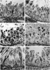
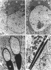
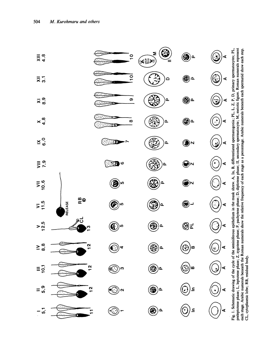
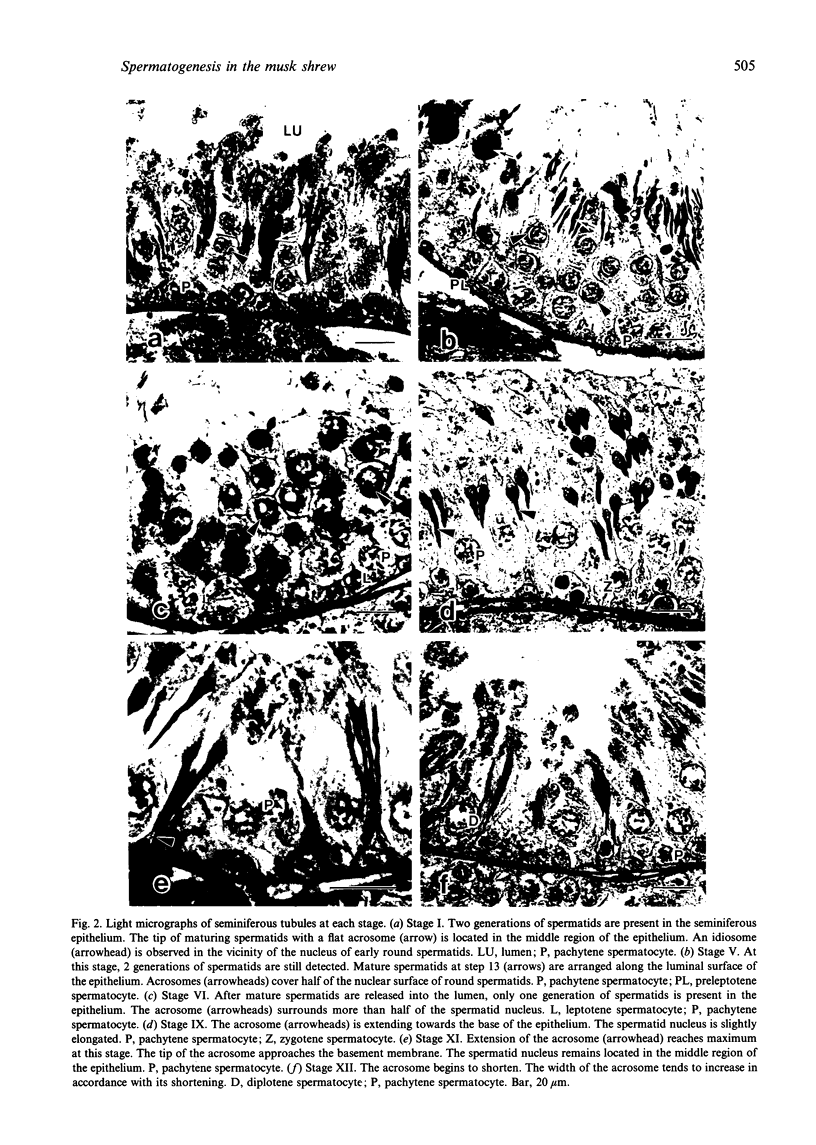
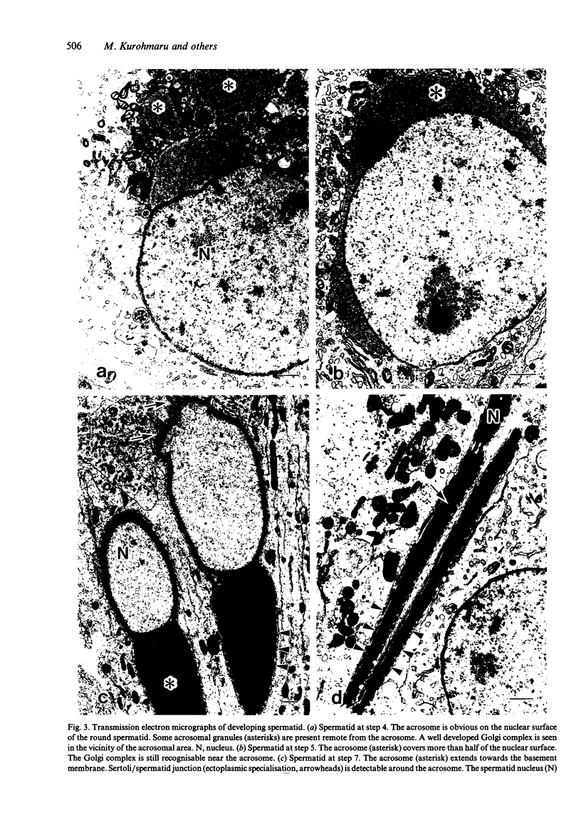
Spermatogenesis and acrosomal formation in the musk shrew, Suncus murinus, were studied by light and transmission electron microscopy. The cycle of the seminiferous epithelium was divided into 13 stages based on the characteristics of acrosomal change and nuclear shape, appearance of meiotic figures, location of spermatids, and period of spermiation. The relative frequencies of stages 1 to 13 were 5.1, 5.9, 10.1, 8.8, 12.5, 11.5, 10.6, 7.9, 6.0, 4.8, 8.9, 3.1 and 4.8, respectively. Additionally, spermatid development was subdivided into 13 steps. Acrosomal formation during spermiogenesis in the musk shrew was quite characteristic. However, in contrast to other mammalian species, the nucleus remained in the middle region of the seminiferous epithelium, and only the acrosome extended towards the basement membrane, beginning at step 7. The extension of the acrosome was conspicuous and reached maximum at step 9. At that time, the tip of the acrosome extended nearly to the basement. The acrosome of maturing spermatids was about 3-fold longer than that of spermatozoa. Thereafter, the acrosome gradually shortened and became flat. The enormous fan-shaped acrosome was completely formed at step 13. The prominent extension and subsequent shortening and flattening of the acrosome in the musk shrew appears to be a unique process to form the enormous fan-shaped acrosome.
Full text
PDF
Images in this article
Selected References
These references are in PubMed. This may not be the complete list of references from this article.
- Adachi Y., Kurohmaru M., Hattori S., Hayashi Y. Spermatogenesis in the Watase's shrew, Crocidura watasei--a light electron microscopic study. Jikken Dobutsu. 1992 Jul;41(3):295–303. [PubMed] [Google Scholar]
- CLERMONT Y., HARVEY S. C. DURATION OF THE CYCLE OF THE SEMINIFEROUS EPITHELIUM OF NORMAL, HYPOPHYSECTOMIZED AND HYPOPHYSECTOMIZED-HORMONE TREATED ALBINO RATS. Endocrinology. 1965 Jan;76:80–89. doi: 10.1210/endo-76-1-80. [DOI] [PubMed] [Google Scholar]
- Chiba T., Ishikawa T., Murakami A. Histologische Untersuchung der Kinetik der Spermatogenese beim Mink (Mustela vison). I. Samenepithelzyklus in der Paarungszeit. Jpn J Vet Res. 1968 Sep;16(2):73–86. [PubMed] [Google Scholar]
- Clermont Y., Antar M. Duration of the cycle of the seminiferous epithelium and the spermatogonial renewal in the monkey Macaca arctoides. Am J Anat. 1973 Feb;136(2):153–165. doi: 10.1002/aja.1001360204. [DOI] [PubMed] [Google Scholar]
- Clermont Y., Bustos-Obregon E. Re-examination of spermatogonial renewal in the rat by means of seminiferous tubules mounted "in toto". Am J Anat. 1968 Mar;122(2):237–247. doi: 10.1002/aja.1001220205. [DOI] [PubMed] [Google Scholar]
- Cooper G. W., Bedford J. M. Asymmetry of spermiation and sperm surface charge patterns over the giant acrosome in the musk shrew Suncus murinus. J Cell Biol. 1976 May;69(2):415–428. doi: 10.1083/jcb.69.2.415. [DOI] [PMC free article] [PubMed] [Google Scholar]
- Ekstedt E., Söderquist L., Plöen L. Fine structure of spermatogenesis and Sertoli cells (Epitheliocytus sustentans) in the bull. Anat Histol Embryol. 1986 Mar;15(1):23–48. doi: 10.1111/j.1439-0264.1986.tb00529.x. [DOI] [PubMed] [Google Scholar]
- Foote R. H., Swierstra E. E., Hunt W. L. Spermatogenesis in the dog. Anat Rec. 1972 Jul;173(3):341–351. doi: 10.1002/ar.1091730309. [DOI] [PubMed] [Google Scholar]
- Green J. A., Dryden G. L. Ultrastructure of epididymal spermatozoa of the Asiatic Musk Shrew, Suncus murinus. Biol Reprod. 1976 Apr;14(3):327–331. doi: 10.1095/biolreprod14.3.327. [DOI] [PubMed] [Google Scholar]
- Grocock C. A., Clarke J. R. Spermatogenesis in mature and regressed testes of the vole (Microtus agrestis). J Reprod Fertil. 1975 Jun;43(3):461–470. doi: 10.1530/jrf.0.0430461. [DOI] [PubMed] [Google Scholar]
- Kennelly J. J. Coyote reproduction. I. The duration of the spermatogenic cycle and epididymal sperm transport. J Reprod Fertil. 1972 Nov;31(2):163–170. doi: 10.1530/jrf.0.0310163. [DOI] [PubMed] [Google Scholar]
- Koehler J. K. Fine structure of spermatozoa of the Asiatic musk shrew, Suncus murinus. Am J Anat. 1977 Jun;149(2):135–151. doi: 10.1002/aja.1001490202. [DOI] [PubMed] [Google Scholar]
- Kurohmaru M., Tiba T., Nishida N., Hayashi Y. Spermatogenesis and ultrastructural changes of spermatids during spermiogenesis in the cotton rat, Sigmodon hispidus. Okajimas Folia Anat Jpn. 1988 Oct;65(4):203–219. doi: 10.2535/ofaj1936.65.4_203. [DOI] [PubMed] [Google Scholar]
- MONESI V. Autoradiographic study of DNA synthesis and the cell cycle in spermatogonia and spermatocytes of mouse testis using tritiated thymidine. J Cell Biol. 1962 Jul;14:1–18. doi: 10.1083/jcb.14.1.1. [DOI] [PMC free article] [PubMed] [Google Scholar]
- Oud J. L., de Rooij D. G. Spermatogenesis in the Chinese hamster. Anat Rec. 1977 Jan;187(1):113–124. doi: 10.1002/ar.1091870109. [DOI] [PubMed] [Google Scholar]
- Plöen L., Ekwall H., Afzelius B. A. Spermiogenesis and the spermatozoa of the European common shrew (Sorex araneus L.). J Ultrastruct Res. 1979 Aug;68(2):149–159. doi: 10.1016/s0022-5320(79)90150-3. [DOI] [PubMed] [Google Scholar]
- Russell L. Observations on rat Sertoli ectoplasmic ('junctional') specializations in their association with germ cells of the rat testis. Tissue Cell. 1977;9(3):475–498. doi: 10.1016/0040-8166(77)90007-6. [DOI] [PubMed] [Google Scholar]
- SWIERSTRA E. E., FOOTE R. H. Cytology and kinetics of spermatogenesis in the rabbit. J Reprod Fertil. 1963 Jun;5:309–322. doi: 10.1530/jrf.0.0050309. [DOI] [PubMed] [Google Scholar]
- Schuler H. M., Gier H. T. Duration of the cycle of the seminiferous epithelium in the prairie vole (Microtus ochrogaster ochrogaster). J Exp Zool. 1976 Jul;197(1):1–11. doi: 10.1002/jez.1401970102. [DOI] [PubMed] [Google Scholar]
- Swierstra E. E. Cytology and duration of the cycle of the seminiferous epithelium of the boar; duration of spermatozoan transit through the epididymis. Anat Rec. 1968 Jun;161(2):171–185. doi: 10.1002/ar.1091610204. [DOI] [PubMed] [Google Scholar]
- Swierstra E. E., Gebauer M. R., Pickett B. W. Reproductive physiology of the stallion. I. Spermatogenesis and testis composition. J Reprod Fertil. 1974 Sep;40(1):113–123. doi: 10.1530/jrf.0.0400113. [DOI] [PubMed] [Google Scholar]


