Abstract
Previously, it was reported that mRNA/cationic liposome complexes (mRNA lipoplexes) composed of the cationic triacyl lipid, 11-((1,3-bis(dodecanoyloxy)-2-((dodecanoyloxy)methyl)propan-2-yl)amino)-N,N,N- trimethyl-11-oxoundecan-1-aminium bromide (TC-1-12), with 1,2-dioleoyl-sn-glycero-3-phosphoethanolamine and poly(ethylene glycol) cholesteryl ether, induce high protein expression in human cervical carcinoma HeLa cells. In the present study, the authors aimed to optimize mRNA transfection using TC-1-12-based mRNA lipoplexes. mRNA lipoplexes were prepared at various charge ratios (+:-) using modified ethanol injection (MEI) and thin-film hydration (TFH) methods and compared the protein expression efficiency after transfection of HeLa cells with the developed mRNA lipoplexes. Firefly luciferase (FLuc) and enhanced green fluorescent protein (EGFP) mRNA lipoplexes prepared using the MEI method exhibited higher Luc and EGFP expression levels in cells than those prepared using the TFH method. Moreover, FLuc mRNA lipoplexes prepared using the MEI and TFH methods at charge ratios of 3:1 and 4:1, respectively, exhibited the highest Luc expression in cells. However, transfection with mRNA lipoplexes using the MEI and TFH methods induced moderate cytotoxicity in HeLa cells (46 and 57% cell viability, respectively). Furthermore, Cy5-labeled mRNA lipoplexes, which were prepared using the MEI method, showed higher cellular uptake of mRNA than those prepared using the TFH method. In the transfection of FLuc mRNA lipoplexes prepared using the MEI method, the storage of the lipid-ethanol solution at 37˚C for 4 months did not decrease Luc expression in HeLa cells. Additionally, FLuc mRNA lipoplexes prepared using the MEI method, induced relatively high Luc expression in human prostate carcinoma PC-3 and human liver cancer HepG2 cells with low cytotoxicity (103 and 81% cell viability, respectively). Overall, the results highlighted the potential of TC-1-12-based mRNA lipoplexes prepared using the MEI method for efficient mRNA delivery to cells.
Keywords: lipoplex, liposome, mRNA delivery
Introduction
mRNAs are single-stranded RNAs encoding protein sequences that are translated into proteins by ribosomes in the cytoplasm. mRNAs are designed and synthesized to immediately and efficiently express therapeutic proteins in the target cells for various therapeutic applications (1). For example, mRNAs are used as vaccines to produce protein antigens to activate the host immune system (2,3) and reprogram differentiated somatic cells into pluripotent stem cells via the expression of transcription factors associated with pluripotency (4). mRNA therapeutics must be delivered to the cytoplasm of the target cells via the cell membrane (5). However, mRNA is unstable and has limited permeability across cell membranes (6), necessitating the development of effective mRNA delivery systems. Among the various mRNA carriers for cells, cationic liposomes have attracted considerable attention (7-9).
Cationic liposomes are often prepared via thin-film hydration (TFH) (10,11). This method involves making a thin lipid film containing cationic and neutral lipids on the surface of a round-bottom flask by evaporating chloroform from the lipids-containing chloroform. Cationic liposomes are prepared by adding a heated aqueous solution to a thin lipid film, followed by sonication and/or extrusion. For mRNA transfection, a cationic liposome suspension is added to an mRNA solution to form mRNA/cationic liposome complexes (mRNA lipoplexes) (12). Therefore, the TFH method requires considerable time and equipment for the preparation of mRNA lipoplexes. Recently, the authors developed a simple one-step procedure to prepare mRNA lipoplexes using a modified ethanol injection (MEI) method. In this method, mRNA-containing phosphate-buffered saline (PBS) is rapidly poured into a small volume of lipid-ethanol solution, resulting in the formation of small and homogeneous mRNA lipoplexes without the need for special equipment (13). The advantage of this method relies on that it does not require the preparation of cationic liposomes in advance, allowing the easy preparation of mRNA lipoplexes by only using the lipid-ethanol solution.
Generally, cationic liposomes consist of cationic and neutral lipids that increase the transfection efficiency and stability (9,11). However, structures of cationic and neutral lipids, such as head group size and charge, acyl chain length, and saturation, affect the transfection efficiency of mRNA lipoplexes into cells (9). Therefore, the determination of the optimal combination of cationic and neutral lipids in liposomal formulations is important for efficient mRNA transfection. Previously, the authors demonstrated that cationic liposomes composed of the cationic triacyl lipid, 11-((1,3-bis (dodecanoyloxy)-2-((dodecanoyloxy)methyl)propan-2-yl)amino)-N,N,N-trimethyl-11-oxoundecan-1-aminium bromide (TC-1-12), with 1,2-dioleoyl-sn-glycero-3-phosphoethanolamine (DOPE) as a neutral lipid and poly (ethylene glycol) cholesteryl ether (PEG-Chol) as a dispersant agent exhibited high mRNA transfection efficiency in cells (9). However, an optimal preparation method for mRNA lipoplexes is needed for their effective transfection into cells. In the present study, in order to determine the optimal preparation method for mRNA lipoplexes, TC-1-12-based mRNA lipoplexes were prepared at various charge ratios (+:-) using the MEI and TFH methods, and the protein expression efficiency and cytotoxicity in cells transfected with these lipoplexes were evaluated.
Materials and methods
Materials
TC-1-12 was obtained from Sogo Pharmaceutical Co., Ltd. DOPE and PEG-Chol (mean molecular weight of PEG =1,600 g/mol) were purchased from NOF Co., Ltd. Firefly luciferase (FLuc) mRNA (CleanCap FLuc mRNA; 1,922 nucleotides; cat. no. L-7602) and enhanced green fluorescent protein (EGFP) mRNA (CleanCap EGFP mRNA; 997 nucleotides; cat. no. L-7601) were purchased from TriLink Biotechnologies. Cyanine 5 (Cy5)-labeled mRNA [EZCap Cyanine 5 FLuc mRNA (5 moUTP); 1,921 nucleotides; cat. no. R1010] was purchased from APeXBIO Technology LLC.
Cell culture
Human cervical carcinoma cell line, HeLa (cat. no. 93021013; CVCL_0030) and human liver cancer cell line, HepG2 (cat. no. 85011430; CVCL_0027) were purchased from the European Collection of Authenticated Cell Cultures. Human prostate carcinoma cell line, PC-3 (cat. no. TKG 0600; CVCL_0035) was obtained from the Cell Resource Center for Biomedical Research, Tohoku University, Miyagi, Japan.
HeLa cells were maintained in the Eagle's minimum essential medium (Wako Pure Chemical Industries, Ltd.) with 10% heat-inactivated fetal bovine serum (FBS; Thermo Fisher Scientific, Inc.) and 100 µg/ml kanamycin (KM) in a humidified incubator at 37˚C with 5% CO2. HepG2 cells were maintained in Dulbecco's modified Eagle's medium (Wako Pure Chemical Industries, Ltd.) with 10% FBS and 100 µg/ml KM under the same conditions. PC-3 cells were maintained in the Roswell Park Memorial Institute-1640 medium (Wako Pure Chemical Industries, Ltd.) with 10% FBS and 100 µg/ml KM under the same conditions.
Preparation of mRNA lipoplexes for transfection
To prepare mRNA lipoplexes using the MEI method, 2 mg TC-1-12, 1.53 mg DOPE and 0.08 mg PEG-Chol (molar ratio of 49.5:49.5:1) were dissolved in 1 ml ethanol, as previously described (9). Briefly, 0.5 µl of a 1 mg/ml mRNA solution (0.5 µg mRNA) was transferred to a tube containing 100 µl of PBS (pH 7.4). The resulting solution was rapidly added to 0.76, 1.52, 2.27, 3.03, 3.79 and 4.55 µl of the lipid-ethanol solution in another tube at charge ratios of 1:1, 2:1, 3:1, 4:1, 5:1 and 6:1, respectively. The charge ratio was calculated as the molar ratio of the quaternary amine in TC-1-12 to the mRNA phosphate.
To prepare mRNA lipoplexes using the TFH method, 10 mg TC-1-12, 7.64 mg DOPE and 0.38 mg PEG-Chol (molar ratio of 49.5:49.5:1) were dissolved in chloroform, as previously described (14). Briefly, chloroform was evaporated under vacuum using a rotary evaporator at 60˚C to form a thin film. The film was then hydrated with 10 ml of sterilized water at 60˚C via vortexing, followed by 10 min of sonication at room temperature in a bath-type sonicator (Bransonic 2510 JMTH; 42 kHz, 100 W; Branson Ultrasonics Co.). To prepare the mRNA lipoplexes, cationic liposome suspensions (1.52, 3.03, 4.55, 6.06, 7.58 and 9.09 µl at charge ratios of 1:1, 2:1, 3:1, 4:1, 5:1 and 6:1, respectively) were mixed with 0.5 µl of 1 mg/ml mRNA solution (0.5 µg mRNA).
Size measurement of mRNA lipoplexes. To prepare mRNA lipoplexes using the MEI method, 5 µg of FLuc mRNA (5 µl of a 1 mg/ml mRNA) was transferred to a tube containing 1,000 µl of PBS (pH 7.4), and the obtained solution was quickly added to the lipid-ethanol solution (7.6, 15.2, 22.7, 30.3, 37.9 and 45.5 µl at charge ratios of 1:1, 2:1, 3:1, 4;1, 5:1 and 6:1, respectively) in another tube. The mRNA lipoplex suspension was diluted three times with water before measurement of particle size and ζ-potential.
To prepare mRNA lipoplexes using the TFH method, cationic liposome suspensions (15.2, 30.3, 45.5, 60.6, 75.8, and 90.9 µl at charge ratios of 1:1, 2:1, 3:1, 4;1, 5:1, and 6:1, respectively) were added to FLuc mRNA solution (5 µg mRNA). The mRNA lipoplex suspension was diluted with the appropriate volume of water before measurement of particle size and ζ-potential.
Next, particle size distribution, polydispersity index (PDI), and ζ-potential of mRNA lipoplexes were determined using a light-scattering photometer (ELS-Z2; Otsuka Electronics Co., Ltd.), as previously described (14).
Luciferase activity in cells transfected with the FLuc mRNA lipoplexes
HeLa, PC-3 and HepG2 cells were seeded in 12-well culture plates at a density of 1x10⁵ cells per well. Following 24 h of incubation at 37˚C, FLuc mRNA lipoplexes (0.5 µg mRNA) were prepared by employing the MEI and TFH methods, then they were diluted with the culture medium containing 10% FBS (final mRNA concentration: 0.5 µg/ml) and lastly, they were added to the cells. Transfection of FLuc mRNA using Lipofectamine MessengerMAX (Thermo Fisher Scientific, Inc.) was performed according to the manufacturer's instructions. Briefly, 0.75 µl of Lipofectamine MessengerMAX transfection reagent was diluted in 25 µl of Opti-MEM medium (Thermo Fisher Scientific, Inc.) and incubated for 10 min at room temperature. FLuc mRNA (0.5 µg) diluted in 25 µl of Opti-MEM medium was added to the reagent solution and incubated for 5 min at room temperature. The mixture was diluted with culture medium containing 10% FBS (final mRNA concentration: 0.5 µg/ml) and introduced into the cells.
A total of 24 h post-transfection, the cells were lysed with 125 µl of cell lysis buffer (Pierce™ Luciferase Cell Lysis Buffer; Thermo Fisher Scientific Inc.), and subjected to one cycle of freezing (-80˚C) and thawing at 37˚C, followed by centrifugation at 15,000 g for 10 sec at 4˚C. A total of 10 µl of supernatant were mixed with 50 µl of PicaGene MelioraStar-LT Luminescence Reagent (Toyo Ink Mfg. Co. Ltd.), and luminescence [counts per sec (cps)] was measured using a chemo-luminometer (ARVO X2; PerkinElmer, Inc.). Protein concentration in the supernatant was measured by BCA assay (Pierce BCA Protein Assay Kit; Pierce; Thermo Fisher Scientific, Inc.), and luciferase (Luc) activity (cps/µg protein) was calculated.
EGFP expression in cells transfected with the EGFP mRNA lipoplexes
HeLa, PC-3 and HepG2 cells were seeded in a 12-well culture plate at a density of 1x10⁵ cells per well. Following 24 h of incubation at 37˚C, EGFP mRNA lipoplexes (0.5 µg mRNA) were prepared using the MEI and TFH methods at charge ratios of 2:1 to 5:1. Next, they were diluted with the culture medium, which contained 10% FBS (final mRNA concentration: 0.5 µg/ml), and were subsequently introduced into the cells. Transfection of EGFP mRNA using Lipofectamine® MessengerMAX was performed as aforementioned. At 24 h post-transfection, the cells were washed two times with PBS and were fixed with 10% neutral buffered formalin (Mildform 10N; Wako Pure Chemical Industries, Ltd.) for 10 min at room temperature. EGFP expression in cells was detected using a fluorescence microscope (Eclipse TS100-F; Nikon Corporation) equipped with an optical filter (excitation, 480/30 nm; dichroic mirror, 505 nm; emission, 535/45 nm; Nikon Corporation).
To calculate the percentage of EGFP-expressing cells using flow cytometry, EGFP mRNA lipoplexes (0.5 µg mRNA) were prepared using the MEI and TFH methods at charge ratios of 2:1 to 5:1. These lipoplexes were diluted in culture medium containing 10% FBS (final mRNA concentration: 0.5 µg/ml) and were subsequently added to the cells. After 24 h of transfection, the cells were detached with TrypLE™ Express Enzyme (Gibco; Thermo Fisher Scientific, Inc.) and resuspended in PBS containing 0.1% bovine serum albumin (BSA) and 1 mM ethylenediaminetetraacetic acid (EDTA). EGFP expression levels were evaluated by measuring EGFP-expressing cells with flow cytometry (BD FACSVerseTM; BD Biosciences) using a 488-nm laser and analyzed with BD FACSuite software ver. 1.0.3 (BD Biosciences). Data for 10,000 fluorescence events were collected, including forward scatter, side scatter, and 527/32 nm fluorescence.
Cytotoxicity in cells transfected with the FLuc mRNA lipoplexes
HeLa, PC-3 and HepG2 cells were seeded in 96-well culture plates at a density of 1x10⁴ cells per well. Following 24 h of incubation at 37˚C, FLuc mRNA lipoplexes (0.05 µg mRNA) were prepared using the MEI and TFH methods at charge ratios of 1:1 to 6:1, were diluted with the culture medium containing 10% FBS (final mRNA concentration: 0.5 µg/ml) and were subsequently added to the cells. Cytotoxicity was assessed 24 h post-transfection using Cell Counting Kit-8 (CCK-8; cat. no. CK04; Dojindo Laboratories, Inc.). Briefly, 10 µl of CCK-8 solution was added to each well and incubated for 1 h at 37˚C. Cell viability was calculated as a percentage of the absorbance of untreated cells at 450 nm using an iMark microplate reader (Bio-Rad Laboratories, Inc).
Cellular uptake of mRNA lipoplexes
HeLa cells were seeded in a 12-well culture plate at a density of 1x10⁵ cells per well. After 24 h of incubation at 37˚C, mRNA lipoplexes containing 0.5 µg of Cy5-labeled mRNA in 1 ml of culture medium were transferred to the cells (final mRNA concentration: 0.5 µg/ml). A total of 3 h post-transfection, the cells were washed two times with PBS and fixed with Mildform 10N for 10 min at room temperature. Cy5-labeled mRNAs in the cells were detected using a fluorescence microscope equipped with optical filter Cy5 HQ (excitation, 620/60 nm; dichroic mirror, 660 nm; emission, 700/75 nm; Nikon Corporation).
To quantify intracellular of Cy5-labeled mRNA in HeLa cells using flow cytometry, mRNA lipoplexes containing 0.5 µg of Cy5-labeled mRNA in 1 ml of culture medium were added to the cells (final mRNA concentration: 0.5 µg/ml). After a 3 h transfection period, the cells were detached with TrypLE™ Express Enzyme, followed by suspension in PBS containing 0.1% BSA and 1 mM EDTA. Cy5-labeled mRNA levels were assessed by flow cytometry (BD FACSVerse™) using a 640-nm laser as aforementioned. Data for 10,000 fluorescence events were obtained by recording the forward scatter, side scatter, and 660/10 nm fluorescence. Geo-mean fluorescence intensity in Cy5-labeled mRNA-transfected cells was calculated.
Evaluation of the stability of the lipid-ethanol solution
TC-1-12, DOPE and PEG-Chol were dissolved in 1 ml ethanol at a molar ratio of 49.5:49.5:1, as aforementioned. The lipid-ethanol solution was stored at -20, 4 and 37˚C. After storage for a total of 4 months, FLuc mRNA lipoplexes were prepared using the lipid-ethanol solution, and the lipoplex size and Luc expression in HeLa cells were measured as aforementioned.
Statistical analysis
Statistical analyses were conducted using an unpaired Student's t-test to compare two groups or one-way analysis of variance, followed by Tukey's post-hoc test to compare multiple groups using the GraphPad Prism software (v.4.0; Dotmatics). The data are presented as the mean ± standard deviation. P<0.05 was considered to indicate a statistically significant difference.
Results
Size and ζ-potential of mRNA lipoplexes
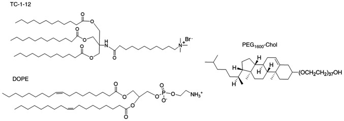
In the present study, TC-1-12 was used as a cationic lipid, DOPE as a neutral lipid, and PEG-Chol as a dispersing agent (Fig. 1). mRNA lipoplexes were prepared using the MEI and TFH methods. It was previously demonstrated that incorporating 1 mol% PEG1600-Chol as a dispersion agent into liposomal formulations effectively reduced lipoplex size after preparation using the MEI method (13). In the MEI method, FLuc mRNA lipoplexes were formed by rapidly mixing FLuc mRNA-containing PBS with a lipid-ethanol solution at various charge ratios. FLuc mRNA lipoplexes prepared at a charge ratio of 2:1 were 238 nm in size (PDI: 0.11). However, an increase in the charge ratio from 2:1 to 6:1 in the mRNA lipoplexes decreased the size to 80 nm (PDI: 0.17; Table I). By contrast, ζ-potential increased from -24.5 to 9.9 mV with an increase in charge ratio from 1:1 to 6:1.
Figure 1.
Structures of TC-1-12, DOPE, and PEG-Chol. TC-1-12, 11-((1,3-bis(dodecanoyloxy)-2-((dodecanoyloxy)methyl)propan-2-yl)amino)-N,N,N-trimethyl-11-oxoundecan-1-aminium bromide; DOPE, 1,2-dioleoyl-sn-glycero-3-phosphoethanolamine; PEG-Chol, poly(ethylene glycol) cholesteryl ether.
Table I.
Size of TC-1-12-based mRNA lipoplexes prepared using MEI and TFH methods.
| Preparation method | Charge ratio | Sizea (nm) | PDI | ζ-potentiala (mV) |
|---|---|---|---|---|
| MEI | 1:1 | 139.1±2.1 | 0.18±0.02 | -24.5±1.0 |
| 2:1 | 237.5±3.9 | 0.11±0.01 | 5.2±0.1 | |
| 3:1 | 115.8±1.1 | 0.11±0.00 | 1.7±3.1 | |
| 4:1 | 80.8±0.7 | 0.20±0.02 | 6.1±0.6 | |
| 5:1 | 86.3±0.1 | 0.19±0.02 | 8.4±0.2 | |
| 6:1 | 80.0±1.1 | 0.17±0.01 | 9.9±0.9 | |
| TFH | Only liposome | 104.7±0.8 | 0.25±0.00 | 54.2±1.6 |
| 1:1 | 275.4±9.6 | 0.24±0.01 | 11.8±0.7 | |
| 2:1 | 186.9±3.7 | 0.24±0.01 | 18.5±1.2 | |
| 3:1 | 183.9±12.3 | 0.21±0.05 | 27.0±1.9 | |
| 4:1 | 165.3±29.1 | 0.18±0.08 | 44.1±3.5 | |
| 5:1 | 216.4±21.1 | 0.11±0.00 | 44.0±0.8 | |
| 6:1 | 198.2±23.1 | 0.13±0.04 | 47.1±1.3 |
aIn MEI method, PBS solution containing FLuc mRNA lipoplexes was diluted three times with water. In TFH method, FLuc mRNA lipoplexes suspended in water were diluted with an appropriate amount of water. Each value represents the mean ± standard deviation (n=3). PDI, polydispersity index; MEI, modified ethanol injection; TFH, thin-film hydration.
In the TFH method, a dry lipid film was formed on the surface of the flask by the evaporation of chloroform containing TC-1-12, DOPE and PEG-Chol, followed by hydration with water and sonication. The cationic liposomes were 105 nm in size (PDI: 0.25). Mixing the cationic liposome suspension with FLuc mRNA at charge ratios of 1:1 to 6:1 increased the size to 165-275 nm (PDI: 0.11-0.24) and ζ-potential from 11.8 to 47.1 mV.
The mRNA lipoplexes were prepared by using the TFH and MEI methods and were suspended in water and 1/3 diluted PBS, respectively, for particle size and ζ-potential measurements. Adsorption of ions to the surface of mRNA lipoplexes causes change of ζ-potential value (15). Therefore, ζ-potential of mRNA lipoplexes that were prepared using the MEI method showed lower values than those prepared using the TFH method with only water. By contrast, the measurement of particle size using dynamic light scattering was influenced by viscosity. The viscosity of PBS is very similar to that of water (dynamic viscosity at 25˚C is 0.8882 mPa·s for PBS and 0.890 mPa·s for water). Therefore, it was appropriate to compare particle sizes obtained from water and 1/3 diluted PBS. Commercially available Lipofectamine MessengerMAX is widely used as a standard for in vitro mRNA transfection (16). In the present study, Lipofectamine MessengerMAX mRNA lipoplexes were 606±35.5 nm in size with 0.32±0.01 in PDI (data not shown).
Effects of the charge ratios of mRNA lipoplexes on the protein expression efficiency and cytotoxicity of mRNA lipoplexes
To examine the effect of the charge ratio of TC-1-12-based mRNA lipoplexes on protein expression, FLuc mRNA lipoplexes were prepared at charge ratios of 1:1 to 6:1 and transfected into HeLa cells. Among the mRNA lipoplexes prepared using the MEI method, FLuc mRNA lipoplexes prepared at a charge ratio of 3:1 exhibited the highest Luc activity, which was 2-fold lower than that of the Lipofectamine MessengerMAX mRNA lipoplexes (Fig. 2). Among the mRNA lipoplexes prepared using the TFH method, FLuc mRNA lipoplexes prepared at a charge ratio of 4:1 exhibited the highest Luc activity, which was 2-fold lower than that of the lipoplexes prepared using the MEI method at a charge ratio of 3:1. As for negative controls, cells were transfected with cationic liposomes (mock transfection) that were prepared using the MEI and TFH methods; however, these liposomes did not induce Luc activity in HeLa cells (Fig. S1). These findings suggested that the mRNA lipoplexes that were prepared using the MEI method induce higher protein expression in HeLa cells than those that were prepared using the TFH method.
Figure 2.
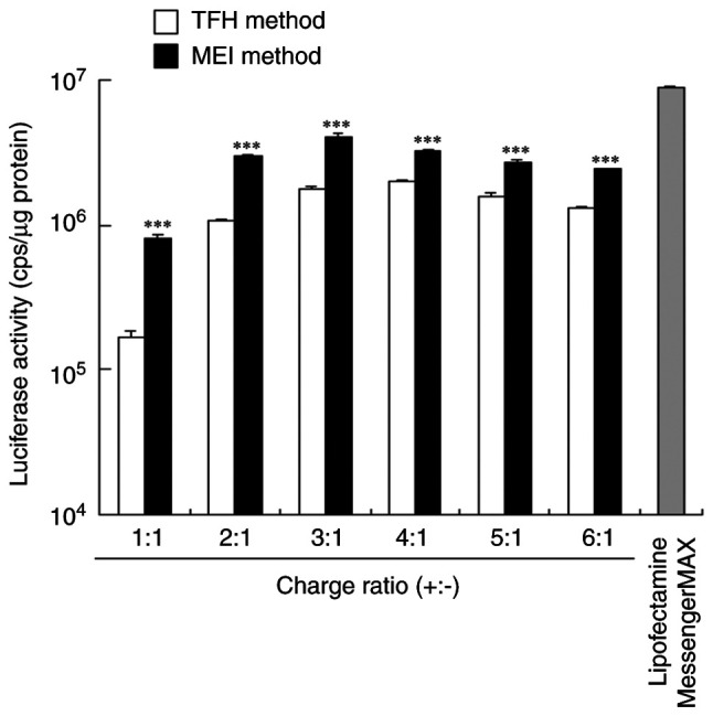
Effects of the charge ratios of TC-1-12-based mRNA lipoplexes on luciferase levels in HeLa cells transfected with the FLuc mRNA lipoplexes. FLuc mRNA lipoplexes were prepared using the MEI and TFH methods at charge ratios of 1:1 to 6:1, transfected into HeLa cells, and incubated for 24 h. Lipofectamine MessengerMAX was used as a control. Each value is represented as the mean ± standard deviation (n=3). ***P<0.001 vs. TFH method. TC-1-12, 11-((1,3-bis(dodecanoyloxy)-2-((dodecanoyloxy)methyl)propan-2-yl)amino)-N,N,N-trimethyl-11-oxoundecan-1-aminium bromide; FLuc, firefly luciferase; MEI, modified ethanol injection; TFH, thin-film hydration.
Next, the effect of the charge ratio of TC-1-12-based mRNA lipoplexes was investigated on the cytotoxicity of HeLa cells 24 h after transfection with the FLuc mRNA lipoplexes. In mRNA lipoplexes which were prepared using both the MEI and TFL methods, cytotoxicity after transfection increased with the increase in the charge ratio of mRNA lipoplexes (Fig. 3). Cytotoxicity of cationic liposomes increases with the increase in ζ-potential (17). Similarly, in the present study, ζ-potential of mRNA lipoplexes was associated with their cytotoxicity in cells. Cell viabilities after transfection with the FLuc mRNA lipoplexes that were prepared using the MEI method at a charge ratio of 3:1 and the TFL method at a charge ratio of 4:1 were 46 and 57%, respectively. By contrast, cell viability after transfection with the Lipofectamine MessengerMAX mRNA lipoplexes was 22%, which is consistent with previously reported results (18). These results suggested that the Lipofectamine MessengerMAX mRNA lipoplexes induce higher protein expression than the TC-1-12-based mRNA lipoplexes with higher cytotoxicity in cells.
Figure 3.
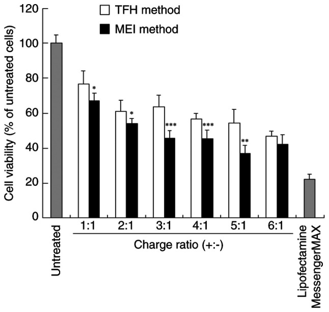
Effects of the charge ratios of TC-1-12-based mRNA lipoplexes on cytotoxicity in HeLa cells transfected with the FLuc mRNA lipoplexes. FLuc mRNA lipoplexes were prepared using the MEI and TFH methods at charge ratios of 1:1 to 6:1, transfected into HeLa cells, and incubated for 24 h. Lipofectamine MessengerMAX was used as a control. Each value is represented as the mean ± standard deviation (n=5 for 5:1 (MEI), 1:1 (TFH), and 3:1 (TFH), and n=6 for other groups). *P<0.05, **P<0.01 and ***P<0.001 vs. TFH method. TC-1-12, 11-((1,3-bis(dodecanoyloxy)-2-((dodecanoyloxy)methyl)propan-2-yl)amino)-N,N,N-trimethyl-11-oxoundecan-1-aminium bromide; FLuc, firefly luciferase; MEI, modified ethanol injection; TFH, thin-film hydration.
EGFP expression in HeLa cells transfected with the EGFP mRNA lipoplexes
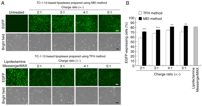
To examine the effect of the charge ratio of TC-1-12-based mRNA lipoplexes on protein expression efficiency, EGFP mRNA lipoplexes were prepared at charge ratios of 2:1 to 5:1 and transfected into HeLa cells. EGFP expression was observed 24 h after incubation (Figs. 4A and B; S2). Transfection with EGFP mRNA lipoplexes prepared using the MEI method resulted in high EGFP levels in most cells (71-83% cells) at charge ratios of 2:1 to 4:1, which were comparable to those observed after transfection with Lipofectamine MessengerMAX mRNA lipoplexes (81% cells). By contrast, transfection with EGFP mRNA lipoplexes that were prepared using the TFH method resulted in lower EGFP levels (58-74% cells) at all charge ratios compared with those observed after transfection with lipoplexes prepared using the MEI method. These results suggested that mRNA lipoplexes prepared using the MEI method effectively induce protein expression in most cells.
Figure 4.
Effects of the charge ratios of TC-1-12-based mRNA lipoplexes on EGFP levels in HeLa cells transfected with the EGFP mRNA lipoplexes. EGFP mRNA lipoplexes were prepared using the MEI and TFH methods at charge ratios of 2:1 to 5:1, transfected into HeLa cells, and incubated for 24 h. Lipofectamine MessengerMAX was used as a control. (A) EGFP expression in cells was observed using a fluorescence microscope. Scale bar, 100 µm. (B) EGFP transfection efficiency was determined using flow cytometry. Each column represents the mean ± standard deviation (n=3). **P<0.01 and ***P<0.001 vs. TFH method. TC-1-12, 11-((1,3-bis(dodecanoyloxy)-2-((dodecanoyloxy)methyl)propan-2-yl)amino)-N,N,N-trimethyl-11-oxoundecan-1-aminium bromide; EGFP, enhanced green fluorescent protein; MEI, modified ethanol injection; TFH, thin-film hydration.
Cellular uptake following mRNA lipoplex transfection
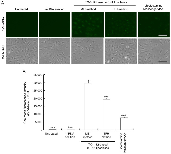
To understand the mechanism by which the TC-1-12-based mRNA lipoplexes, which were prepared using the MEI method, showed high protein expression levels, the cellular uptake of Cy5-labeled mRNA 3 h post-transfection was investigated. For this experiment, TC-1-12-based mRNA lipoplexes that were prepared using the MEI method at a charge ratio of 3:1 and the TFH method at a charge ratio of 4:1 were used; because these charge ratios resulted in the highest protein expression (Fig. 2). Stronger fluorescent signals of Cy5-labeled mRNA were detected in cells transfected with the mRNA lipoplexes that were prepared using the MEI method than in those transfected with the mRNA lipoplexes that were prepared using the THF method (Fig. 5A and B; Fig. S3). Fluorescent signals of Cy5-labeled mRNA in the cells transfected with the Lipofectamine MessengerMAX mRNA lipoplexes were weak, indicating that the size of lipoplexes (~600 nm) affects the cellular uptake of mRNA lipoplexes. As a negative control, Cy5-labeled mRNA solution did not introduce mRNA into the cells. Regarding the TC-1-12-based mRNA lipoplexes, fluorescence levels of Cy5-labeled mRNA in the cells closely corresponded to the efficacy of Luc expression following cell transfection with FLuc mRNA lipoplexes. Therefore, TC-1-12-based mRNA lipoplexes which were prepared using the MEI method at a charge ratio of 3:1 were used in subsequent experiments.
Figure 5.
Cellular uptake by HeLa cells after transfection with the TC-1-12-based Cy5-labeled mRNA lipoplexes. Cy5-labeled mRNA lipoplexes were prepared at charge ratios of 3:1 and 4:1 using the MEI and TFH methods, respectively, and added to HeLa cells at 0.5 μg/mL mRNA. As controls, Cy5-labeled mRNA solution (free mRNA) and Lipofectamine MessengerMAX mRNA lipoplexes were added to the HeLa cells. In (A), localization of Cy5-labeled mRNA (green) was observed 3 h after incubation. Scale bar, 100 μm. In (B), geo-mean fluorescence intensity of Cy5-labeled mRNA was measured using flow cytometry 3 h after incubation. Each column represents the mean ± standard deviation (n=3). ***P<0.001 vs. MEI method. TC-1-12, 11-((1,3-bis(dodecanoyloxy)-2-((dodecanoyloxy)methyl)propan-2-yl)amino)-N,N,N-trimethyl-11-oxoundecan-1-aminium bromide; Cy5, cyanine 5; MEI, modified ethanol injection; TFH, thin-film hydration.
Effect of lipid-ethanol solution storage on the size and protein expression efficiency of mRNA lipoplexes
To examine the stability of lipids in the ethanol solution, the lipid-ethanol solution was stored at -20, 4, and 37˚C for 4 months. Subsequently, TC-1-12-based mRNA lipoplexes were prepared by mixing the lipid-ethanol solution with a PBS solution of mRNA. The size of mRNA lipoplexes increased to 191, 151 and 221 nm (PDI: 0.10, 0.08 and 0.11, respectively) after 4 months of storage at -20, 4, and 37˚C, respectively (Table II). However, Luc expression levels after transfection of the FLuc mRNA lipoplexes were not affected by the 4-month storage at -20, 4 and 37˚C (Fig. 6). These results suggested that the lipid-ethanol solution used for the preparation of mRNA lipoplexes in the MEI method can be stored at 37˚C for at least 4 months.
Table II.
Size of mRNA lipoplexes prepared using modified ethanol injection method with a lipid-ethanol solution stored at -20, 4 and 37˚C.
| 4-month storage | ||
|---|---|---|
| Temperature (˚C) | Lipoplex sizea (nm) | Polydispersity index |
| -20 | 191.6±4.3 | 0.10±0.01 |
| 4 | 151.5±2.0 | 0.08±0.02 |
| 37 | 221.5±2.5 | 0.11±0.02 |
aCharge ratio = 3:1. Each value represents the mean ± standard deviation (n=3).
Figure 6.
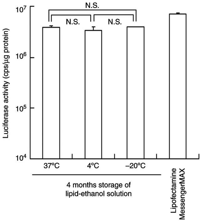
Effect of the storage of the lipid-ethanol solution on Luc expression in HeLa cells transfected with the TC-1-12-based FLuc mRNA lipoplexes. Lipid-ethanol solution was stored at -20, 4, and 37˚C for 4 months. FLuc mRNA lipoplexes were prepared at a charge ratio of 3:1 using the MEI method, transfected into HeLa cells, and incubated for 24 h. Lipofectamine MessengerMAX was used as a control. Each column represents the mean ± standard deviation (n=3). TC-1-12, 11-((1,3-bis(dodecanoyloxy)-2-((dodecanoyloxy)methyl)propan-2-yl)amino)-N,N,N-trimethyl-11-oxoundecan-1-aminium bromide; N.S., not significant; FLuc, firefly luciferase; MEI, modified ethanol injection.
Effects of cell type on the protein expression efficiency and cytotoxicity of mRNA lipoplexes
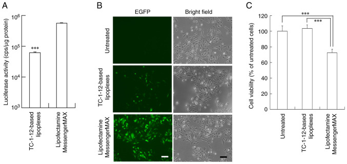
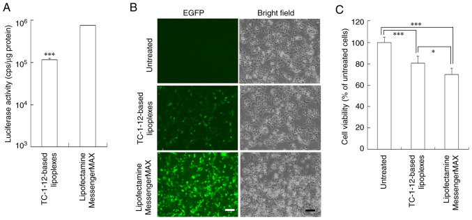
To examine the effect of cell type on the protein expression efficiency of TC-1-12-based mRNA lipoplexes that were prepared using the MEI method, FLuc and EGFP mRNA lipoplexes formed at a charge ratio of 3:1 were transfected into PC-3 (Fig. 7) and HepG2 cells (Fig. 8). HepG2 cells are known as a hard-to-transfect-cell line (19). TC-1-12-based FLuc mRNA lipoplexes exhibited 9- and 6.6-fold lower Luc expression in PC-3 and HepG2 cells, respectively, than the Lipofectamine MessengerMAX mRNA lipoplexes (Figs. 7A and 8A). However, TC-1-12-based EGFP mRNA lipoplexes induced moderate EGFP expression in both cell lines (Figs. 7B and 8B) with lower cytotoxicity (103 and 81% cell viability, respectively) compared with the Lipofectamine MessengerMAX mRNA lipoplexes (72 and 70% cell viability, respectively; Figs. 7C and 8C).
Figure 7.
Protein expression and cytotoxicity in PC-3 cells transfected with the mRNA lipoplexes. (A and C) FLuc mRNA and (B) EGFP mRNA lipoplexes were prepared at a charge ratio of 3:1 using the MEI method, transfected into PC-3 cells, and incubated for 24 h. Lipofectamine MessengerMAX was used as a control. (A) Luc activity was measured 24 h after transfection. Each column represents the mean ± standard deviation (n=3). ***P<0.001. (B) EGFP expression in cells was observed 24 h after transfection using a fluorescence microscope. Scale bar, 100 µm. (C) Cell viability was measured 24 h after transfection. Each column represents the mean ± standard deviation (n=4 for TC-1-12-based lipoplexes; n=5 for other groups). ***P<0.001. Luc, luciferase; FLuc, firefly luciferase; EGFP, enhanced green fluorescent protein; MEI, modified ethanol injection.
Figure 8.
Protein expression and cytotoxicity in HepG2 cells transfected with the mRNA lipoplexes. (A and C) FLuc mRNA and (B) EGFP mRNA lipoplexes were prepared at a charge ratio of 3:1 using the MEI method, transfected into HepG2 cells, and incubated for 24 h. Lipofectamine MessengerMAX was used as a control. (A) Luc activity was measured 24 h after transfection. Each column represents the mean ± standard deviation (n=3). ***P<0.001. (B) EGFP expression in cells was observed 24 h after transfection using a fluorescence microscope. Scale bar, 100 µm. (C) cell viability was measured 24 h after transfection. Each column represents the mean ± standard deviation (n=6 for Lipofectamine MessengerMAX; n=7 for other groups). *P<0.05 and ***P<0.001. Luc, luciferase; FLuc, firefly luciferase; EGFP, enhanced green fluorescent protein; MEI, modified ethanol injection
Discussion
It was previously reported that mRNA lipoplexes containing the cationic triacyl lipid, TC-1-12, exhibit high mRNA transfection efficiency in cells (9). In the present study, the preparation method for mRNA lipoplexes was further optimized to ensure their efficient delivery to cells. FLuc and EGFP mRNA lipoplexes prepared using the MEI method exhibited higher Luc activity and EGFP expression, respectively, than those prepared using the TFH method. Moreover, mRNA lipoplexes prepared using the MEI method exhibited higher cellular uptake than those prepared using the TFH method. It was previously reported that the presence of sodium chloride in the formation of cationic lipoplexes of plasmid DNA or small interfering RNA enhances the transfection of cells (20-22). Moderate neutralization of cationic charges on the surface of mRNA lipoplexes using sodium chloride in PBS might stabilize the TC-1-12-based mRNA lipoplexes and increase their cellular association and transfection efficiency.
TC-1-12 is a cationic lipid with short triacyl chains (C12) and long head groups. Cationic lipids with short dialkyl chains (C14-C16) efficiently internalize mRNA lipoplexes into cells compared with those with long dialkyl chains (C18) (9). Koulov et al (23) reported that vesicles containing cationic trialkyl chains promote greater membrane fusion than those with structurally related cationic dialkyl and monoalkyl chains. These studies indicate that TC-1-12-based mRNA lipoplexes are effective mRNA carriers with fusogenic activity. Numerous studies have developed the diacyl cationic lipids 1,2-dioleoyl-3-trimethylammonium-propane (DOTAP)- or dialkyl cationic lipid dimethyl-dioctadecyl-ammonium bromide (DDAB)-based cationic liposomes, which are combined with helper lipids for efficient mRNA delivery (24,25). It was previously revealed that TC-1-12-based mRNA lipoplexes induce higher protein expression than DOTAP- or DDAB-based lipoplexes (9). These findings suggest TC-1-12-based mRNA lipoplexes as useful transfection reagents for cultured cells.
In the present study, commercially available Lipofectamine MessengerMAX mRNA lipoplexes exhibited higher transfection efficiencies in HeLa, PC-3 and HepG2 cells than the TC-1-12-based mRNA lipoplexes; however, they demonstrated high cytotoxicity in these cell lines. Moreover, Lipofectamine MessengerMAX mRNA lipoplexes are not suitable for in vivo transfection. It was previously reported that intravenous injection of TC-1-12-based ovalbumin mRNA lipoplexes prepared using the MEI method induces ovalbumin-specific IgG1 antibodies in the serum (26). Therefore, TC-1-12-based mRNA lipoplexes can be used for both in vitro and in vivo transfection.
The MEI method only requires the preparation of an ethanol solution containing lipids to form mRNA lipoplexes, thus eliminating the need for cationic liposome preparation before mRNA transfection. Additionally, the lipid-ethanol solution used for preparing mRNA lipoplexes in the MEI method can be stored at 37˚C for at least 4 months. In the present study, mRNA lipoplexes prepared using the MEI method at a charge ratio of 3:1 contained 0.23% (v/v) ethanol in the culture medium, and this ethanol concentration was well-tolerated by the cultured cells (Fig. S4). Overall, the present findings highlight the potential of TC-1-12-based mRNA lipoplexes prepared using a lipid-ethanol solution for effective mRNA transfection.
Supplementary Material
Acknowledgements
Not applicable.
Funding Statement
Funding: No funding was received.
Availability of data and materials
The data generated in the present study may be requested from the corresponding author.
Authors' contributions
YH conceptualized the study, developed methodology, conducted investigation, curated data, preformed formal analysis, prepared the original draft, wrote, reviewed and edited the manuscript. RS conducted investigation. YH and RS confirm the authenticity of all the raw data. All authors read and approved the final version of the manuscript.
Ethics approval and consent to participate
Not applicable.
Patient consent for publication
Not applicable.
Competing interests
The authors declare that they have no competing interests.
References
- 1.Sahin U, Karikó K, Türeci Ö. mRNA-based therapeutics-developing a new class of drugs. Nat Rev Drug Discov. 2014;13:759–780. doi: 10.1038/nrd4278. [DOI] [PubMed] [Google Scholar]
- 2.Liu T, Liang Y, Huang L. Development and delivery systems of mRNA vaccines. Front Bioeng Biotechnol. 2021;9(718753) doi: 10.3389/fbioe.2021.718753. [DOI] [PMC free article] [PubMed] [Google Scholar]
- 3.Barbier AJ, Jiang AY, Zhang P, Wooster R, Anderson DG. The clinical progress of mRNA vaccines and immunotherapies. Nat Biotechnol. 2022;40:840–854. doi: 10.1038/s41587-022-01294-2. [DOI] [PubMed] [Google Scholar]
- 4.Warren L, Manos PD, Ahfeldt T, Loh YH, Li H, Lau F, Ebina W, Mandal PK, Smith ZD, Meissner A, et al. Highly efficient reprogramming to pluripotency and directed differentiation of human cells with synthetic modified mRNA. Cell Stem Cell. 2010;7:618–630. doi: 10.1016/j.stem.2010.08.012. [DOI] [PMC free article] [PubMed] [Google Scholar]
- 5.Ramachandran S, Satapathy SR, Dutta T. Delivery strategies for mRNA vaccines. Pharmaceut Med. 2022;36:11–20. doi: 10.1007/s40290-021-00417-5. [DOI] [PMC free article] [PubMed] [Google Scholar]
- 6.Yen A, Cheng Y, Sylvestre M, Gustafson HH, Puri S, Pun SH. Serum nuclease susceptibility of mRNA cargo in condensed polyplexes. Mol Pharm. 2018;15:2268–2276. doi: 10.1021/acs.molpharmaceut.8b00134. [DOI] [PubMed] [Google Scholar]
- 7.Chen H, Ren X, Xu S, Zhang D, Han T. Optimization of lipid nanoformulations for effective mRNA delivery. Int J Nanomedicine. 2022;17:2893–2905. doi: 10.2147/IJN.S363990. [DOI] [PMC free article] [PubMed] [Google Scholar]
- 8.Malone RW, Felgner PL, Verma IM. Cationic liposome- mediated RNA transfection. Proc Natl Acad Sci USA. 1989;86:6077–6081. doi: 10.1073/pnas.86.16.6077. [DOI] [PMC free article] [PubMed] [Google Scholar]
- 9.Tang M, Sagawa A, Inoue N, Torii S, Tomita K, Hattori Y. Efficient mRNA delivery with mRNA lipoplexes prepared using a modified ethanol injection method. Pharmaceutics. 2023;15(1141) doi: 10.3390/pharmaceutics15041141. [DOI] [PMC free article] [PubMed] [Google Scholar]
- 10.Bangham AD, Standish MM, Watkins JC. Diffusion of univalent ions across the lamellae of swollen phospholipids. J Mol Biol. 1965;13:238–252. doi: 10.1016/s0022-2836(65)80093-6. [DOI] [PubMed] [Google Scholar]
- 11.Hattori Y, Nakamura M, Takeuchi N, Tamaki K, Shimizu S, Yoshiike Y, Taguchi M, Ohno H, Ozaki KI, Onishi H. Effect of cationic lipid in cationic liposomes on siRNA delivery into the lung by intravenous injection of cationic lipoplex. J Drug Target. 2019;27:217–227. doi: 10.1080/1061186X.2018.1502775. [DOI] [PubMed] [Google Scholar]
- 12.Barichello JM, Ishida T, Kiwada H. Complexation of siRNA and pDNA with cationic liposomes: The important aspects in lipoplex preparation. Methods Mol Biol. 2010;605:461–472. doi: 10.1007/978-1-60327-360-2_32. [DOI] [PubMed] [Google Scholar]
- 13.Hattori Y, Saito H, Nakamura K, Yamanaka A, Tang M, Ozaki KI. In vitro and in vivo transfections using siRNA lipoplexes prepared by mixing siRNAs with a lipid-ethanol solution. J Drug Deliv Sci Technol. 2022;75(103635) [Google Scholar]
- 14.Hattori Y, Tamaki K, Sakasai S, Ozaki KI, Onishi H. Effects of PEG anchors in pegylated siRNA lipoplexes on in vitro genesilencing effects and siRNA biodistribution in mice. Mol Med Rep. 2020;22:4183–4196. doi: 10.3892/mmr.2020.11525. [DOI] [PMC free article] [PubMed] [Google Scholar]
- 15.Smith MC, Crist RM, Clogston JD, McNeil SE. Zeta potential: A case study of cationic, anionic, and neutral liposomes. Anal Bioanal Chem. 2017;409:5779–5787. doi: 10.1007/s00216-017-0527-z. [DOI] [PubMed] [Google Scholar]
- 16.Jarzebska NT, Frei J, Lauchli S, French LE, Guenova E, Gouttefangeas C, Kündig TM, Mellett M, Pascolo S. Lipofection with synthetic mRNA as a simple method for T-cell immunomonitoring. Viruses. 2021;13(1232) doi: 10.3390/v13071232. [DOI] [PMC free article] [PubMed] [Google Scholar]
- 17.Wei X, Shao B, He Z, Ye T, Luo M, Sang Y, Liang X, Wang W, Luo S, Yang S, et al. Cationic nanocarriers induce cell necrosis through impairment of Na(+)/K(+)-ATPase and cause subsequent inflammatory response. Cell Res. 2015;25:237–253. doi: 10.1038/cr.2015.9. [DOI] [PMC free article] [PubMed] [Google Scholar]
- 18.Opsomer L, Jana S, Mertens I, Cui X, Hoogenboom R, Sanders NN. Efficient in vitro and in vivo transfection of self-amplifying mRNA with linear poly(propylenimine) and poly(ethylenimine-propylenimine) random copolymers as non-viral carriers. J Mater Chem B. 2024;12:3927–3946. doi: 10.1039/d3tb03003b. [DOI] [PubMed] [Google Scholar]
- 19.Ahlemeyer B, Vogt JF, Michel V, Hahn-Kohlberger P, Baumgart-Vogt E. Microporation is an efficient method for siRNA-induced knockdown of PEX5 in HepG2 cells: Evaluation of the transfection efficiency, the PEX5 mRNA and protein levels and induction of peroxisomal deficiency. Histochem Cell Biol. 2014;142:577–591. doi: 10.1007/s00418-014-1254-6. [DOI] [PubMed] [Google Scholar]
- 20.Hattori Y, Yoshizawa T, Koga K, Maitani Y. NaCl induced high cationic hydroxyethylated cholesterol-based nanoparticle-mediated synthetic small interfering RNA transfer into prostate carcinoma PC-3 cells. Biol Pharm Bull. 2008;31:2294–2301. doi: 10.1248/bpb.31.2294. [DOI] [PubMed] [Google Scholar]
- 21.Hattori Y, Hagiwara A, Ding W, Maitani Y. NaCl improves siRNA delivery mediated by nanoparticles of hydroxyethylated cationic cholesterol with amido-linker. Bioorg Med Chem Lett. 2008;18:5228–5232. doi: 10.1016/j.bmcl.2008.08.070. [DOI] [PubMed] [Google Scholar]
- 22.Hattori Y, Ding WX, Maitani Y. Highly efficient cationic hydroxyethylated cholesterol-based nanoparticle-mediated gene transfer in vivo and in vitro in prostate carcinoma PC-3 cells. J Control Release. 2007;120:122–130. doi: 10.1016/j.jconrel.2007.04.012. [DOI] [PubMed] [Google Scholar]
- 23.Koulov AV, Vares L, Jain M, Smith BD. Cationic triple-chain amphiphiles facilitate vesicle fusion compared to double-chain or single-chain analogues. Biochim Biophys Acta. 2002;1564:459–465. doi: 10.1016/s0005-2736(02)00496-0. [DOI] [PubMed] [Google Scholar]
- 24.Ma X, Wu F, Peng C, Chen H, Zhang D, Han T. Exploration of mRNA nanoparticles based on DOTAP through optimization of the helper lipids. Biotechnol J. 2023;18(e2300123) doi: 10.1002/biot.202300123. [DOI] [PubMed] [Google Scholar]
- 25.Anderluzzi G, Lou G, Gallorini S, Brazzoli M, Johnson R, O'Hagan DT, Baudner BC, Perrie Y. Investigating the impact of delivery system design on the efficacy of self-amplifying RNA vaccines. Vaccines (Basel) 2020;8(212) doi: 10.3390/vaccines8020212. [DOI] [PMC free article] [PubMed] [Google Scholar]
- 26.Hattori Y, Tang M. Effect of cationic and neutral lipids in cationic liposomes on antibody production induced by systemic administration of mRNA lipoplexes into mice. J Drug Deliv Sci Technol. 2024;100(106034) [Google Scholar]
Associated Data
This section collects any data citations, data availability statements, or supplementary materials included in this article.
Supplementary Materials
Data Availability Statement
The data generated in the present study may be requested from the corresponding author.


