Abstract
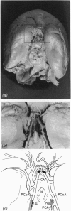
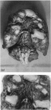
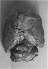
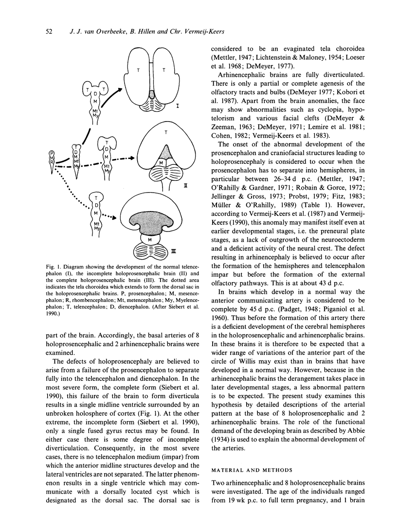
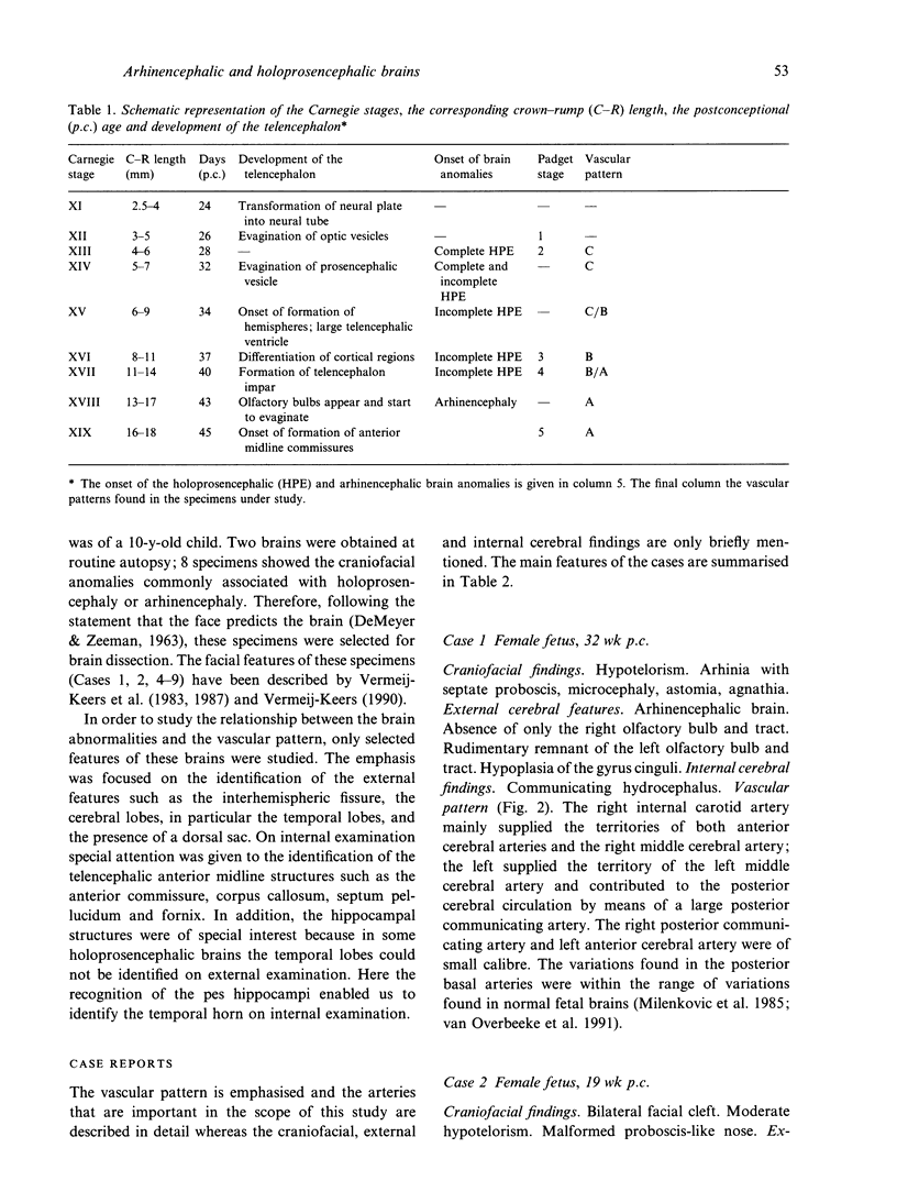
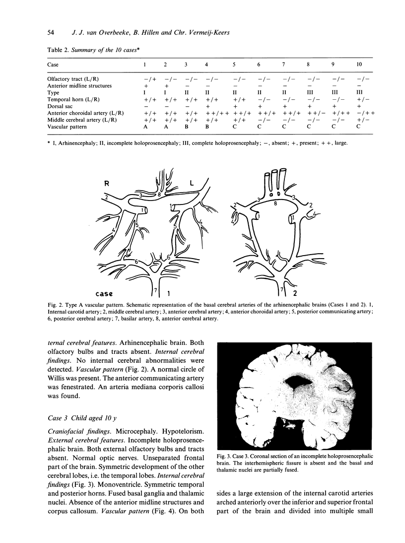
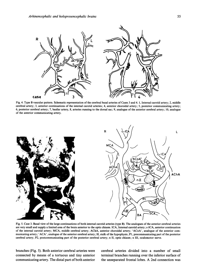
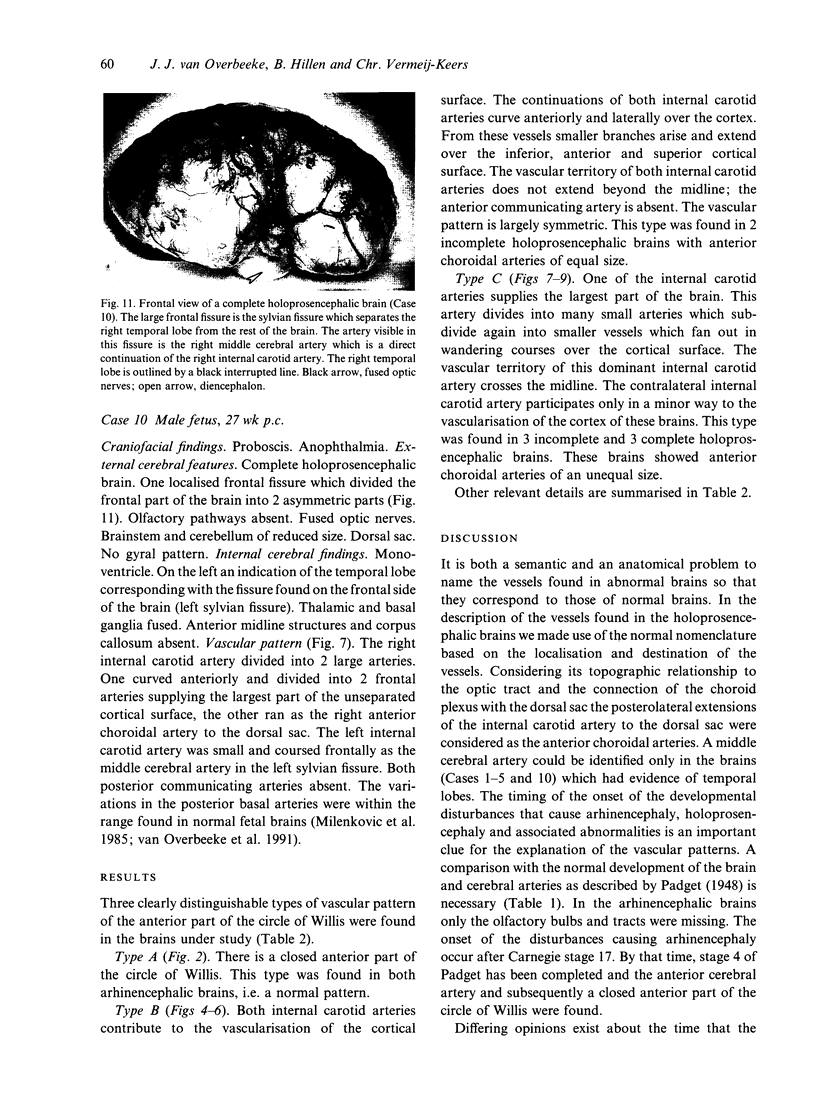
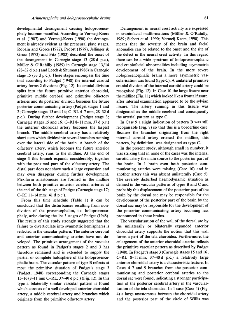
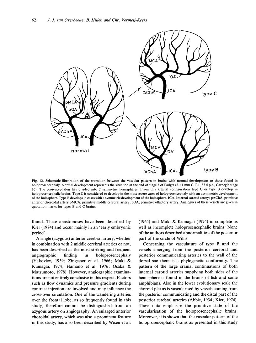
The mechanisms by which the anatomical variations of the circle of Willis develop is considered to be related to haemodynamic factors, i.e. the differential growth of the various parts of the brain will continuously change the haemodynamic demands and consequently the flow patterns in the cerebral arteries. It is therefore to be expected that, if a selected part of the brain does not develop, the change in the haemodynamic demand will affect the development of some cerebral arteries. Consequently the arteries at the base of 2 arhinencephalic and 8 holoprosencephalic brains were studied in conjunction with the brain malformations. The defects of holoprosencephaly are believed to arise from a failure of the prosencephalon to separate fully into the telencephalon and diencephalon and become manifest at the time that the prosencephalon normally starts to separate into the hemispheres, i.e. 28-34 d p.c. Arhinencephalic brains are fully diverticulated. There is only a partial or complete agenesis of the olfactory tracts and bulbs. The defect causing arhinencephaly starts at 43 d p.c. In the arhinencephalic brains no particular vascular abnormalities were found. However, at the base of the holoprosencephalic brains no complete circle of Willis was present; the anterior part was lacking and was replaced by anterior branches which emerged unilaterally or bilaterally from the internal carotid artery. The choroidal arteries were of very large calibre and ran to the highly vascularised wall of the dorsal cyst which is usually present in holoprosencephalic brains. In contrast to the anterior part, the posterior arterial pattern was almost identical to the posterior part of the circle of Willis of normal brains. The basic vascular patterns found in the holoprosencephalic brains displayed the features of Padget's developmental stages 2 and 3 of the cerebral vasculature, i.e. the pattern that has normally developed within 28-40 d p.c. The further modification of this pattern could largely be understood from the functional demand imposed on the circulation by the enlarged anterior choroidal arteries. Because the development of the anterior part of the circle of Willis precedes the developmental derangement causing arhinencephaly, a complete circle was found in these brains.
Full text
PDF
Images in this article
Selected References
These references are in PubMed. This may not be the complete list of references from this article.
- Abbie A. A. The Morphology of the Fore-Brain Arteries, with Especial Reference to the Evolution of the Basal Ganglia. J Anat. 1934 Jul;68(Pt 4):433–470. [PMC free article] [PubMed] [Google Scholar]
- Cohen M. M., Jr An update on the holoprosencephalic disorders. J Pediatr. 1982 Nov;101(5):865–869. doi: 10.1016/s0022-3476(82)80349-1. [DOI] [PubMed] [Google Scholar]
- DEMYER W., ZEMAN W. Alobar holoprosencephaly (arhinencephaly) with median cleft lip and palate: clinical, electroencephalographic and nosologic considerations. Confin Neurol. 1963;23:1–36. doi: 10.1159/000104278. [DOI] [PubMed] [Google Scholar]
- DeMyer W. Classification of cerebral malformations. Birth Defects Orig Artic Ser. 1971 Feb;7(1):78–93. [PubMed] [Google Scholar]
- Fitz C. R. Holoprosencephaly and related entities. Neuroradiology. 1983;25(4):225–238. doi: 10.1007/BF00540235. [DOI] [PubMed] [Google Scholar]
- Jellinger K., Gross H. Congenital telencephalic midline defects. Neuropadiatrie. 1973 Dec;4(4):446–452. doi: 10.1055/s-0028-1091760. [DOI] [PubMed] [Google Scholar]
- Kobori J. A., Herrick M. K., Urich H. Arhinencephaly. The spectrum of associated malformations. Brain. 1987 Feb;110(Pt 1):237–260. doi: 10.1093/brain/110.1.237. [DOI] [PubMed] [Google Scholar]
- LICHTENSTEIN B. W., MALONEY J. E. Malformation of the forebrain with comments on the so-called dorsal cyst, the corpus callosum and the hippocampal structures. J Neuropathol Exp Neurol. 1954 Jan;13(1):117–128. doi: 10.1093/jnen/13.1.117. [DOI] [PubMed] [Google Scholar]
- Leech R. W., Shuman R. M. Holoprosencephaly and related midline cerebral anomalies: a review. J Child Neurol. 1986 Jan;1(1):3–18. doi: 10.1177/088307388600100102. [DOI] [PubMed] [Google Scholar]
- Lemire R. J., Cohen M. M., Jr, Beckwith J. B., Kokich V. G., Siebert J. R. The facial features of holoprosencephaly in anencephalic human specimens. I. Historical review and associated malformations. Teratology. 1981 Jun;23(3):297–303. doi: 10.1002/tera.1420230304. [DOI] [PubMed] [Google Scholar]
- Loeser J. D., Alvord E. C., Jr Agenesis of the corpus callosum. Brain. 1968 Sep;91(3):553–570. doi: 10.1093/brain/91.3.553. [DOI] [PubMed] [Google Scholar]
- MOFFAT D. B. The embryology of the arteries of the brain. Ann R Coll Surg Engl. 1962 Jun;30:368–382. [PMC free article] [PubMed] [Google Scholar]
- Maki Y., Kumagai K. Angiographic features of alobar holoprosencephaly. Neuroradiology. 1974;6(5):270–276. doi: 10.1007/BF00345787. [DOI] [PubMed] [Google Scholar]
- Milenković Z., Vucetić R., Puzić M. Asymmetry and anomalies of the circle of Willis in fetal brain. Microsurgical study and functional remarks. Surg Neurol. 1985 Nov;24(5):563–570. doi: 10.1016/0090-3019(85)90275-7. [DOI] [PubMed] [Google Scholar]
- Müller F., O'Rahilly R. Mediobasal prosencephalic defects, including holoprosencephaly and cyclopia, in relation to the development of the human forebrain. Am J Anat. 1989 Aug;185(4):391–414. doi: 10.1002/aja.1001850404. [DOI] [PubMed] [Google Scholar]
- O'Rahilly R., Gardner E. The timing and sequence of events in the development of the human nervous system during the embryonic period proper. Z Anat Entwicklungsgesch. 1971;134(1):1–12. doi: 10.1007/BF00523284. [DOI] [PubMed] [Google Scholar]
- Osaka K., Matsumoto S. Holoprosencephaly in neurosurgical practice. J Neurosurg. 1978 May;48(5):787–803. doi: 10.3171/jns.1978.48.5.0787. [DOI] [PubMed] [Google Scholar]
- PIGANIOL G., SEDAN R., TOGA M., PAILLAS J. E. [The anterior communicating artery. Embryological and anatomical study]. Neurochirurgie. 1960 Jan-Mar;6:3–19. [PubMed] [Google Scholar]
- Robain O., Gorce F. Arhinencéphalie. Etude clinique, anatomique et étiologique de 13 cas. Arch Fr Pediatr. 1972 Oct;29(8):861–879. [PubMed] [Google Scholar]
- Van Overbeeke J. J., Hillen B., Tulleken C. A. A comparative study of the circle of Willis in fetal and adult life. The configuration of the posterior bifurcation of the posterior communicating artery. J Anat. 1991 Jun;176:45–54. [PMC free article] [PubMed] [Google Scholar]
- Vermeij-Keers C., Mazzola R. F., Van der Meulen J. C., Strickler M. Cerebro-craniofacial and craniofacial malformations: an embryological analysis. Cleft Palate J. 1983 Apr;20(2):128–145. [PubMed] [Google Scholar]
- WISEN M., DEMYER W., CAMPBELL R. UNIQUE ANGIOGRAPHIC AND VENTRICULOGRAPHIC PATTERN OF ALOBAR HOLOPROSENCEPHALY (ARHINENCEPHALY). Radiology. 1965 May;84:945–949. doi: 10.1148/84.5.945. [DOI] [PubMed] [Google Scholar]
- YAKOVLEV P. I. Pathoarchitectonic studies of cerebral malformations. III. Arrhinencephalies (holotelencephalies). J Neuropathol Exp Neurol. 1959 Jan;18(1):22–55. doi: 10.1097/00005072-195901000-00003. [DOI] [PubMed] [Google Scholar]
- Zingesser L. H., Schechter M. M., Medina A. Angiographic and pneumoencephalographic features of holoprosencephaly. Am J Roentgenol Radium Ther Nucl Med. 1966 Jul;97(3):561–574. doi: 10.2214/ajr.97.3.561. [DOI] [PubMed] [Google Scholar]


