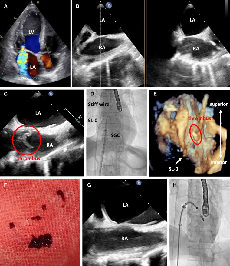
An 86-year-old male underwent MitraClip (Abbott Vascular, CA) implantation through the right femoral vein for significant ventricular functional mitral regurgitation (MR) because of his high surgical risk (Figure 1A). Despite no evidence of structure in right atrium (RA) immediately after transseptal puncture through 8.5 Fr SL-0 sheath (St. Jude Medical, MN) (Figure 1B), when 24 Fr steerable guide catheter (SGC) was advanced to RA along Amplatz Super Stiff guidewire (Boston Scientific, MA), transoesophageal echocardiography (TEE) demonstrated a floating structure with 24 mm length in RA (Figure 1C and Supplementary material). It appeared to get caught in the gap between the tip of SGC and its inner sheath. We confirmed adequate anticoagulation [activated clotting time (ACT) > 250 s] and attempted to maintain ACT > 300 s. Considering the possibility of the air contamination through the valve of SGC if aspiration was directly performed through the side-port of SGC in this condition, we aspirated this abnormal structure using the SL-0 sheath through the left femoral vein under TEE and fluoroscopic guidance (Figure 1D–F). Transoesophageal echocardiography revealed no structure in RA after aspiration (Figure 1G), and then SGC was removed for the purpose of washing out a potential thrombus inside it. Subsequently, MitraClip G4-NTW was successfully placed at A2-P2 without further device-related complications, resulting in less-than-mild degree of residual MR (Figure 1H). The pathological diagnosis of this aspirated structure showed red blood clots.
Figure 1.
(A) Transthoracic echocardiography on heart failure admission showed significant mitral regurgitation. (B) Transoesophageal echocardiography (TEE) with x-plane imaging before advancing steerable guide catheter (SGC) to right atrium (RA) revealed no structure in RA. (C) TEE immediately after advancing SGC to RA demonstrated a floating abnormal structure in RA. (D, E) Fluoroscopic imaging and 3D TEE during the structure aspiration in RA, respectively. (F) The aspirated structure was pathologically proven as thrombus later. (G) TEE after thrombus aspiration showed no structure in RA. (H) Fluoroscopic imaging after MitraClip implantation. Red circle indicates the thrombus in RA.
Large-bore venous sheath or catheter-derived thrombus formation, which leads to RA thrombus, is a known phenomenon, albeit rare, despite adequate anticoagulation. In left-sided structural heart disease interventions, RA thrombus is fraught with the risk of not only pulmonary but also systemic embolization. Regarding MitraClip procedure, a limited number of case reports on thrombus formation have been published, and no report on RA thrombus immediately after transseptal puncture. On the other hand, an observational study reported a higher than expected incidence of intraoperative thrombus detected on the transseptal sheath and in the left atrium after transseptal puncture during pulmonary vein isolation,1 and in all the cases of thrombus detection, successful retrieval of the clots with vigorous aspiration was achieved.1,2 In the present case, it is assumed that large-bore sheath or perhaps transseptal puncture caused RA thrombus, and we also successfully aspirated RA thrombus through transseptal sheath. This case underscores an optimal bailout strategy when thrombus is dragged to RA during MitraClip procedure.
Supplementary Material
Acknowledgements
The authors would like to thank Akiho Seno, Riyo Ogura, and Keitaro Mahara for assistance during the procedure.
Consent: The patient and his family gave consent to appear in the publication in accordance with the COPE guidelines.
Funding: None declared.
Contributor Information
Hiroaki Yokoyama, Department of Cardiology, Tokushima Red Cross Hospital, 103 Irinokuchi, Komatsushima, Tokushima 7730001, Japan.
Tatsuya Kokawa, Department of Cardiology, Tokushima Red Cross Hospital, 103 Irinokuchi, Komatsushima, Tokushima 7730001, Japan.
Tomoko Izumi, Department of Cardiology, Tokushima Red Cross Hospital, 103 Irinokuchi, Komatsushima, Tokushima 7730001, Japan.
Shinobu Hosokawa, Department of Cardiology, Tokushima Red Cross Hospital, 103 Irinokuchi, Komatsushima, Tokushima 7730001, Japan.
Supplementary material
Supplementary material is available at European Heart Journal – Case Reports online.
Data availability
The data underlying this article will be shared on reasonable request to the corresponding author.
References
- 1. Maleki K, Mohammadi R, Hart D, Cotiga D, Farhat N, Steinberg JS. Intracardiac ultrasound detection of thrombus on transseptal sheath: incidence, treatment, and prevention. J Cardiovasc Electrophysiol 2005;16:561–565. [DOI] [PubMed] [Google Scholar]
- 2. Blendea D, Barrett CD, Heist EK, Ruskin JN, Mansour MC. Right atrial thrombus aspiration guided by intracardiac echocardiography during catheter ablation for atrial fibrillation. Circ Arrhythm Electrophysiol 2009;2:e18–e20. [DOI] [PubMed] [Google Scholar]
Associated Data
This section collects any data citations, data availability statements, or supplementary materials included in this article.
Supplementary Materials
Data Availability Statement
The data underlying this article will be shared on reasonable request to the corresponding author.


