



Abstract
Thoracic descending aortic perforation with overlap syndrome (systemic sclerosis and dermatomyositis) is a rare, unreported vascular pathology. We describe the case of a 75-year-old woman who presented with chest tightness, back pain, and dyspnea. Computed tomography revealed a pinhole rupture in the descending aorta. The patient underwent a thoracic endovascular aortic repair and left chest drainage. Her postoperative course was uneventful.
Keywords: thoracic aortic rupture, pinhole, overlap syndrome
Introduction
Thoracic aortic pinhole ruptures are uncommon. Although open repair is the conventional treatment, thoracic endovascular aortic repair (TEVAR) is an effective alternative for patients with emergency conditions. Herein, we present a rare case of thoracic descending aortic rupture, not an aneurysm, in a patient with systemic sclerosis (SSc) and dermatomyositis.
Case Report
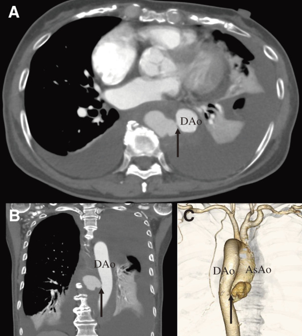
A 75-year-old woman with a history of hypertension, dyslipidemia, and overlap syndrome (SSc and dermatomyositis) presented with chief complaints of chest tightness, back pain, and dyspnea for the past 3 hours. The patient’s hypertension and dyslipidemia were well controlled. She also had a history of gastroesophageal reflux disease, which was well controlled with an oral proton pump inhibitor. Although she had been diagnosed with overlap syndrome based on biopsy results and skin symptoms at the dermatology department of our hospital and the rheumatology department of another hospital 1 year ago, she did not regularly follow up with her physician and thus did not receive timely oral medication. Her overlap syndrome was not well managed. Her antinuclear antibody level was 2560 times higher than normal. Acute myocardial infarction was suspected; however, coronary angiography revealed no significant stenosis. After treatment for heart failure in our department for 5 days, her blood oxygenation level decreased gradually. The computed tomography (CT) scan taken 5 days after admission showed pleural effusion, but no clear leakage of contrast agent was observed. Therefore, the cardiologist concluded that it was not a rupture. In retrospect, there were some areas with high attenuation values within the pleural effusion. The right pleural drainage was performed, and the patient was treated for heart failure for another 5 days. Her hematocrit level was 34.2% on the day of admission. Despite decreasing to 26.1% the next day, her hematocrit level did not decrease thereafter (27.5%–27.8%). The cardiologist re-examined the CT scan and found a pinhole rupture in the descending aorta (Fig. 1). They consulted us about this rupture.
Fig. 1 Preoperative enhanced CT showing thoracic descending pinhole aortic rupture (arrow). (A) Axial view. (B) Coronal view. (C) Three-dimensional-CT. AsAo: ascending aorta; CT: computed tomography; DAo: descending aorta.
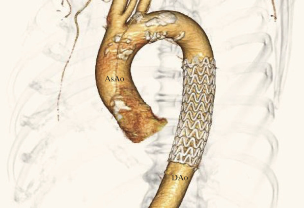
The anatomy of the artery was suitable for endovascular repair, and we subsequently performed the repair. After TEVAR, left pleural drainage was performed. The pleural effusion culture showed the absence of bacteria, fungi, and viruses. No complications occurred during the perioperative period, and the patient was discharged 15 days later. Follow-up CT revealed no aortic abnormalities (Fig. 2).
Fig. 2 Three-dimensional CT of the aorta prior to patient discharge. AsAo: ascending aorta; DAo: descending aorta; CT: computed tomography.
Discussion
Overlap syndrome is diagnosed if at least two connective tissue diseases are concurrently present. Connective tissue diseases include SSc, Sjogren’s syndrome, dermatomyositis, polymyositis, rheumatoid arthritis, and systemic lupus erythematosus. Whether overlap syndrome is related to aortic rupture is unclear; however, overlap syndrome and aortic stiffness may be associated. Moyssakis et al.1) showed that aortic stiffness increases in SSc patients regardless of the disease state. Timár et al.2) showed that arterial stiffness may be correlated with disease duration and patient age in SSc. In a study on mice, it was shown that mice with SSc presented with vessel wall fibrosis and altered vascular smooth muscle cell composition.3) Although the rupture site of the descending aorta showed minimal calcification in our case, the thoracic aorta may have been stiff. Even reports of pinhole ruptures of the thoracic aorta accompanied by arteriosclerosis or aneurysms are rare4,5); however, in this case, a pinhole rupture formed in an area with minimal atherosclerosis. There are few reports of thoracic aortic rupture with little or no arteriosclerosis, except for traumatic ruptures. Ohashi et al.6) reported on an aortic rupture due to co-localizing aortic intimal myofibroblastic sarcoma and urothelial carcinoma. To the best of our knowledge, no such case of connective tissue disease and aortic rupture has been reported.
The Society for Vascular Surgery recently recommended TEVAR over open repair surgery for treating a ruptured descending thoracic aorta when anatomically feasible.7) Descending thoracic aortic ruptures can be repaired through open repair surgery or an endovascular approach. Open repair surgery with artificial vascular grafts has been well established for resecting tissues around the fistula and reconstructing systemic perfusion. However, a recent meta-analysis showed that open repair surgery was associated with higher perioperative mortality, 1-year mortality, renal insufficiency rates, and pulmonary complications than TEVAR.8) In contrast, a Swedish report showed that TEVAR has acceptable short-term survival but poor long-term survival. This report showed that the postoperative complication rate was high, and re-interventions were needed in 20% of the cases.9) Goodney et al.10) indicated that better short-term survival after TEVAR compared with open repair surgery for treating thoracic aortic aneurysms disappeared within 2 years after surgery. It is possible that some high-risk patients who could not undergo open repair surgery were offered endovascular repair. Thus, careful interpretation of results is essential when comparing open and endovascular repair approaches.
When patients with a ruptured descending aorta are transported to a hospital, they might be in a state of shock. In contrast to open surgery, emergent TEVAR rapidly stabilizes these patients’ conditions. Therefore, TEVAR is a suitable first-choice treatment for ruptures. However, many anatomical cases are unsuitable for endovascular repair. After TEVAR, it is vital to closely follow up and consider re-intervention as needed. In the present case, the rupture was caused by a small perforation. We obtained adequate proximal and distal landing zones for TEVAR. Thus, we believe complications may rarely occur if the procedure is completed safely.
Conclusion
We successfully treated a patient with a pinhole rupture in the descending thoracic aorta with a history of overlap syndrome. Careful follow-up is necessary because the long-term results after TEVAR in such cases are not known.
Declarations
Ethical considerations and approval
The study was conducted with the approval of the ethics committee at the institution to which the first author belongs (Approval No. 202424).
Consent
The patient provided consent for the publication of this report and accompanying images.
Acknowledgments
We thank Dr. Yasushige Shingu and Editage (www.editage.jp) for manuscript preparation and English language editing. This study received no funding from any institution.
Disclosure statement
The authors declare that they have no competing interests.
Author contributions
Manuscript preparation: NA
Critical review and revision: all authors
Final approval of the article: all authors
Accountability for all aspects of the work: all authors.
References
- 1).Moyssakis I, Gialafos E, Vassiliou V, et al. Aortic stiffness in systemic sclerosis is increased independently of the extent of skin involvement. Rheumatology (Oxford) 2005; 44: 251–4. [DOI] [PubMed] [Google Scholar]
- 2).Timár O, Soltesz P, Szamosi S, et al. Increased arterial stiffness as the marker of vascular involvement in systemic sclerosis. J Rheumatol 2008; 35: 1329–33. [PubMed] [Google Scholar]
- 3).Derrett-Smith EC, Dooley A, Khan K, et al. Systemic vasculopathy with altered vasoreactivity in a transgenic mouse model of scleroderma. Arthritis Res Ther 2010; 12: R69. [DOI] [PMC free article] [PubMed] [Google Scholar]
- 4).Warisawa T, Kotoku A, Miyauchi R, et al. Pulmonary artery occlusion due to abrupt pinhole rupture of aortic arch aneurysm. JACC Cardiovasc Interv 2021; 14: e15–6. [DOI] [PubMed] [Google Scholar]
- 5).Sako H, Hadama T, Shigemitsu O, et al. Pinhole rupture of aortic root aneurysm with severe atherosclerotic change. Ann Thorac Surg 2004; 77: 1075–7. [DOI] [PubMed] [Google Scholar]
- 6).Ohashi Y, Yamamoto Y, Yamada A, et al. Aortic rupture due to co-localization of aortic intimal myofibroblastic sarcoma and urothelial carcinoma: a unique case report. Intern Med 2021; 60: 269–74. [DOI] [PMC free article] [PubMed] [Google Scholar]
- 7).Upchurch GR Jr, Escobar GA, Azizzadeh A, et al. Society for Vascular Surgery clinical practice guidelines of thoracic endovascular aortic repair for descending thoracic aortic aneurysms. J Vasc Surg 2021; 73 1S: 55S–83S. [DOI] [PubMed] [Google Scholar]
- 8).Xie X, Shu X, Zhang W, et al. A comparison of clinical outcomes of endovascular repair versus open surgery for ruptured descending thoracic aorta. J Endovasc Ther 2022; 29: 307–18. [DOI] [PubMed] [Google Scholar]
- 9).Hammo S, Larzon T, Hultgren R, et al. Outcome after endovascular repair of ruptured descending thoracic aortic aneurysm: a national multicentre study. Eur J Vasc Endovasc Surg 2019; 57: 788–94. [DOI] [PubMed] [Google Scholar]
- 10).Goodney PP, Travis L, Lucas FL, et al. Survival after open versus endovascular thoracic aortic aneurysm repair in an observational study of the Medicare population. Circulation 2011; 124: 2661–9. [DOI] [PMC free article] [PubMed] [Google Scholar]