Abstract
Background
Congenital radioulnar synostosis (CRUS) is a rare upper limb deformity characterized by impaired rotational movement of the forearm. Rotational osteotomy is a commonly employed surgical procedure for treatment. This study aimed to analyze its surgical efficacy in treating CRUS in children.
Methods
22 children (24 limbs) with CRUS from January 2010 to December 2023 were retrospectively collected. Rotational osteotomy of proximal ulna and distal radius was performed. Forearm function was evaluated using Failla scores and hygiene and self-care scores in Activities of daily Living score (ADL score). In addition, patients were further grouped and compared according to type of ulnar internal fixation and age at surgery.
Results
22 patients (14 males, 8 females), with an average age of 6.0 years and an average follow-up time of 56 months. The mean pronation angle before surgery was 75.0 ± 11.3°, the mean postoperative pronation angle was 3.8 ± 7.1°, and the mean correction degree was 78.8 ± 12.9°. The average Failla scores were 5.6 ± 2.1 points before operation and 14.0 ± 1.0 points after operation. The average scores of hygiene and self-care scores were 19.0 ± 5.1 points before surgery and 36.0 ± 3.9 points after surgery. No child developed complications such as osteofascial compartment syndrome or infection. The correction angle in the plate fixation group was 86.8 ± 10.6°, while in the K-wires group was 72.0 ± 10.7°. The postoperative Failla scores in the older age group were 13.0 ± 1.1 points, and in the younger age group were 14.3 ± 0.8 points.
Conclusion
Rotational osteotomy of forearm bones is safe and effective in the treatment of CRUS in children. Ulnar plate fixation has better correction than K—wires. Furthermore, younger children have better surgical outcomes than older ones.
Keywords: Rotational osteotomy, Children, Congenital radioulnar synostosis
Introduction
CRUS, a rare congenital upper—limb anomaly from embryonic issues, shows as abnormal proximal ulnar—radius fusion. It was initially reported by Sandifort in 1793 [1]. Due to the low prevalence of CRUS, most studies are case reports or small cohort studies [2], therefore large—sample detailed epidemiological information is lacking. The clinical presentation of CRUS mainly depends on the fixed position of the forearm deformity, and less on factors like radial head stability and the child’s age [3]. Mild pronation of the forearm may mask the diagnosis as daily functions can be compensated by shoulder and wrist joints. However, extreme pronation deformity of the forearm affects personal hygiene and daily life and work, reducing the quality of life.
There is no consensus on CRUS treatment. For patients with unilateral onset and mild functional limitation of the forearm with rotational fixation, the most common treatment is conservative [4]. Surgery is the main treatment for children with severe functional limitation of the forearm with a more generalized pronation deformity angle. Although the controversy over surgical indications remains unsettled, the relative consensus is to fixate pronation beyond 60° [3, 5]. Surgical treatments are mainly divided into two major categories [3]: mobilization procedures (such as Kanaya procedure) and positioning procedures (such as rotational osteotomy). Rotational osteotomy is the main surgical procedure, accounting for more than 90% of surgical treatment [6]. This procedure does not restore the rotational function of the forearm, but only moves the forearm from an over-rotated forward position to an appropriate position that facilitates the function of the hand, but it makes it easier for the patient to accomplish daily activities.
Rotational osteotomy for the treatment of CRUS has been reported in the relevant literature, and most of them have achieved more satisfactory results, but there are also some limitations [7–9]. The process of osteotomy at the fusion site is complex and prone to complications such as neurovascular injury and compartment syndrome [10]; The unilateral osteotomy of the radius presents issues such as insufficient correction angle and postoperative loss of angle [9]. Bilateral osteotomies of the radius and ulna have certain advantages over osteotomies at other sites, but there is relatively little research on this topic.
Therefore, the purpose of this study is to retrospectively analyze the surgical efficacy of rotational osteotomy of the radius and ulna in the treatment of CRUS in children, and to summarize clinical experience. In addition, the effects of type of ulnar internal fixation and age at surgery on surgical outcomes were further explored.
Materials and methods
Patients
This study has been approved by the Ethics Committee of the First Affiliated Hospital of Guangxi Medical University. The research retrospectively collected patients diagnosed with CRUS at the First and Second Affiliated Hospitals of Guangxi Medical University from January 2010 to December 2023. The inclusion criteria were children who met the diagnostic criteria for CRUS and underwent rotational osteotomy for treatment upon admission. Furthermore, the preoperative forearm pronation angle on the affected side was greater than 60°. Exclusion criteria were the presence of other upper limb deformities or neuromuscular disorders.
There were 14 males and 8 females among the included cases, with a mean age at surgery of 6.0 ± 2.8 years (3–14 years). The mean follow-up duration was 56.8 months. Nineteen cases underwent left forearm surgery, one case of right forearm surgery, and two cases of bilateral surgery. According to Cleary—Omer classification, 18 limbs that underwent surgery were type III, 3 limbs were type II, and 1 limb was type IV.
According to the difference of ulnar internal fixation, all children were divided into two groups: K-wires internal fixation group and plate internal fixation group, and the clinical data and surgical outcomes of the two groups were compared. In addition, based on the difference of surgical age, with 8 years old as the boundary, the differences in surgical outcomes between older and younger children were explored.
Surgical technique
Supine positioning was applied to the patient, and after successful anesthesia, tourniquet application was performed intraoperatively to arrest bleeding. The distal radius and proximal ulna were exposed respectively. Perform a subperiosteal radial osteotomy at the level of the insertion point of the pronator teres muscle, and select the location of the ulnar osteotomy close to the fusion site. The forearm was manually rotated to a suitable angle, and the blood flow of each finger end was observed to be acceptable. The osteotomy ends were temporarily fixed with K-wires, and fluoroscopic examination was performed to check whether the alignment of the osteotomy ends was satisfactory. The proximal osteotomy of the ulna was fixed with plate or K-wires, and the distal osteotomy of the radius was fixed with plate.
Postoperative management
Postoperatively, reduce swelling, pain relief and symptomatic supportive treatment were given, and the forearm was immobilized with a long arm cast for 4–6 weeks. Observe the blood circulation of the fingertips and swelling of the limb to prevent osteofascial compartment syndrome. During the period of plaster immobilization, children were instructed to perform active grasping activities of metacarpophalangeal joints and interphalangeal joints to promote swelling reduction, and elbow joint functional exercises were actively carried out after the plaster was removed.
Clinical and radiographic measurement
The position of forearm rotational fixation was determined by measuring the angle formed between the line connecting the styloid processes of the radius and ulna and the longitudinal axis of the humeral shaft [11]. The method for calculating the correction angle is to compare the difference between the angles of forearm rotation fixed before and after surgery. The pre- operative and post-operative forearm function was assessed using the Failla scoring system [8, 12, 13] and hygiene and self-care scores in ADL score. The bone healing status was evaluated by X-ray examination. Observation of postoperative complications such as osteofascial compartment syndrome, nonunion of osteotomy end, and wound infection.
Statistical analysis
Statistical analysis was performed using SPSS 22 statistical software (IBM, USA). The numerical data were presented as the mean, complemented by the standard deviation. Comparisons of children’s scores before and after surgery were performed by paired-samples t-test or nonparametric test, and comparisons between different groups were performed by independent-samples t-test or rank-sum test. A p value of less than 0.05 was considered statistically significant.
Results
Twenty-two pediatric patients (24 limbs) had a mean preoperative pronation angle of 75.0 ± 11.3° (range 60°-90°). The overall condition is shown in Table 1. All pediatric patients achieved bony union. No cases of compartment syndrome, nonunion at the osteotomy site, or infection were observed as complications.
Table 1.
Overall condition of patients
| Item | values |
|---|---|
| Surgical Side (Left/Right/Bilateral) | 19/1/2 |
| Sex (Male/Female) | 14/8 |
| Age at Surgery (years) | 6.0 ± 2.8 |
| Cleary–Omer Classification(II/III/IV) | 3/20/1 |
| Preoperative Pronation Angle (degrees) | 75.0 ± 11.3 |
| Postoperative Supination Angle (degrees) | 3.8 ± 7.1 |
| Correction Angle (degrees) | 78.8 ± 12.9 |
| Preoperative Failla Scores | 5.6 ± 2.1 |
| Postoperative Failla Scores | 14.0 ± 1.0 |
| Preoperative hygiene and self-care scores | 19.0 ± 5.1 |
| Postoperative hygiene and self-care scores | 36.0 ± 3.9 |
| Bone Healing Time (weeks) | 5.8 ± 1.6 |
| Complications (number of cases) | 0 |
According to the difference of ulnar internal fixation, it was divided into two groups: K-wires internal fixation (13 limbs) and plate internal fixation (11 limbs). Failla scores and hygiene and self-care scores in ADL scores before and after surgery in both groups showed statistical differences. It suggests that both forms of internal fixation have significant corrective effects. The clinical data comparison between the two groups showed that there were no differences in preoperative pronation angle, bone healing time, postoperative Failla scores and hygiene and self-care scores between the two groups. Although the average age of patients undergoing surgery in the plate group (6.7 ± 3.4) was higher than that in the K-wires group (5.4 ± 2.1), there was no statistically significant difference between the two groups (P = 0.424). The correction angle in the plate fixation group was 86.8 ± 10.6°, while in the K-wires group was 72.0 ± 10.7°, and this difference was statistically significant (P = 0.003). This indicates that the plate is more corrective than the K-wires and that there is no significant difference between the two internal fixation modalities in terms of postoperative outcome.
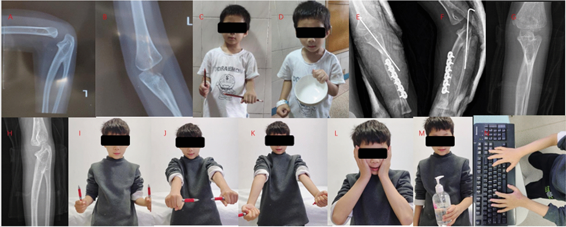
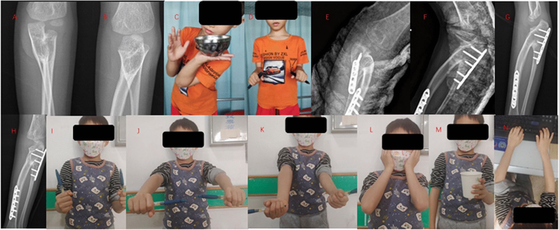
Based on the accepted age cut-off of 8 years for older children [14], the age at surgery is divided into a younger age group (less than 8 years) and an older age group (more than or equal to 8 years). Failla scores and hygiene and self-care scores before and after surgery in both groups suggest that both older and younger children can benefit from the procedure. The comparison of clinical data between the older and younger age groups demonstrates that there is no significant difference in the degree of preoperative deformity, the angle of surgical correction and bone healing time between the two groups. The postoperative Failla scores in the older age group were 13.0 ± 1.1 points, and in the younger age group were 14.3 ± 0.8 points, with a statistically significant difference (P = 0.015). This indicates to some extent that early surgery is more effective. Two typical case is shown in Figs. 1, 2.
Fig. 1.

Typical case 1, male, bilateral CRUS, age at surgery 4 years. A-B: preoperative left forearm anteroposterior and lateral x-rays, C: preoperative elbow-flexed neutral appearance, D: preoperative left-handed bowl holding, E–F: 1 day postoperative left forearm anteroposterior and lateral x-rays, G-H: 3.5 years postoperative left forearm anteroposterior and lateral x-rays. I-N: 3.5 years postoperative follow-up appearance photographs. The forearm can perform pronation and supination movements with compensation at the shoulder and wrist joints
Fig. 2.

Typical case 2. male, left CRUS, age at surgery 6 years old. A-B: Preoperative anteroposterior and lateral X-rays of the left forearm, C-D: Preoperative gross examination. E–F: Postoperative anteroposterior and lateral X-rays of the left forearm on the first day, G-H: Postoperative anteroposterior and lateral X-rays of the left forearm at 4 months. I-N: Follow-up photographs of the appearance at 4 months postoperatively. The forearm can perform pronation and supination movements with compensation at the shoulder and wrist joints
Discussion
The predominant pathological feature of CRUS is the failure of longitudinal segmentation of the cartilaginous primordia of the ulna and radius to form abnormally rigid junctions as a result of misrouting of signals during embryonic development [3, 15]. The superior ulnar-radial joint (PRUJ) is an important structural basis for forearm rotation [16], and previous studies have shown that the radius rotates around the ulna with little ulnar motion during forearm rotational movements [16–18]. Fusion of the PRUJ limits rotational motion of the joint.
Rotational osteotomy is the main procedure for the treatment of CRUS, but the position of the forearm after surgery has been a hot topic of research and has not been standardized yet [19, 20]. In Western countries, due to economic and social development, the high demand for computers, and the habit of eating with a knife and fork, they tend to maintain the pronation for the postoperative position of the nondominant limb. In China and other East Asian countries, traditional dining etiquette involves using the non-dominant forearm to hold the bowl and the dominant hand to operate chopsticks. Therefore, it is customary to correct the non-dominant side to a neutral or slightly pronated position, while the dominant side is positioned in a slightly supinated position. Based on these reasons, in this study we corrected the nondominant limb to a neutral to mildly rotated posterior position and the dominant limb to a mildly rotated pronation status postoperatively. Postoperative follow-up showed that all the children and their families reported a significant improvement in their daily activities compared to the preoperative period, and they were all satisfied with the results of the surgery.
Currently, there are various methods for fixation following rotational osteotomy, such as cast immobilization [21]、K-wire fixation [22]、plate and screw fixation [12], and Ilizarov external fixation [23]. In this research, we comprehensively considered factors such as the child’s age, skeletal size, the angle to be corrected, and the actual situation of the fixed equipment prepared, and employed two methods of ulnar fixation. The results showed that ulnar plate fixation was more corrective than ulnar K-wire fixation, which suggests that ulnar-radial plate fixation should be chosen when a large degree of correction is expected preoperatively.
Choosing the right time for surgery is also an important element in achieving a desirable outcome. However, there is no consensus on the timing of surgery [24, 25]. In this research we found that postoperative Failla scores were significantly higher in the younger age group, which suggests to some extent that early surgery is more effective. As the age of the pediatric patient at the time of surgery increases, there is a longer duration of soft tissueand interosseous membrane contracture, and atrophy of the supinator muscle [11, 26]. Moreover, early surgery allows for the initiation of forearm motion exercises earlier, which is beneficial for enhancing the compensatory capabilities of the shoulder and wrist joints. Regrettably, the specific optimal age for surgery still requires research with a larger sample size.
Previously reported Pei used fusion site osteotomy to treat 31 children with a high complication rate of 9.7% [8]. Horii used radial diaphysis osteotomy to treat 26 children, but one limb developed ulnar-radial cross-fertilization postoperatively and one limb showed loss of correction [25]. In this study, proximal ulnar and distal radial rotational osteotomies were employed. Compared with single osteotomy, the advantages of this surgical technique lie in that the rotation of the osteotomy ends is easier, the correction ability is enhanced, and more severe pronation deformities can be corrected safely. There were no complications such as compartment syndrome and neurovascular compromise in this study. In comparison with other studies, this research further explores the impact of the type of ulnar internal fixation devices and the age at the time of surgery on the surgical outcomes, which contributes a certain degree of innovation to the study results.
This study also has some limitations. First, this study was a retrospective analysis and the sample size needs to be further expanded; second, this study was not controlled with other rotational osteotomies; and lastly, due to the young age of most of the children, the functional assessment was performed by both parents and children, and the scores may be biased.
Conclusions
The results of this study indicate that the rotational osteotomy of the radius and ulna for the treatment of CRUS is safe and effective, with ulnar plate fixation offering superior corrective force compared to K-wire fixation. Furthermore, the surgical outcomes are superior in younger children compared to older children.
Acknowledgements
We are grateful to The First Affiliated Hospital of Guangxi Medical University’s medical record query system for providing data.
Author contributions
All authors have contributed significantly to the work of the report in terms of concept, study design, execution, data collection, analysis, and interpretation; Participate in the drafting, revision, or critical review of clauses; The main manuscript was written by L.X.L. and L.Z.B., T.S.P. and Z.X.D. wrote the figure and tables., Y.X.J. and C.Y.J were mainly responsible for collecting the data. D.X.F. and L.S.J. are primarily responsible for the design guidance of the project. All authors read and approved the final manuscript.
Funding
The authors received no external funding to support this project.
Availability of data and materials
No datasets were generated or analysed during the current study.
Declarations
Ethics approval and consent to participate
The ethics committee of The First Affiliated Hospital of Guangxi Medical University approved this study.
Consent for publication
Not applicable.
Competing interests
The authors declare no competing interests.
Footnotes
Publisher's Note
Springer Nature remains neutral with regard to jurisdictional claims in published maps and institutional affiliations.
Xiaolin Luo and Zhenbiao Li have equally contributed to this work.
Contributor Information
Shijie Liao, Email: gxliaoshijie@163.com.
Xiaofei Ding, Email: dxfeicsgk2014@163.com.
References
- 1.Fujimoto M, Kato H, Minami A. Rotational osteotomy at the diaphysis of the radius in the treatment of congenital radioulnar synostosis[J]. J Pediatr Orthop. 2005;25(5):676–9. [DOI] [PubMed] [Google Scholar]
- 2.Hong P, Tan W, Zhou WZ, et al. The relation between radiographic manifestation and clinical characteristics of congenital radioulnar synostosis in children: a retrospective study from multiple centers[J]. Front Pediatr. 2023. 10.3389/fped.2023.1117060. [DOI] [PMC free article] [PubMed] [Google Scholar]
- 3.Rutkowski PT, Samora JB. Congenital radioulnar synostosis[J]. J Am Acad Orthop Surg. 2021;29(13):563–70. [DOI] [PubMed] [Google Scholar]
- 4.Kepenek-Varol B, Hosbay Z. Is short-term hand therapy effective in a child with congenital radioulnar synostosis? A case report[J]. J Hand Ther. 2020;33(3):435–42. [DOI] [PubMed] [Google Scholar]
- 5.Bo H, Xu J, Lin J, et al. Outcomes of two-stage double-level rotational osteotomy in treating patients with congenital proximal radioulnar synostosis[J]. World J Pediatr Surg. 2023;6(2): e000578. [DOI] [PMC free article] [PubMed] [Google Scholar]
- 6.Barik S, Farr S, Gallone G, et al. Results after treatment of congenital radioulnar synostosis: a systematic review and pooled data analysis[J]. J Pediatr Orthop B. 2021;30(6):593–600. [DOI] [PMC free article] [PubMed] [Google Scholar]
- 7.Tan W, Yuan ZK, Lin YC, et al. Rotational osteotomy with single incision and elastic fixation for congenital radioulnar synostosis in children: a retrospective cohort study[J]. Transl Pediatr. 2022;11(5):687–95. [DOI] [PMC free article] [PubMed] [Google Scholar]
- 8.Pei XJ, Han JH. Efficacy and feasibility of proximal radioulnar derotational osteotomy and internal fixation for the treatment of congenital radioulnar synostosis[J]. J Orthop Surg Res. 2019. 10.1186/s13018-019-1130-0. [DOI] [PMC free article] [PubMed] [Google Scholar]
- 9.Satake H, Kanauchi Y, Kashiwa H, et al. Long-term results after simple rotational osteotomy of the radius shaft for congenital radioulnar synostosis[J]. J Shoulder Elbow Surg. 2018;27(8):1373–9. [DOI] [PubMed] [Google Scholar]
- 10.Simcock X, Shah AS, Waters PM, et al. Safety and efficacy of derotational osteotomy for congenital radioulnar synostosis[J]. J Pediatr Orthop. 2015;35(8):838–43. [DOI] [PubMed] [Google Scholar]
- 11.Martínez-Alvarez S, González-Codó S, Vara-Patudo I, et al. Double-level intraperiosteal derotational osteotomy for congenital radioulnar synostosis[J]. J Pediatr Orthop. 2022;42(7):E756–61. [DOI] [PubMed] [Google Scholar]
- 12.Hamiti Y, Yushan M, Yalikun A, et al. Derotational osteotomy and plate fixation of the radius and ulna for the treatment of congenital proximal radioulnar synostosis[J]. Front Surg. 2022;9:888916. [DOI] [PMC free article] [PubMed] [Google Scholar]
- 13.Shingade VU, Shingade RV, Ughade SN. Results of single-staged rotational osteotomy in a child with congenital proximal radioulnar synostosis: subjective and objective evaluation[J]. J Pediatr Orthop. 2014;34(1):63–9. [DOI] [PubMed] [Google Scholar]
- 14.Liu F, Tang K, Zheng PF, et al. Performance of Tonnis triple osteotomy in older children with developmental dysplasia of the hip (DDH) assisted by a 3D printing navigation template[J]. Bmc Musculoskelet Disord. 2022;23(1):712. [DOI] [PMC free article] [PubMed] [Google Scholar]
- 15.Simmons BP, Southmayd WW, Riseborough EJ. Congenital radioulnar synostosis[J]. J Hand Surg Am. 1983;8(6):829–38. [DOI] [PubMed] [Google Scholar]
- 16.Lastayo PC, Lee MJ. The forearm complex: anatomy, biomechanics and clinical considerations[J]. J Hand Ther. 2006;19(2):137–44. [DOI] [PubMed] [Google Scholar]
- 17.Rahman AM, Montero-Lopez N, Hinds RM, et al. Assessment of forearm rotational control using 4 upper extremity immobilization constructs[J]. Hand (N Y). 2018;13(2):202–8. [DOI] [PMC free article] [PubMed] [Google Scholar]
- 18.Stroyan M, Wilk KE. The functional anatomy of the elbow complex[J]. J Orthop Sports Phys Ther. 1993;17(6):279–88. [DOI] [PubMed] [Google Scholar]
- 19.Green WT, Mital MA. Congenital radio-ulnar synostosis: surgical treatment[J]. J Bone Joint Surg Am. 1979;61(5):738–43. [PubMed] [Google Scholar]
- 20.Ogino T, Hikino K. Congenital radio-ulnar synostosis: compensatory rotation around the wrist and rotation osteotomy[J]. J Hand Surg Br. 1987;12(2):173–8. [DOI] [PubMed] [Google Scholar]
- 21.El-Adl W. Two-stage double-level rotational osteotomy in the treatment of congenital radioulnar synostosis[J]. Acta Orthop Belg. 2007;73(6):704–9. [PubMed] [Google Scholar]
- 22.Bishay SNG. Minimally invasive single-session double-level rotational osteotomy of the forearm bones to correct fixed pronation deformity in congenital proximal radioulnar synostosis[J]. J Child Orthop. 2016;10(4):295–300. [DOI] [PMC free article] [PubMed] [Google Scholar]
- 23.Rubin G, Rozen N, Bor N. Gradual correction of congenital radioulnar synostosis by an osteotomy and ilizarov external fixation[J]. J Hand Surg-Am. 2013;38a(3):447–52. [DOI] [PubMed] [Google Scholar]
- 24.Hung NN. Derotational osteotomy of the proximal radius and the distal ulna for congenital radioulnar synostosis[J]. J Child Orthop. 2008;2(6):481–9. [DOI] [PMC free article] [PubMed] [Google Scholar]
- 25.Horii E, Koh S, Hattori T, et al. Single osteotomy at the radial diaphysis for congenital radioulnar synostosis[J]. J Hand Surg-Am. 2014;39(8):1553–7. [DOI] [PubMed] [Google Scholar]
- 26.Chen CL, Kao HK, Chen CC, et al. Long-term follow-up of microvascular free tissue transfer for mobilization of congenital radioulnar synostosis[J]. J Plast Reconstr Aesthet Surg. 2012;65(12):E363–5. [DOI] [PubMed] [Google Scholar]
Associated Data
This section collects any data citations, data availability statements, or supplementary materials included in this article.
Data Availability Statement
No datasets were generated or analysed during the current study.


