Abstract
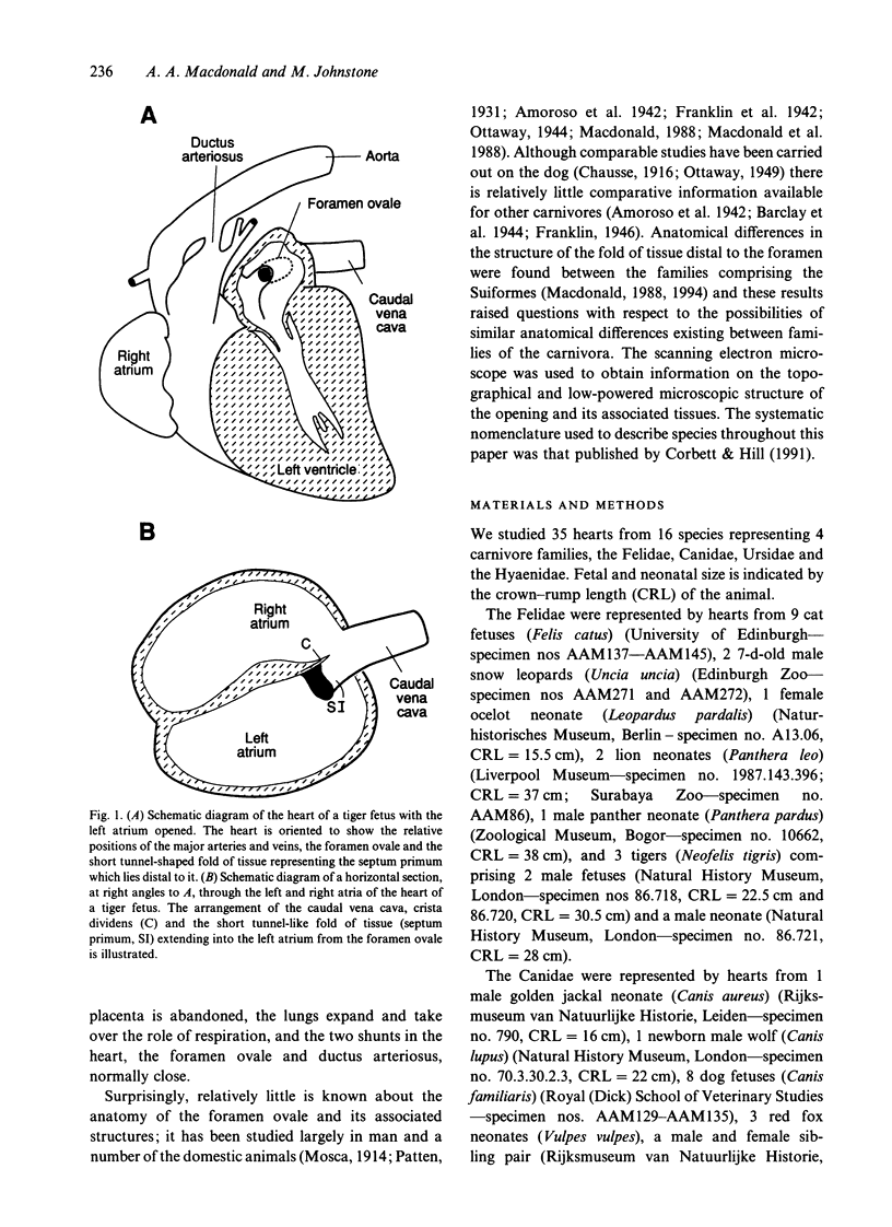
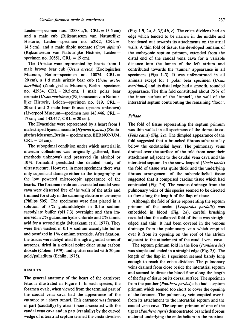
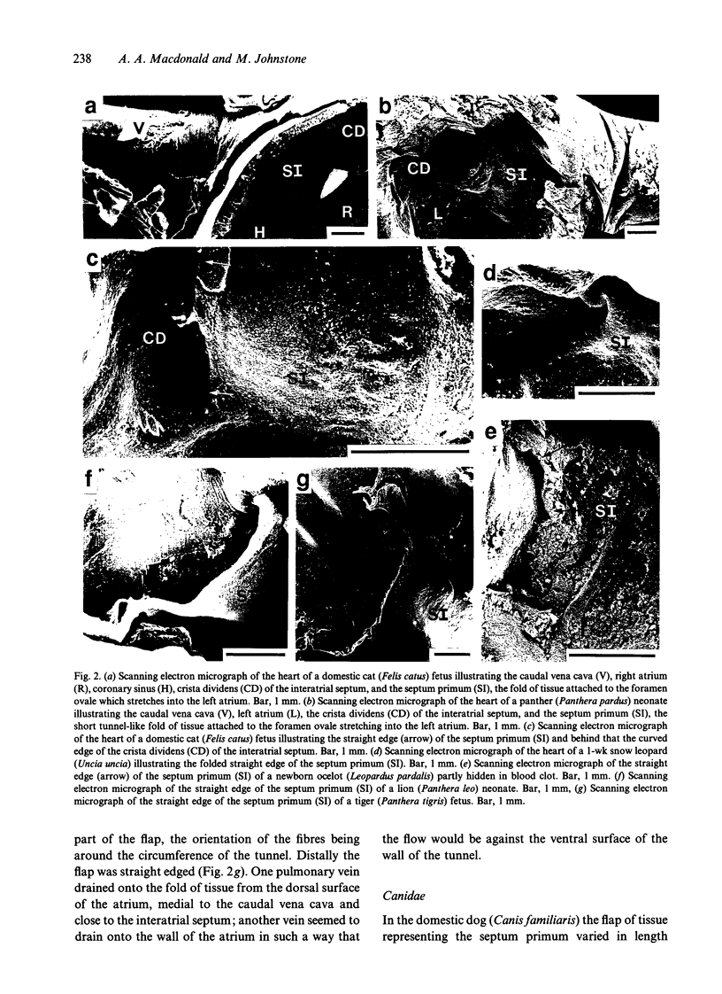
The structure of the foramen ovale from 16 species representing 4 carnivore families, the Felidae, Canidae, Ursidae and Hyaenidae, was studied using the scanning electron microscope. The Felidae were represented by 9 domestic cat fetuses (Felis catus), 2 snow leopard neonates (Uncia uncia), an ocelot neonate (Leopardus pardalis), 2 lion neonates (Panthera leo), a panther neonate (Panthera pardus) and 3 tigers (Neofelis tigris), comprising 2 fetuses and a neonate. The Canidae were represented by a golden jackal neonate (Canis aureus), a newborn wolf (Canis lupus), 8 domestic dog fetuses (Canis familiaris), 3 red fox neonates (Vulpes vulpes) and a dhole neonate (Cuon alpinus). The Ursidae were represented by a brown bear neonate (Ursus arctos), a day-old grizzly bear cub (Ursus arctos horribilis), a polar bear neonate (Ursus maritimus), and 2 additional bear fetuses (species unknown). The Hyaenidae were represented by a striped hyaena neonate (Hyaena hyaena). In each species, the foramen ovale, when viewed from the terminal part of the caudal vena cava, had the appearance of a short tunnel. A thin fold of tissue, the developed remains of the embryonic septum primum, extended from the distal end of the caudal vena cava for a variable distance into the lumen of the left atrium and contributed towards the 'tunnel' appearance in all specimens. It constituted a large proportion of the tube, and its distal end was straight-edged. There was fibrous material underlying the endothelium of the flap, the apparent morphology of which suggested that it comprised cardiac muscle.(ABSTRACT TRUNCATED AT 250 WORDS)
Full text
PDF
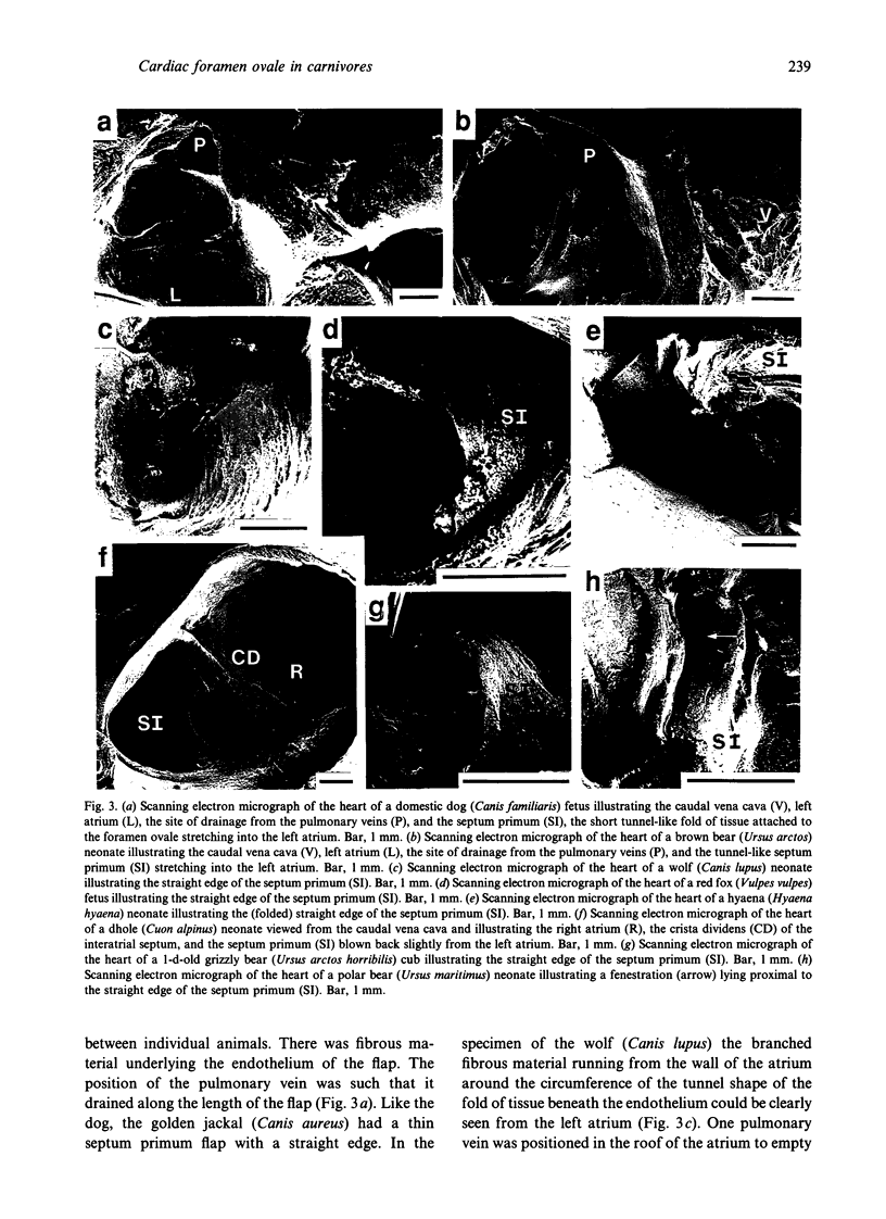

Images in this article
Selected References
These references are in PubMed. This may not be the complete list of references from this article.
- Amoroso E. C., Barclay A. E., Franklin K. J., Prichard M. M. The bifurcation of the posterior caval channel in the eutherian foetal heart. J Anat. 1942 Apr;76(Pt 3):240–247. [PMC free article] [PubMed] [Google Scholar]
- Barclay A. E., Franklin K. J. The time of functional closure of the foramen ovale in the lamb. J Physiol. 1938 Nov 14;94(2):256–258. doi: 10.1113/jphysiol.1938.sp003678. [DOI] [PMC free article] [PubMed] [Google Scholar]
- Edelstone D. I., Rudolph A. M. Preferential streaming of ductus venosus blood to the brain and heart in fetal lambs. Am J Physiol. 1979 Dec;237(6):H724–H729. doi: 10.1152/ajpheart.1979.237.6.H724. [DOI] [PubMed] [Google Scholar]
- Franklin K. J., Barclay A. E., Prichard M. M. Some observations on the cardio-vascular system in the viable foetal lamb. J Anat. 1940 Oct;75(Pt 1):75–87. [PMC free article] [PubMed] [Google Scholar]
- Franklin K. J. Patent foramen ovale and ductus arteriosus in a lion cub. J Anat. 1946 Oct;80(Pt 4):209–209. [PMC free article] [PubMed] [Google Scholar]
- Kiserud T., Eik-Nes S. H., Blaas H. G., Hellevik L. R. Foramen ovale: an ultrasonographic study of its relation to the inferior vena cava, ductus venosus and hepatic veins. Ultrasound Obstet Gynecol. 1992 Nov 1;2(6):389–396. doi: 10.1046/j.1469-0705.1992.02060389.x. [DOI] [PubMed] [Google Scholar]
- MacDonald A. A., Fowden A. L., Silver M., Ousey J., Rossdale P. D. The foramen ovale of the foetal and neonatal foal. Equine Vet J. 1988 Jul;20(4):255–260. doi: 10.1111/j.2042-3306.1988.tb01517.x. [DOI] [PubMed] [Google Scholar]
- Macdonald A. A. Comparative anatomy of the foramen ovale in the Suina. Anat Embryol (Berl) 1988;178(1):53–57. doi: 10.1007/BF00305014. [DOI] [PubMed] [Google Scholar]
- Momma K., Ito T., Mori Y., Yamamura Y. Perinatal adaptation of the cardiovascular system. Early Hum Dev. 1992 Jun-Jul;29(1-3):167–170. doi: 10.1016/0378-3782(92)90133-2. [DOI] [PubMed] [Google Scholar]
- Murakami T., Vamamoto K., Itoshima T., Irino S. Modified tannin-osmium conductive staining method for non-coated scanning electron microscope specimens. Its application to microdissection scanning electron microscopy of the spleen. Arch Histol Jpn. 1977 Feb;40(1):35–40. doi: 10.1679/aohc1950.40.35. [DOI] [PubMed] [Google Scholar]
- Oliveira M. C., Pinto e Silva P., Orsi A. M., Mello Dias S., Define R. M. Observaciones anatómicas sobre el cierre del foramen oval en el perro (Canis familiaris). Anat Histol Embryol. 1980;9(4):321–324. doi: 10.1111/j.1439-0264.1980.tb00919.x. [DOI] [PubMed] [Google Scholar]
- Rudolph A. M. Distribution and regulation of blood flow in the fetal and neonatal lamb. Circ Res. 1985 Dec;57(6):811–821. doi: 10.1161/01.res.57.6.811. [DOI] [PubMed] [Google Scholar]


