

Abstract
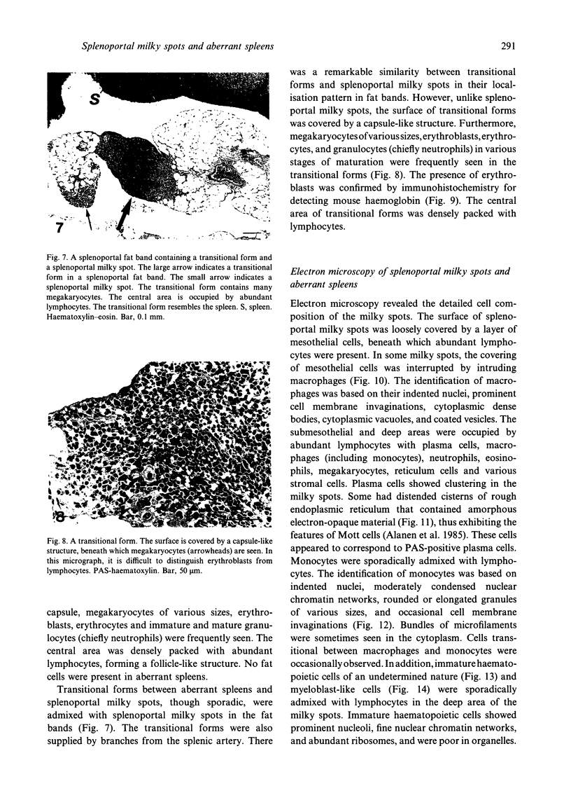
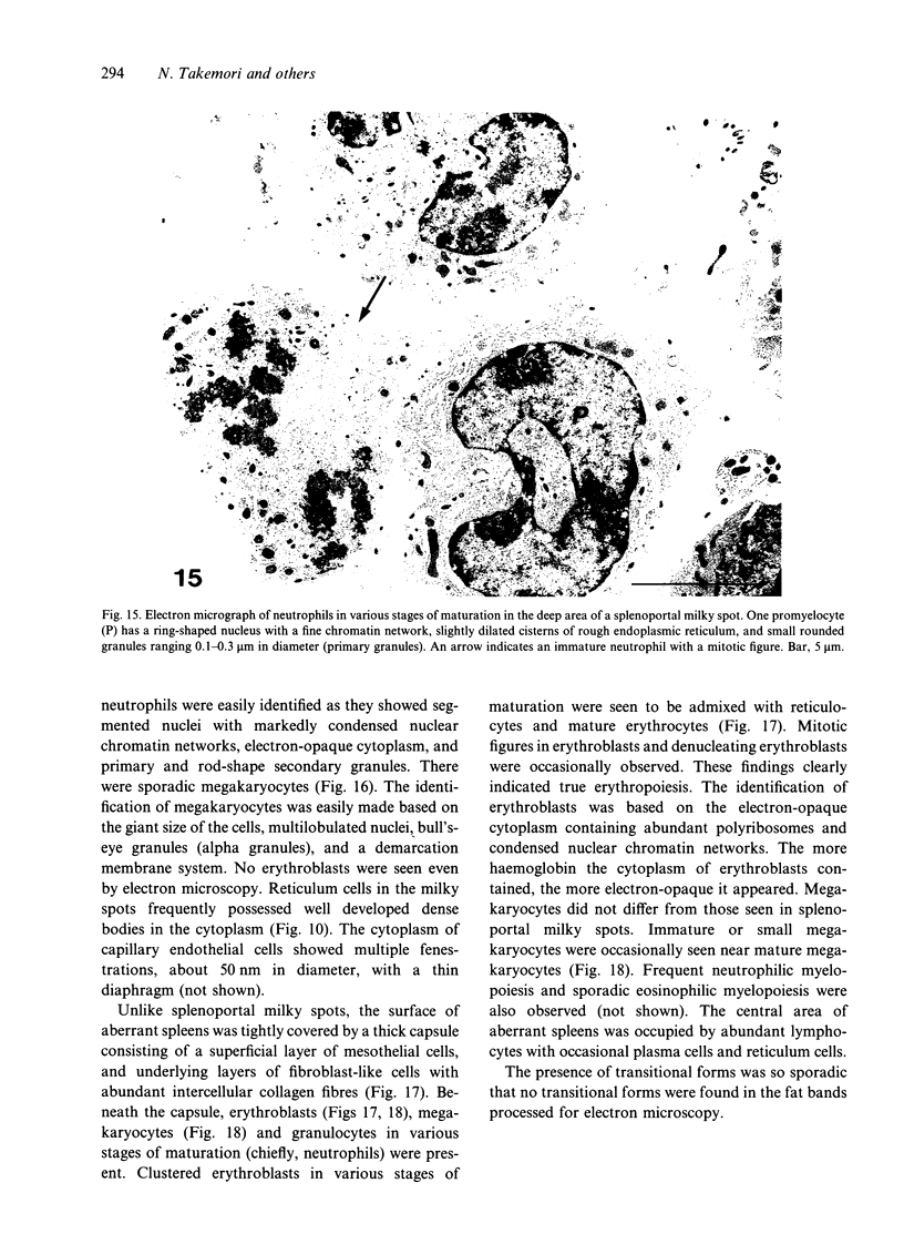
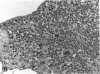
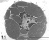
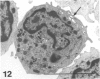
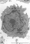
The omentum contains peculiar lymphoid tissues termed omental milky spots. In mice, similar milky spots (splenoportal milky spots) are present in splenoportal fat bands developing along the splenic artery. We found that New Zealand Black (NZB) mice, which are known to develop spontaneous autoimmune diseases, have well developed splenoportal milky spots. However, little is known about these milky spots. Thus we investigated splenoportal fat bands in NZB mice by light and electron microscopy. Splenoportal fat bands contained sporadic aberrant spleens as well as abundant milky spots. In addition, transitional forms between splenoportal milky spots and aberrant spleens, although sporadic, were present in the fat bands. Splenoportal milky spots were supplied with offshoots from the splenic artery and were composed of abundant lymphocytes with macrophages, plasma cells, granulocytes, megakaryocytes and various stromal cells. In addition, they showed active neutrophilic myelopoiesis and probable megakaryopoiesis. Aberrant spleens were also supplied by branches from the splenic artery. They showed active granulopoiesis, megakaryopoiesis, and erythropoiesis. The transitional forms resembled splenoportal milky spots in structure, but the former showed extramedullary haematopoiesis of three cell lineages. The morphological transition from aberrant spleens, via transitional forms, to splenoportal milky spots seems to indicate that splenoportal milky spots represent splenoid lymphoid tissues.
Full text
PDF


Images in this article


Selected References
These references are in PubMed. This may not be the complete list of references from this article.
- Alanen A., Pira U., Lassila O., Roth J., Franklin R. M. Mott cells are plasma cells defective in immunoglobulin secretion. Eur J Immunol. 1985 Mar;15(3):235–242. doi: 10.1002/eji.1830150306. [DOI] [PubMed] [Google Scholar]
- Bartoszewicz W., Dux K. Badania mikroskopowo-elektronowe plamek mlecyncych omentum maius normalnych myszy. Nowotwory. 1968 Jul-Sep;18(3):225–230. [PubMed] [Google Scholar]
- Beelen R. H., Fluitsma D. M., Hoefsmit E. C. Peroxidatic activity of mononuclear phagocytes developing in omentum milky spots. J Reticuloendothel Soc. 1980 Dec;28(6):601–609. [PubMed] [Google Scholar]
- Beelen R. H., Fluitsma D. M., Hoefsmit E. C. The cellular composition of omentum milky spots and the ultrastructure of milky spot macrophages and reticulum cells. J Reticuloendothel Soc. 1980 Dec;28(6):585–599. [PubMed] [Google Scholar]
- Beelen R. H. The greater omentum: physiology and immunological concepts. Neth J Surg. 1991 Oct;43(5):145–149. [PubMed] [Google Scholar]
- Cranshaw M. L., Leak L. V. Milky spots of the omentum: a source of peritoneal cells in the normal and stimulated animal. Arch Histol Cytol. 1990;53 (Suppl):165–177. doi: 10.1679/aohc.53.suppl_165. [DOI] [PubMed] [Google Scholar]
- DeLellis R. A., Sternberger L. A., Mann R. B., Banks P. M., Nakane P. K. Immunoperoxidase technics in diagnostic pathology. Report of a workshop sponsored by the National Cancer Institute. Am J Clin Pathol. 1979 May;71(5):483–488. doi: 10.1093/ajcp/71.5.483. [DOI] [PubMed] [Google Scholar]
- Dux K., Rouse R. V., Kyewski B. Composition of the lymphoid cell populations from omental milky spots during the immune response in C57BL/Ka mice. Eur J Immunol. 1986 Aug;16(8):1029–1032. doi: 10.1002/eji.1830160828. [DOI] [PubMed] [Google Scholar]
- HELYER B. J., HOWIE J. B. Renal disease associated with positive lupus erythematosus tests in a cross-bred strain of mice. Nature. 1963 Jan 12;197:197–197. doi: 10.1038/197197a0. [DOI] [PubMed] [Google Scholar]
- Izui S., McConahey P. J., Dixon F. J. Increased spontaneous polyclonal activation of B lymphocytes in mice with spontaneous autoimmune disease. J Immunol. 1978 Dec;121(6):2213–2219. [PubMed] [Google Scholar]
- Koten J. W., den Otter W. Are omental milky spots an intestinal thymus? Lancet. 1991 Nov 9;338(8776):1189–1190. doi: 10.1016/0140-6736(91)92043-2. [DOI] [PubMed] [Google Scholar]
- Minoda M., Horiuchi A. The function of thymic reticuloepithelial cells in New Zealand mice. Thymus. 1983 Sep;5(5-6):363–374. [PubMed] [Google Scholar]
- Phillips J. A., Mehta K., Fernandez C., Raveché E. S. The NZB mouse as a model for chronic lymphocytic leukemia. Cancer Res. 1992 Jan 15;52(2):437–443. [PubMed] [Google Scholar]
- Raveche E. S., Laskin C. A., Rubin C., Tjio J. H., Steinberg A. D. Comparison of stem-cell recovery in autoimmune and normal strains. Cell Immunol. 1983 Jul 1;79(1):56–67. doi: 10.1016/0008-8749(83)90050-3. [DOI] [PubMed] [Google Scholar]
- Reininger L., Shibata T., Schurmans S., Merino R., Fossati L., Lacour M., Izui S. Spontaneous production of anti-mouse red blood cell autoantibodies is independent of the polyclonal activation in NZB mice. Eur J Immunol. 1990 Nov;20(11):2405–2410. doi: 10.1002/eji.1830201107. [DOI] [PubMed] [Google Scholar]
- Seifert M. F., Marks S. C., Jr The regulation of hemopoiesis in the spleen. Experientia. 1985 Feb 15;41(2):192–199. doi: 10.1007/BF02002613. [DOI] [PubMed] [Google Scholar]
- Shimotsuma M., Takahashi T., Kawata M., Dux K. Cellular subsets of the milky spots in the human greater omentum. Cell Tissue Res. 1991 Jun;264(3):599–601. doi: 10.1007/BF00319049. [DOI] [PubMed] [Google Scholar]
- Takemori N., Hirai K., Onodera R., Saito N., Namiki M. Light and electron microscopic study of omental milky spots in New Zealand black mice, with special reference to the extramedullary hematopoiesis. Anat Embryol (Berl) 1994 Mar;189(3):215–226. doi: 10.1007/BF00239009. [DOI] [PubMed] [Google Scholar]
- Takemori N., Ito T. [Response of omental milk spots to colloidal saccharated ferric oxide in the mouse: light and electron microscopic study (author's transl)]. Hokkaido Igaku Zasshi. 1981 Mar;56(2):199–216. [PubMed] [Google Scholar]
- Takemori N., Ito T. [Uptake of horseradish peroxidase (HRP) in omental milk spot cells of the mouse: an electron microscope study (author's transl)]. Hokkaido Igaku Zasshi. 1980 Sep;55(5):409–418. [PubMed] [Google Scholar]
- Takemori N. [Histogenesis of the omental milk spot of the mouse (author's transl)]. Hokkaido Igaku Zasshi. 1980 May;55(3):223–234. [PubMed] [Google Scholar]
- Takemori N. [Milk spots on the parietal peritoneum over the pancreas in the mouse (author's transl)]. Hokkaido Igaku Zasshi. 1979 Jul;54(4):379–385. [PubMed] [Google Scholar]
- Takemori N. [Morphological studies of the omental milk spots in the mouse: light and electron microscopy (author's transl)]. Hokkaido Igaku Zasshi. 1979 May;54(3):265–283. [PubMed] [Google Scholar]