Abstract
Background
Oxygen therapy is commonly administered to patients with acute cardiovascular conditions during hospitalisation. Both hypoxaemia and hyperoxia can cause harm, making it essential to maintain oxygen saturation (SpO2) within a target range. Traditionally, oxygen administration is manually controlled by nursing staff, guided by intermittent pulse oximetry readings. This study aimed to compare standard manual oxygen administration with automated oxygen administration (AOA) using the O2matic device.
Methods
In this randomised controlled trial, 60 patients admitted to a cardiac department with an acute cardiovascular condition requiring oxygen therapy were randomised to either standard care (manual oxygen administration) or AOA via the O2matic device. The primary outcome was the percentage of time spent within the desired SpO2 range (92%–96% or 94%–98%) over 24 hours.
Results
Patients had a mean age of 75.8±12.4 years, with an average SpO2 of 93%. Those in the AOA group (n=25) spent significantly more time within the target SpO2 range (median 87.0% vs 60.6%, p<0.001) compared with the standard care group (n=28). Time spent below the desired SpO2 range was significantly lower in the AOA group (7.9% vs 33.6%, p<0.001). No significant differences in time spent above the desired SpO2 range were observed between the two groups.
Conclusions
AOA with the O2matic device is superior to standard manual control in maintaining SpO2 within the target range in patients hospitalised with acute cardiovascular conditions. The automated systems significantly reduce the time spent in hypoxaemia without increasing hyperoxia.
Trial registration number
Keywords: heart failure
WHAT IS ALREADY KNOWN ON THIS TOPIC
Oxygen administration is a critical component in the management of acute cardiovascular conditions; however, both insufficient oxygen (hypoxaemia) and excessive oxygen (hyperoxia) can be harmful, and maintaining normoxaemia is recommended.
Traditionally, oxygen delivery is controlled manually by nursing staff, but this method can result in variations in oxygen levels between measurements, potentially leading to episodes of hypoxaemia or hyperoxia.
WHAT THIS STUDY ADDS
This randomised controlled trial demonstrates that automated oxygen administration (AOA) using the O2matic device is superior to manual control in maintaining SpO2 within the desired range.
Patients in the AOA group spent significantly more time within the target SpO2 range and had less time in hypoxaemia compared with those receiving standard manual care.
AOA effectively minimises fluctuations in oxygen levels, ensuring more precise oxygen delivery without increasing the risk of hyperoxia.
HOW THIS STUDY MIGHT AFFECT RESEARCH, PRACTICE OR POLICY
The use of AOA systems like O2matic might reduce the burden on nursing staff and minimise the risk of hypoxaemia, potentially leading to better clinical outcomes and more efficient oxygen therapy management in hospital settings.
Introduction
The treatment of hypoxaemia with oxygen supplementation is considered an essential part of the treatment of patients with acute cardiac conditions.1 Oxygen is frequently administered during in-hospital treatment of patients admitted with cardiac conditions. Hypoxaemia is associated with adverse outcomes in patients with myocardial infarction.2 Hyperoxia causes vasoconstriction and may induce direct damage in conditions such as acute coronary syndrome, heart failure and ischaemic stroke.3,6
Clinical evidence on how to control oxygen supply to avoid hypoxia and hyperoxia is limited.1 Supplemental oxygen is often guided by a non-invasive method such as pulse oximetry (peripheral capillary oxygen saturation, SpO2) or by blood gas analyses. Early warning score (EWS) is calculated from measured values of blood pressure, pulse, respiratory frequency, temperature and peripheral saturation. It is often used to determine the level of monitoring of the patients, including SpO2 measurement.7 The control frequency increases with increasing EWS score. This method with observation and adjustment is time-consuming for the nursing staff and leaves room for severe hypoxaemia or hyperoxia between measurements, which are only snapshots of the patient’s condition. On this background, several research groups have worked on methods to adjust oxygen supply automatically based on SpO2 measurements.8,10
O2matic is a closed-loop automated oxygen administration (AOA) system, where oxygen is administered through the device based on continuous SpO2 measurement. This both ensures minute-to-minute titration of oxygen to reach the desired interval and limits the need for nurses to measure and adjust oxygen manually. Previous studies have demonstrated that O2matic is feasible and superior at keeping the saturation within the desired interval.11 The device is CE marked and manufactured in a version operating in conformity with the demands stated by the Medical Device Directives. O2matic was tested in a crossover trial, comparing manually controlled oxygen treatment with AOA in patients admitted with chronic obstructive pulmonary disease (COPD) exacerbations. The study demonstrated a significantly better oxygen regulation with O2matic (85% vs 47% of the time within the predefined interval).11 In a parallel designed study on patients admitted with COPD exacerbation, treatment with O2matic reduced breathing discomfort and physical perception of dyspnoea compared with nurse-administered oxygen therapy.12 However, no randomised controlled trial has compared O2matic with standard nurse-titrated oxygen administration in hospitalised patients with cardiovascular disease.
The primary aim of this study was to investigate whether AOA with O2matic is superior to conventional control at keeping the oxygen saturation within the desired interval. Secondarily, the study aimed to investigate whether O2matic reduces the time spent in significant hypoxaemia or hyperoxia in acutely hospitalised cardiac patients with oxygen demand.
Our hypothesis was that O2matic significantly increases the duration of time where SpO2 is within the selected saturation interval.
Methods
Study design, setting and population
The study is a prospective, investigator-initiated, parallel-group, randomised, clinical trial. Patients were included in the Department of Cardiology at Hvidovre Hospital, Copenhagen, Denmark, after acute admission with a primary cardiac disease and in need of oxygen. We included patients admitted with heart disease in need of oxygen supplementation, defined as an SpO2 <92%. National and international guidelines recommend initiation of oxygen treatment at different levels of SpO2, between 90% and 94%, depending on the medical condition of the patient.14 13,15 We excluded patients with risk of hypercapnia, unstable patients and pregnant women. Non-compliant patients (defined as an intervention period <2 hours) were excluded from the main analysis. A complete list of inclusion and exclusion criteria can be found in detail in the protocol previously published (onlinesupplemental files 4 5).16 Inclusion began on 1 April 2022, and concluded after 11 months, after the last patient was included. The study is registered at ClinicalTrials.gov (identifier: NCT05452863).
Randomisation and masking
The randomisation module in Research Electronic Data Capture (REDCap) (REDCap Consortium, Nashville, Tennessee, USA) was used for randomisation and all data were registered in their scientific database.17 The database was hosted on the servers of the Capital Region of Copenhagen, with secured back up and double protected with a two-factor authentication. A computer-generated randomisation sequence was created by one of the main investigators and uploaded to REDCap before trial initiation. It was concealed from other investigators, patients and clinical personnel until randomisation. Subinvestigators enrolled and randomised patients consecutively after the trial initiation.
It was not possible to blind the investigators or the clinical staff regarding allocation, as one group depended on nurses to titrate the oxygen supply. The screen was turned off for as much time as possible in the control group during the trial to mimic clinical practice.
Study procedure
The patients were included during admission and randomised in a 1:1 ratio to conventional oxygen treatment or O2matic oxygen treatment for 24 hours. The O2matic device was applied to all patients, but in the control group a manual mode was selected to allow for a usual care oxygen titration by the nurses in the department. Oxygen flow rate, SpO2 and pulse rate was registered every second by the device. All patients were monitored using pulse oximetry connected to the O2matic.18 In the active group, oxygen supply was adjusted according to the measurements made by pulse oximetry. Oxygen was adjusted from 0 to 10 L/min to reach a predefined target saturation interval of either 92%–96% or 94%–98%, as determined by the treating physician. As standard practice, we used a nasal cannula without humidifying the oxygen.
Alarms for saturation, pulse and oxygen flow were turned off in the control group, while technical alarms were still active. In the control group, oxygen treatment was performed with manual saturation measurements with a standard pulse oximeter, via the EWS standard and the oxygen supply was thereafter adjusted on the O2matic device, according to EWS guidelines19 and clinical judgement by doctors and nursing staff. Manual override was possible for patients in the active group, if automatic adjustments were considered inappropriate by the clinicians, for example, in acute need for higher oxygen supply than the set interval.
Patients in both groups were manually monitored for saturation, pulse and other vital signs according to the EWS guidelines by the nursing staff.7 The frequency of measurements and optional medical assessment was directed by the EWS guidelines.19
Monitoring
At the time of inclusion, we registered baseline characteristics, including smoking history, X-ray from the current admission, biochemistry and arterial blood gas. A detailed list of data recorded can be found in the previously published protocol.16 Our main goal was to test the feasibility of O2matic in relation to maintaining the saturation within the preferred SpO2 interval. Clinical end points were not the focus of this study but was recorded as measures for safety. We stopped the intervention after one of the following: after 24 hours, if the patient was moved to another department, if the patient was discharged or died, if the patient withdrew consent, if the patient was not compliant (defined as a treatment period under 2 hours) or if any serious adverse event was suspected.
Outcome measures
The primary outcome was time within the desired saturation interval (SpO2 92%–96% or 94%–98%) when using O2matic, compared with manual oxygen treatment. The primary outcome was changed from SpO2 92%–96% to either SpO2 92%–96% or 94%–98% for each patient, to accommodate different preferences in target saturation among the treating physicians before enrolment was concluded.
Secondary outcomes:
Time with hypoxaemia below the desired interval.
Time with hyperoxia above the desired interval.
Time with severe hypoxaemia (saturation <85%).
Time with significant hypoxaemia (saturation 85%–90%).
Pulse rate.
Subgroups
We compared patients with and without systolic heart failure, defined as left ventricular ejection fraction (LVEF) <45%, and patients with and without supraventricular arrythmias.
Ethics
Eight papers previously published have not revealed any health risks for patients on similar equipment.8,1020 The patient’s mobility was slightly restricted because they were connected to the O2matic, but we considered this inconvenience minor compared with the potential therapeutic benefits of secure and optimised oxygen treatment.
Patient and public involvement
Our research group has a patient group dedicated to give feedback and suggestions in the design and conduct for all clinical studies carried out. They are involved in topics such as trial information material and clinical end points.
Sample size
We calculated the sample size based on an expected 20% improvement in the primary outcome to ensure clinical relevance. In a Danish study, the SD for this parameter was 25%.11 A power of 80% and a level of significance at 0.05 required 25 participants in each group. We chose to include 30 patients in each arm to allow for dropouts.
Statistical analysis plan
Categorical data were compared with Fisher’s exact test. Continuous variables were tested for normality and analysed with unpaired t-test when normally distributed, or Wilcoxon-Mann-Whitney test, in case of a non-normal distribution. Normally distributed data are presented as mean±SD, non-normally distributed data are presented as median (IQR). The primary analysis was defined as a modified intention-to-treat analysis, as non-compliant patients were excluded postrandomisation. We did not have power to assess clinical end points, which is why these must be considered exploratory.
Between-group differences in SpO2, oxygen administration and pulse rate measured for 24 hours were assessed by repeated-measures mixed models with an unstructured covariance structure. Group and time point, defined as the median values for each hour of the intervention, were treated as fixed effects, while time was considered a continuous variable. The interaction term of group with time was included in the model. The output was used to illustrate SpO2 and oxygen administration during the intervention phase. Fraction of inspired oxygen (FiO2) was calculated as 20+4×oxygen supplementation (L/min).25 P values were denoted as p-group. Statistical analyses were made using R, V.4.3.0 and SAS statistical software, V.9.4 (SAS Institute, Cary, North Carolina, USA). All tests were two-tailed, and statistical significance was defined as p<0.05.
Results
A total of 60 patients admitted to the Department of Cardiology were included in the study between 1 April 2022 and 17 March 2023, and were randomised to receive AOA or standard care for 24 hours (n=29 and n=31, respectively). Seven patients were excluded from the primary outcome analysis due to lack of compliance with the O2matic (treated via the device for <2 hours, median 1.2 (0.4–1.5) hours). Four of these patients were in the AOA group. The median intervention period for the remaining group was 19.3 (12.2–22.7) hours, 21.9 (16.7–23.2) hours in the AOA group and 17.5 (11.7–20.0) hours in the standard care group (p=0.04). Reasons for cessation are listed in table 1. The mean age was 75.8±12.4 and 56% were women. Acute heart failure was the most frequent admission diagnosis (65% of patients). The average SpO2 was 93%±2.47% and patients received an average of 2.4±1.2 L/min of supplemental oxygen. The baseline characteristics, including primary diagnosis and comorbidities, were balanced and only the presence of pleural effusion on X-ray prior to inclusion differed with 4 (13.8%) in the AOA group vs 0 in the standard care group, p=0.049. All baseline characteristics are presented in table 2.
Table 1. Reasons for cessation of the therapy.
| Reason for cessation | Number of patients |
| Intervention period completed | 37 (62%) |
| Patient no longer wanted to participate | 2 (3%) |
| Patient was non-compliant | 6 (10%) |
| Patient was transferred to another department | 2 (3%) |
| Unknown | 13 (22%) |
Data are n (%).
Table 2. Baseline characteristics of the intention-to-treat population.
| AOAN=29 | Standard careN=31 | |
| Age (years) | 77.2±12.4 | 74.5±12.5 |
| Gender female | 16 (57.1%) | 17 (54.8%) |
| Height (cm) | 171.4±11.2 | 168.4±10.0 |
| Weight (kg) | 85.8±31.2 | 80.4±20.7 |
| BMI (kg/m2) | 28.7±8.5 | 28.1±6.4 |
| LVEF (%) | 45 (31–60) | 55 (40–60) |
| Primary diagnosis at admission | ||
| Heart failure | 21 (72.4%) | 18 (58.1%) |
| Lung embolism | 2 (6.9%) | 3 (9.7%) |
| Other | 6 (20.7%) | 10 (32.2%) |
| Comorbidities | ||
| None | 6 (20.7%) | 4 (12.9%) |
| AF | 9 (31.0%) | 8 (25.8%) |
| IHD | 4 (13.8%) | 7 (22.6%) |
| HF | 15 (51.7%) | 10 (32.3%) |
| DM | 5 (17.2%) | 3 (9.7%) |
| Cancer | 2 (6.9%) | 3 (9.7%) |
| COPD | 3 (10.3%) | 1 (3.2%) |
| Asthma | 1 (3.5%) | 0 |
| Other lung disease | 0 | 2 (6.5%) |
| SpO2 at inclusion time | 93.1±2.3 | 93.0±2.7 |
| O2 supplement at inclusion (L/min) | 2.3±1.2 | 2.4±1.2 |
| Rhythm at intervention start | ||
| Sinus rhythm | 21 (72.4%) | 17 (56.7%) |
| Supraventricular tachycardia | 6 (20.7%) | 10 (33.3%) |
| Other | 2 (6.9%) | 3 (10.0%) |
| Chest X-ray findings | ||
| Pulmonary congestion | 14 (48.3%) | 7 (22.6%) |
| Pleural effusion | 4 (13.8%) | 0 |
Data are n (%), median (IQR) or mean±SD.
Other primary diagnosis: Aaortic dissection, pneumonia, endocarditis, infection, arrythmias, COPD exacerbation. Other lung disease: PAH, sleep apnoea. Other rhythms: Ppaced rhythms or bradycardias.
AF, atrial fibrillation or flutter; AOAautomated oxygen administrationBMI, body mass index; COPD, chronic obstructive pulmonary diseaseDM, diabetes mellitus; HF, heart failure; IHD, ischaemic heart disease; LVEF, left ventricular ejection fractionPAHpulmonary arterial hypertension
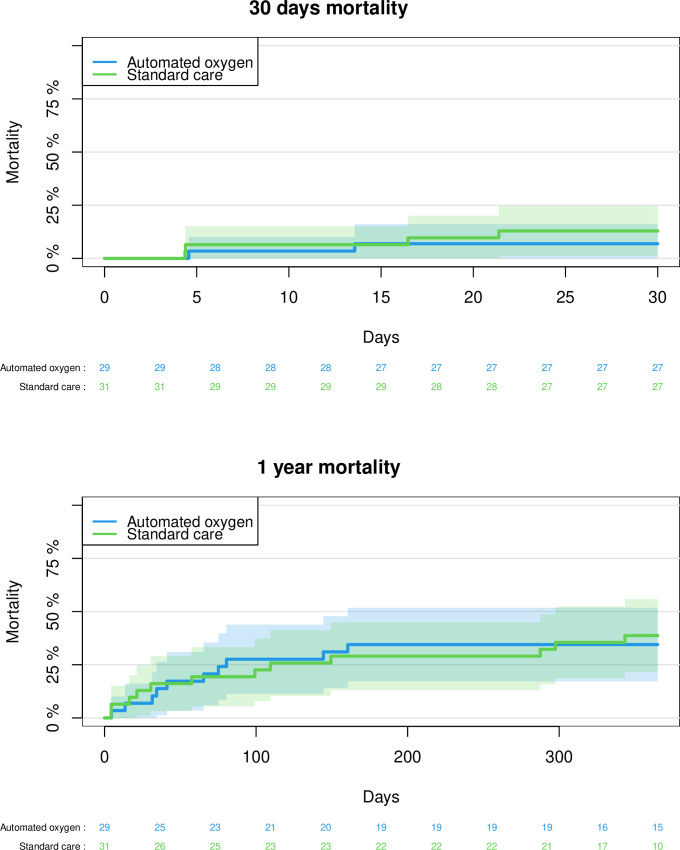
To assess the safety of AOA, we did analysis on mortality, hospitalisation and complications (table 3). There was no difference in mortality, readmission rate or days alive and out of hospital. Figure 1 demonstrates Kaplan-Maier curves for 30 and 365 days all-cause mortality. There was no increase in the rate of pneumonia or the need for assisted ventilation during admission. We also assessed vital signs after the 24-hour intervention without finding a difference between the two groups.
Table 3. Clinical end points.
| AOAN=29 | Standard careN=31 | P value | |
| Death within 30 days | 2 (6.9%) | 5 (16.1%) | 0.43 |
| Readmission within 30 days | 7 (28%) | 7 (25%) | 1 |
| Days alive and out of hospital 30 days | 24.0 (15–26) | 21.0 (17–26) | 0.80 |
| EWS | 3 (2–4) | 2 (2–4) | 0.47 |
| Systolic BP (mm Hg) | 128.2±21.0 | 135.9±21.4 | 0.16 |
| Diastolic BP (mm Hg) | 74.9±12.3 | 73.0±11.9 | 0.55 |
| RF (/min) | 18 (17–20) | 18 (17–20) | 1 |
| Pneumonia during admission | 3 (10.3%) | 3 (10.0%) | 1 |
| Ventilatory assistance during admission | 2 (6.9%) | 1 (3.2%) | 0.61 |
| Δ Haemoglobin | −0.04±0.46 | −0.05±0.44 | 0.95 |
| Δ C reactive protein | 4.20±46.38 | 1.05±40.64 | 0.86 |
Data are n (%), median (IQR) or mean±SD. P values were calculated using an unpaired t-test for normally distributed data, the Wilcoxon-Mann-Whitney test for non-normally distributed data and Fisher’s exact test for categorical dataP-values was calculated with unpaired t-test when normally distributed, Wilcoxon-Mann-Whitney test, when non-normally distributed and Fishers exact test when categorical.
EWS, ssystolic BP, ddiastolic BP and RF was recorded after the 24-hour intervention.
AOAautomated oxygen administrationBPblood pressureEWSearly warning scoreRFrespiratory frequency
Figure 1. Kaplan-Maier mortality analysis.
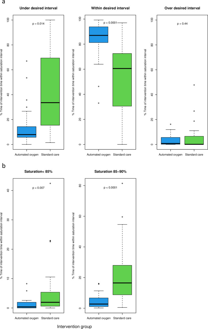
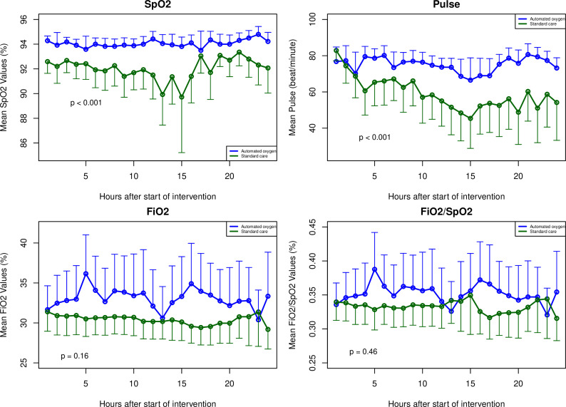
In the AOA group, SpO2 was maintained within the prespecified interval for a median of 87.0% (81.3%–93.8%) of the time, compared with 60.6% (32.4%–71.8%) of the time in the standard care group (p<0.0001) (figure 2a). In an intention-to-treat analysis, we found similar results for time within the desired interval (online supplemental table I). The top left graph in figure 4 shows the mean SpO2 at every hour of the intervention in the two groups. SpO2 was significantly higher in the intervention group over time (p=0.0001), but there was no significant difference in FiO2 or FiO2/SpO2 over time. Time with SpO2 below the desired interval was significantly longer in the standard care group than in the AOA group (33.6% (15.5%–67.6%) vs 7.9% (5.7%–14.3%) of the time, p=0.0007). There was no statistically significant difference in time above the desired interval (median 0.8% (0.14%–5.65%) vs 0.1% (0.0%–6.49%), p=0.27). We found similar results in an intention-to-treat analysis for both time below and time above the desired interval (online supplemental table I).
Figure 2. (a) Time within and outside desired saturation interval. Automated oxygen: n=25, standard care: n=28. P values were calculated using the Wilcoxon-Mann-Whitney test. (b) Time with clinically significant desaturation. Automated oxygen: n=25, standard care: n=28. P values were calculated using the Wilcoxon-Mann-Whitney test.
Time within different saturation levels is demonstrated in figure 2b. Time with both severe hypoxaemia (defined as saturation <85%) and significant hypoxaemia (defined as saturation 85%–90%) was significantly longer in the standard care group compared with AOA (table 4). Figure 3 shows the mean pulse, FiO2 and FiO2/SpO2 at every hour of the intervention in the two groups. Pulse was significantly lower in the standard care group over time (p=0.0003).
Table 4. Time within saturation intervals (% time of the total intervention time).
| AOAN=25 | Standard careN=28 | P value | |
| Saturation <85% | 0.37% (0.01%–1.95%) | 1.9% (0.62%–4.2%) | 0.0069 |
| Saturation 85%–90% | 2.7% (1.1%–6.71%) | 16.5% (9.35%–27.5%) | <0.0001 |
P- values wereas calculated withusing the Wilcoxon-Mann-Whitney test.
AOAautomated oxygen administration
Figure 3. Mean values of SpO2, pulse, FiO2 and FiO2/SpO2 at each hour of the intervention. FiO2, fraction of inspired oxygen; SpO2, oxygen saturation. Automated oxygen: n=25, standard care: n=28. P values were calculated using repeated-measures mixed models with an unstructured covariance structure.
When performing the main analysis on the subgroup with supraventricular tachycardia (SVT) (n=15), time within desired interval remained significantly longer in the AOA group compared with the standard care group, while time under desired interval was not significantly shorter in the AOA group (online supplemental table II).
When performing the main analysis on the subgroup with LVEF <45% (n=17), there was no difference in time within, time below or time above the desired interval between the AOA group and the standard care group (online supplemental table II). See onlinesupplemental figures I IIundefined for box plots of time within the different intervals. We did not find a significant interaction when fitting the variables into a linear model (p=0.24 for SVT and p=0.08 for LVEF <45%).
Discussion
In this randomised clinical trial, we demonstrated that oxygen treatment with an automated feedback system (O2matic) is superior to standard care at keeping saturation within a predefined interval. Patients in the AOA group had significantly less time with hypoxia and severe hypoxia compared with the standard care group. There was no difference in the rate of hyperoxia between the groups. Furthermore, we found no evidence of increased harm, as death and readmission were numerically higher in the control group. In the interval below 90%, omitting fluctuations around the lower limit of the desired saturation interval, we also found a highly significant difference. AOA is not used routinely and there are still no clinical trials with enough power to evaluate parameters of health economics or clinical end points. A closed loop system (FreeO2), similar to O2matic, was tested in a study on patients with COPD exacerbations, with 25 patients in each group. In accordance with our study, FreeO2 was significantly superior at keeping the saturation within the desired interval (81% vs 51%).21 They also found a non-significant reduction in the duration of the hospital stay (2.6 days, p=0.051) and the duration of oxygen treatment (1.8 days, p=0.14).
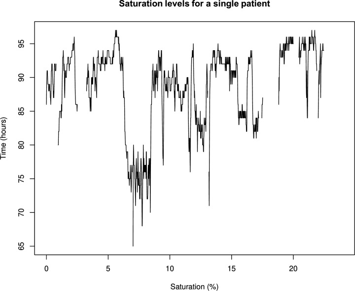
In the absence of arterial hypoxaemia, patients are sometimes still treated with oxygen due to the belief that it will improve organ oxygenation.26 Although the indication for oxygen treatment in the absence of severe hypoxaemia is unclear, there is consensus on the treatment of severe hypoxaemia.1 In the control group in our study, one patient experienced severe hypoxaemia for 2 hours during the night (figure 4), with saturation levels between 65% and 85% (mean 76%). Such prolonged and potentially harmful events would be less likely to occur with an AOA system, as the oxygen supply would automatically increase, and an alarm would alert the nursing staff to the situation.
Figure 4. Example of a prolonged period of nocturnal hypoxaemia.
Preventing hyperoxia could be pivotal for individuals with cardiac disease, as it is thought to induce systemic vasoconstriction, affecting the myocardium and thereby reducing cardiac output.27 Several physiological studies have highlighted the detrimental effects of hyperoxia.28 In this study, we found no significant difference in hyperoxia between the groups.
The primary limitation of our study is the sample size, which limited our power to test relevant clinical outcomes. Second, the study was not blinded, as the nurses needed to know which patients required standard care. Third, our population comprised a heterogeneous group of patients with different cardiac conditions. We did not find the same results in the subgroups with SVT and LVEF <45%. In both subgroups, the small sample size should be taken into consideration. The primary end points were analysed using a modified intention-to-treat approach, excluding seven patients who were included but did not provide sufficient data for the primary end point analysis, which poses a potential risk of selection bias.
Future trials should investigate whether the use of AOA systems like O2matic could reduce the burden on nursing staff or lead to better clinical outcomes and more efficient oxygen therapy management in hospital settings. In this trial, we did not collect data to evaluate whether nurses saved time when using O2matic. AOA has a potential to improve oxygen treatment for individual patients by delivering personalised treatment that allows for adjustments based on fluctuations in oxygen requirements from minute to minute—something that can be challenging for the nursing staff to manage. Furthermore, whether the improved oxygenation and avoidance of hypoxaemia translates to clinical important outcomes needs to be validated in larger studies powered for clinically relevant end points.
Conclusion
In this randomised clinical trial, we demonstrated that oxygen treatment with an automated feedback system (O2matic) is superior to standard of care at keeping saturation within a predefined desired interval in patients hospitalised due to an acute cardiovascular condition. We also found significantly fewer events with clinically significant and severe hypoxaemia.
supplementary material
The funder of the study had no role in study design, data collection, data analysis, data interpretation or writing of the report.
Footnotes
Funding: JG’s salary during this project is supported by a research grant from the Danish Cardiovascular Academy funded by Novo Nordisk Foundation (grant number NNF20SA0067242) and by The Danish Heart Foundation. IAT’s salary during this project is supported by a research grant from Gangstedfonden (grant number A43194).
Provenance and peer review: Not commissioned; externally peer reviewed.
Patient consent for publication: Not applicable.
Ethics approval: The study was approved by the Scientific Ethical Committee (H-19033702) and the Data Protection Authorities in Denmark (P-2019-369). Participants gave informed consent to participate in the study before taking part. Participants could at any point withdraw from the study and were covered by patient insurance at Hvidovre Hospital.
Data availability free text: Deidentified participant data can be provided from the main author (orchid id: 0000-0002-3574-243X).
Patient and public involvement: Patients and/or the public were involved in the design, or conduct, or reporting, or dissemination plans of this research. Refer to the 'Methods' section for further details.
Contributor Information
Ida Arentz Taraldsen, Email: ida.arentz.taraldsen.01@regionh.dk.
Johannes Grand, Email: johannes.grand@regionh.dk.
Jasmin Dam Lukoschewitz, Email: jasmin.dam.lukoschewitz@regionh.dk.
Ekim Seven, Email: ekim.seven@regionh.dk.
Ulrik Dixen, Email: Ulrik.Dixen@regionh.dk.
Morten Petersen, Email: morten.petersen@regionh.dk.
Laura Rytoft, Email: laura.amalie.rytoft@regionh.dk.
Marie Munk Jakobsen, Email: mariemunk_jakobsen@hotmail.com.
Ejvind Frausing Hansen, Email: Ejvind.Frausing.Hansen@regionh.dk.
Jens Dahlgaard Hove, Email: Jens.Dahlgaard.Hove@regionh.dk.
Data availability statement
Data are available on reasonable request.
References
- 1.McDonagh TA, Metra M, Adamo M, et al. 2021 ESC Guidelines for the diagnosis and treatment of acute and chronic heart failure. Eur Heart J. 2021;42:3599–726. doi: 10.1093/eurheartj/ehab368. [DOI] [PubMed] [Google Scholar]
- 2.James SK, Erlinge D, Herlitz J, et al. Effect of Oxygen Therapy on Cardiovascular Outcomes in Relation to Baseline Oxygen Saturation. JACC Cardiovasc Interv. 2020;13:502–13. doi: 10.1016/j.jcin.2019.09.016. [DOI] [PubMed] [Google Scholar]
- 3.Cornet AD, Kooter AJ, Peters MJL, et al. The potential harm of oxygen therapy in medical emergencies. Crit Care . 2013;17:313. doi: 10.1186/cc12554. [DOI] [PMC free article] [PubMed] [Google Scholar]
- 4.Siemieniuk RAC, Chu DK, Kim LH-Y, et al. Oxygen therapy for acutely ill medical patients: a clinical practice guideline. BMJ. 2018;363:k4169. doi: 10.1136/bmj.k4169. [DOI] [PubMed] [Google Scholar]
- 5.Chu DK, Kim LH-Y, Young PJ, et al. Mortality and morbidity in acutely ill adults treated with liberal versus conservative oxygen therapy (IOTA): a systematic review and meta-analysis. Lancet. 2018;391:1693–705. doi: 10.1016/S0140-6736(18)30479-3. [DOI] [PubMed] [Google Scholar]
- 6.Hofmann R, James SK, Jernberg T, et al. Oxygen Therapy in Suspected Acute Myocardial Infarction. N Engl J Med. 2017;377:1240–9. doi: 10.1056/NEJMoa1706222. [DOI] [PubMed] [Google Scholar]
- 7.Nielsen PB, Pedersen NE, Schultz M, et al. Review of Early Warning Score in preventing sudden critical illness and death. Ugeskr Laeger . 2018;180:V02180135. [PubMed] [Google Scholar]
- 8.Rice KL, Schmidt MF, Buan JS, et al. AccuO2 oximetry-driven oxygen-conserving device versus fixed-dose oxygen devices in stable COPD patients. Respir Care. 2011;56:1901–5. [PubMed] [Google Scholar]
- 9.Cirio S, Nava S. Pilot study of a new device to titrate oxygen flow in hypoxic patients on long-term oxygen therapy. Respir Care. 2011;56:429–34. doi: 10.4187/respcare.00983. [DOI] [PubMed] [Google Scholar]
- 10.Lellouche F, L’her E. Automated oxygen flow titration to maintain constant oxygenation. Respir Care. 2012;57:1254–62. doi: 10.4187/respcare.01343. [DOI] [PubMed] [Google Scholar]
- 11.Hansen EF, Hove JD, Bech CS, et al. Automated oxygen control with O2matic® during admission with exacerbation of COPD. Int J Chron Obstruct Pulmon Dis. 2018;13:3997–4003. doi: 10.2147/COPD.S183762. [DOI] [PMC free article] [PubMed] [Google Scholar]
- 12.Sandau C, Hansen EF, Ringbæk TJ, et al. Automated Oxygen Administration Alleviates Dyspnea in Patients Admitted with Acute Exacerbation of COPD: A Randomized Controlled Trial. Int J Chron Obstruct Pulmon Dis. 2023;18:599–614. doi: 10.2147/COPD.S397782. [DOI] [PMC free article] [PubMed] [Google Scholar]
- 13.O’Driscoll BR, Howard LS, Earis J, et al. British Thoracic Society Guideline for oxygen use in adults in healthcare and emergency settings. BMJ Open Respir Res. 2017;4:e000170. doi: 10.1136/bmjresp-2016-000170. [DOI] [PMC free article] [PubMed] [Google Scholar]
- 14.National Klinisk Retningslinje for iltbehandling til den akut syge voksne patient - ikke gældende. https://app.magicapp.org/#/guideline/6899 n.d. Available.
- 15.Beasley R, Chien J, Douglas J, et al. Thoracic Society of Australia and New Zealand oxygen guidelines for acute oxygen use in adults: “Swimming between the flags.”. Respirology. 2015;20:1182–91. doi: 10.1111/resp.12620. [DOI] [PMC free article] [PubMed] [Google Scholar]
- 16.Taraldsen IA, Grand J, Lukoschewitz JD, et al. Automated versus manual oxygen administration for patients admitted with acute cardiovascular disease - a study protocol of a randomised controlled trial. Dan Med J. 2023;70:A12220784. [PubMed] [Google Scholar]
- 17.Dunn WD, Cobb J, Levey AI, et al. REDLetr: Workflow and tools to support the migration of legacy clinical data capture systems to REDCap. Int J Med Inform. 2016;93:103–10. doi: 10.1016/j.ijmedinf.2016.06.015. [DOI] [PMC free article] [PubMed] [Google Scholar]
- 18.Bernard SL, An D, Glenny RW. Validation of the Nonin 8600V Pulse Oximeter for heart rate and oxygen saturation measurements in rats. Contemp Top Lab Anim Sci. 2004;43:43–5. [PubMed] [Google Scholar]
- 19.RCP London National Early Warning Score (NEWS) 2. 2017. https://www.rcplondon.ac.uk/projects/outputs/national-early-warning-score-news-2 Available.
- 20.Johannigman JA, Branson R, Lecroy D, et al. Autonomous Control of Inspired Oxygen Concentration During Mechanical Ventilation of the Critically Injured Trauma Patient. J Trauma Acute Care Surg. 2009;66:386–92. doi: 10.1097/TA.0b013e318197a4bb. [DOI] [PubMed] [Google Scholar]
- 21.Lellouche F, Bouchard P-A, Roberge M, et al. Automated oxygen titration and weaning with FreeO2 in patients with acute exacerbation of COPD: a pilot randomized trial. Int J Chron Obstruct Pulmon Dis. 2016;11:1983–90. doi: 10.2147/COPD.S112820. [DOI] [PMC free article] [PubMed] [Google Scholar]
- 22.Lellouche F, L’Her E, Bouchard P-A, et al. Automatic Oxygen Titration During Walking in Subjects With COPD: A Randomized Crossover Controlled Study. Respir Care. 2016;61:1456–64. doi: 10.4187/respcare.04406. [DOI] [PubMed] [Google Scholar]
- 23.L’Her E, Dias P, Gouillou M, et al. Automatic versus manual oxygen administration in the emergency department. Eur Respir J. 2017;50:1602552. doi: 10.1183/13993003.02552-2016. [DOI] [PubMed] [Google Scholar]
- 24.Huynh Ky MT, Bouchard P-A, Morin J, et al. Closed-loop adjustment of oxygen flowrate with freeo2 in patients with acute coronary syndrome: comparison of automated titration with freeo2 (set at two spo2 target) and of manual titration. a randomized controlled study. B54. Critical Care: Goldilocks Syndrome - Getting the Pressure and Vent ‘Just Right’ in ARDs and Acute Respiratory Failure; 2017. [DOI] [Google Scholar]
- 25.FAARC RMKPR, FCCP JKSMMF, FAARC AJHPMRR. Egan’s Fundamentals of Respiratory Care. Missouri: Mosby, St. Louis; 2016. [Google Scholar]
- 26.Sepehrvand N, Ezekowitz JA. Oxygen Therapy in Patients With Acute Heart Failure: Friend or Foe? JACC Heart Fail. 2016;4:783–90. doi: 10.1016/j.jchf.2016.03.026. [DOI] [PubMed] [Google Scholar]
- 27.Park JH, Balmain S, Berry C, et al. Potentially detrimental cardiovascular effects of oxygen in patients with chronic left ventricular systolic dysfunction. Heart . 2010;96:533–8. doi: 10.1136/hrt.2009.175257. [DOI] [PubMed] [Google Scholar]
- 28.Farquhar H, Weatherall M, Wijesinghe M, et al. Systematic review of studies of the effect of hyperoxia on coronary blood flow. Am Heart J. 2009;158:371–7. doi: 10.1016/j.ahj.2009.05.037. [DOI] [PubMed] [Google Scholar]
Associated Data
This section collects any data citations, data availability statements, or supplementary materials included in this article.
Supplementary Materials
Data Availability Statement
Data are available on reasonable request.


