Abstract
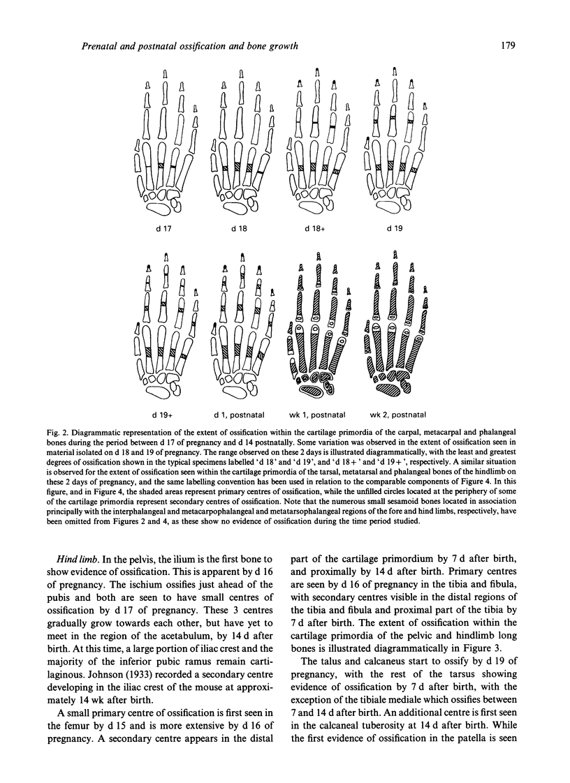
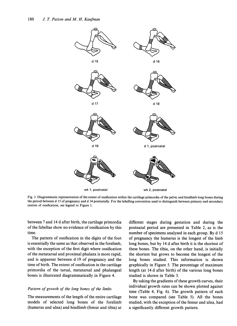
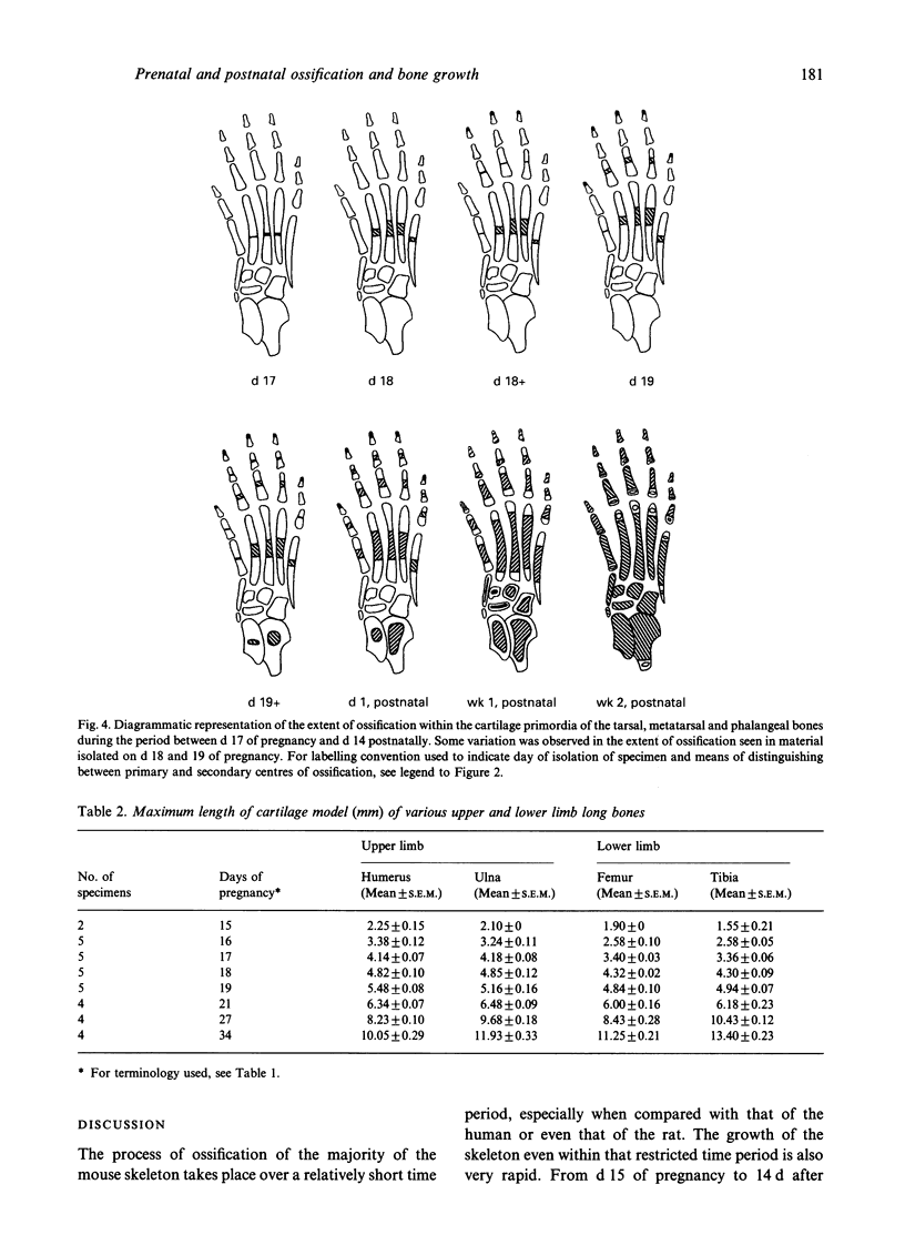
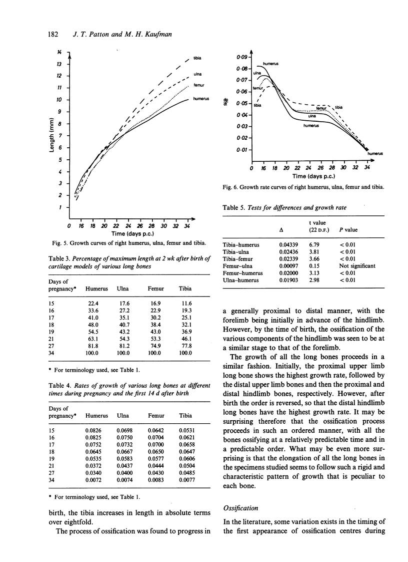
In order to study the pattern of ossification of the skeletal components of the fore and hind limb of the mouse, intact embryos were isolated between days (d) 15 and 19 of pregnancy (the morning of finding a vaginal plug is termed d 1 of pregnancy), and postnatal animals isolated on d 1 (newborns), 7 and 14 after birth. The total number of fore and hind limbs studied for each day of pregnancy or postnatal day for the bone growth study is given in parentheses: d 15 (2), d 16, 17, 18 and 19 of pregnancy (5 specimens for each of these days), d 1 (newborn), wk 1 and 2, postnatal (4 specimens analysed at each of these times), since only the right limbs were studied. For the study involving the time of first appearance of ossification centres, either the right or the left limb of each of these prenatal and postnatal specimens was analysed. All specimens were fixed in 80% ethanol, bulk-stained using alizarin and Alcian blue, in order to stain ossification centres and cartilage, respectively, and cleared. The limbs were then disarticulated from the axial skeleton at the sternoclavicular and sacroiliac joints to facilitate (1) the determination of the sequential pattern of ossification in the various cartilage primordia analysed, and (2) the analysis of the pattern of growth of the humerus, ulna, femur and tibia. The latter values were plotted graphically, and the individual growth rate of each of the long bones studied was then deduced and also plotted graphically. The findings demonstrated that, with the exception of the femur and ulna, all of the long bones studied had significantly different growth patterns. The time of appearance of the various centres of ossification in the skeletal elements studied proceeded in a similar order to that described by previous authors, though there was some discrepancy in the exact time of first appearance of certain ossification centres. Of particular interest was the somewhat unusual pattern of ossification of the first digits of both the fore and hind limb compared with that of the other digits. The data presented here provide useful baseline information on the normal sequential pattern of ossification in the fore and hind limb, and the characteristic growth pattern of the individual long bones of the limbs in this species.
Full text
PDF
Selected References
These references are in PubMed. This may not be the complete list of references from this article.
- Bagnall K. M., Harris P. F., Jones P. R. A radiographic study of the longitudinal growth of primary ossification centers in limb long bones of the human fetus. Anat Rec. 1982 Jun;203(2):293–299. doi: 10.1002/ar.1092030211. [DOI] [PubMed] [Google Scholar]
- MARESH M. M. Linear growth of long bones of extremities from infancy through adolescence; continuing studies. AMA Am J Dis Child. 1955 Jun;89(6):725–742. [PubMed] [Google Scholar]
- MEYER D. B., O'RAHILLY R. Multiple techniques in the study of the onset of prenatal ossification. Anat Rec. 1958 Oct;132(2):181–193. doi: 10.1002/ar.1091320207. [DOI] [PubMed] [Google Scholar]
- NOBACK C. R., ROBERTSON G. G. Sequences of appearance of ossification centers in the human skeleton during the first five prenatal months. Am J Anat. 1951 Jul;89(1):1–28. doi: 10.1002/aja.1000890102. [DOI] [PubMed] [Google Scholar]
- O'Rahilly R., Gardner E. The initial appearance of ossification in staged human embryos. Am J Anat. 1972 Jul;134(3):291–301. doi: 10.1002/aja.1001340303. [DOI] [PubMed] [Google Scholar]
- Seeds J. W., Cefalo R. C. Relationship of fetal limb lengths to both biparietal diameter and gestational age. Obstet Gynecol. 1982 Dec;60(6):680–685. [PubMed] [Google Scholar]


