Abstract
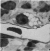
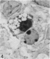
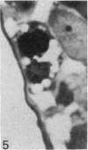
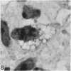
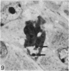
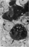
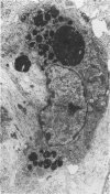
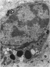
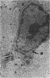
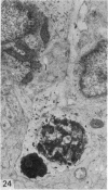
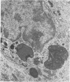
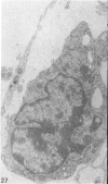
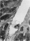
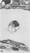
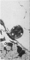
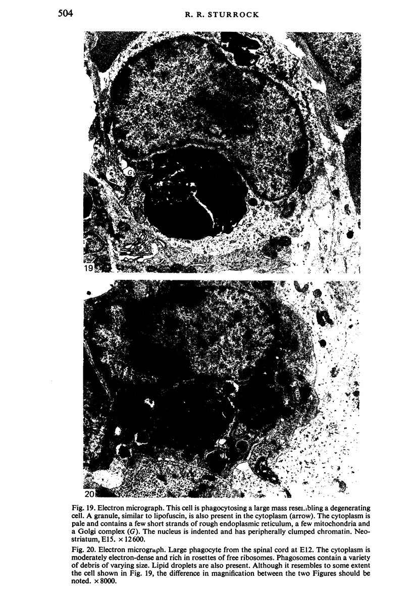
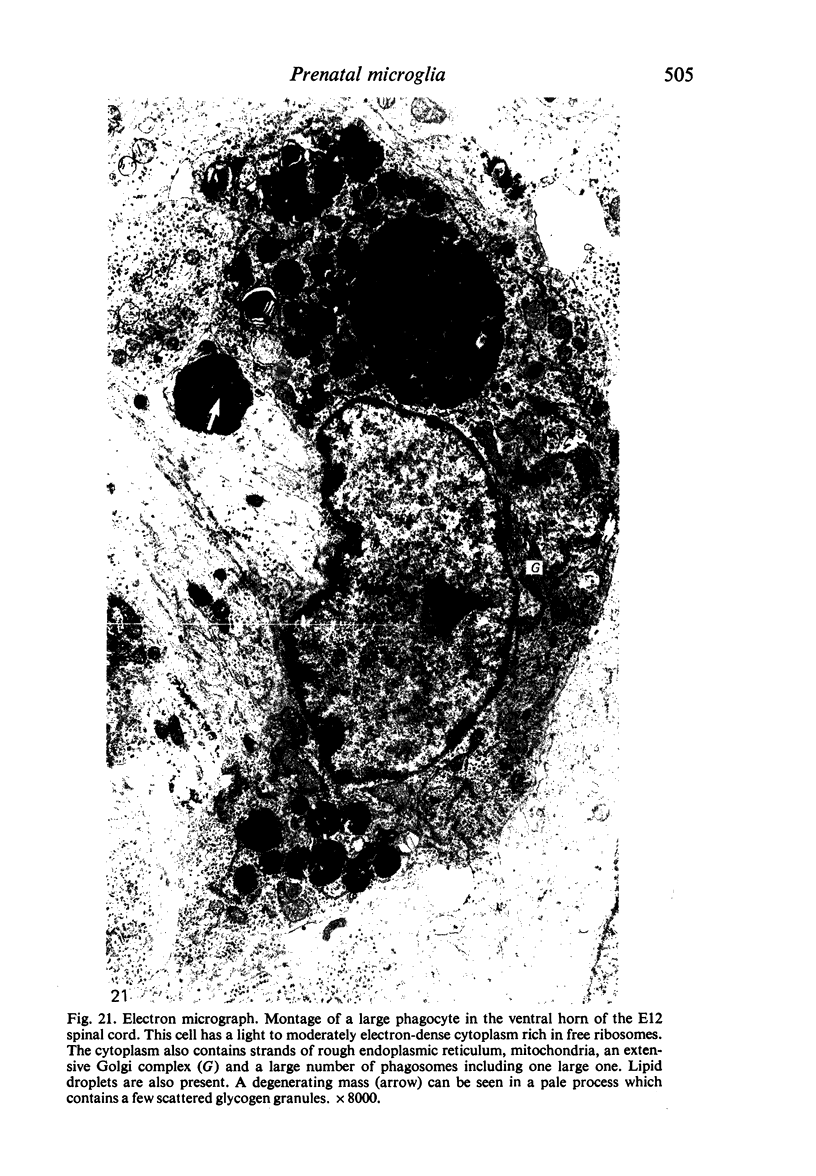
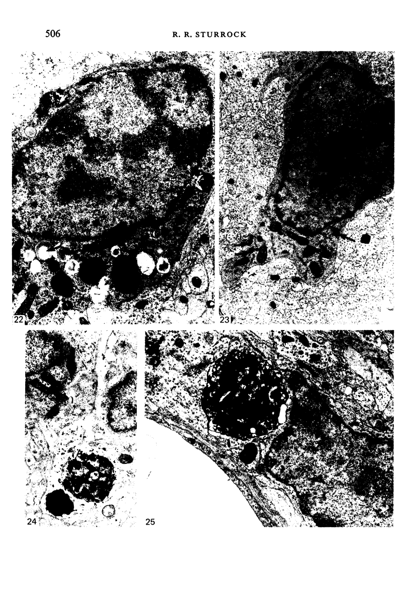
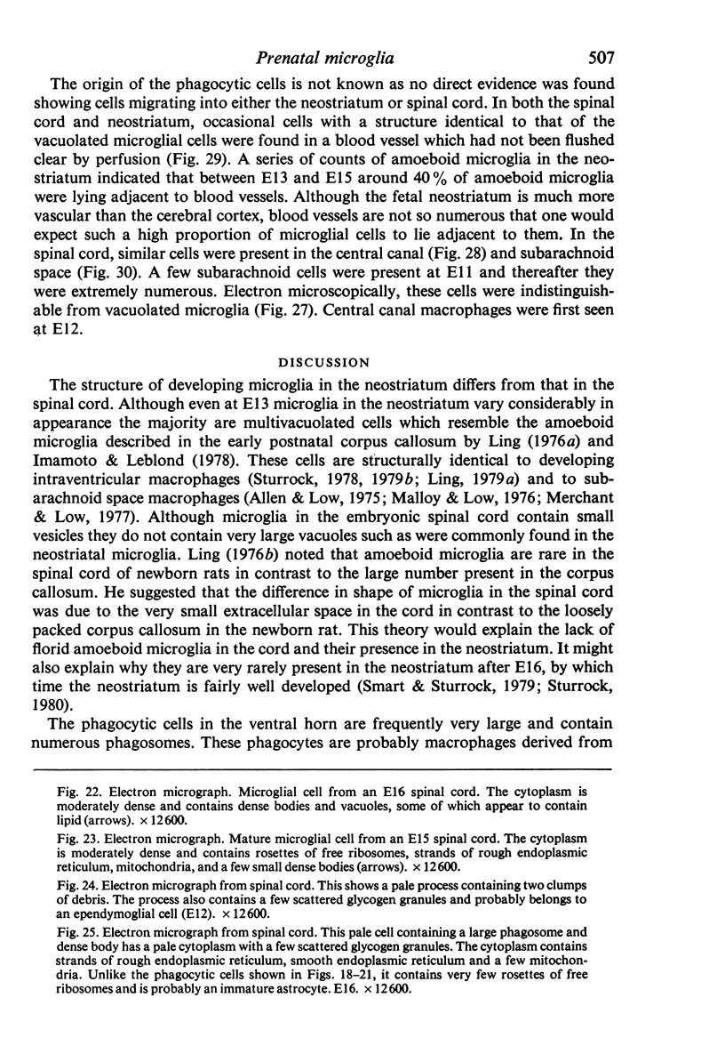
The neostriatum and spinal cord of embryonic and fetal mice were examined to determine the identity of prenatal phagocytes and the differentiation of microglia. In the neostriatum amoeboid microglia were present from E13 and were phagocytosing cellular debris. A wide variety of forms was present, from microglia with extremely vacuolated cytoplasm to some with virtually no vacuoles. Both types were observed in the process of mitotic division. In the spinal cord microglia could be identified in both white and grey matter from E 12 onwards. Between E12 and E15 large phagocytic cells were present, especially in the ventral horns. These appear to be macrophages, probably derived from blood monocytes. It was possible to construct a structural sequence of differentiation of these large macrophages into microglia. Amoeboid microglia with large vacuoles were not present in the prenatal spinal cord. Both actively phagocytic macrophages and microglia were found undergoing mitosis.
Full text
PDF
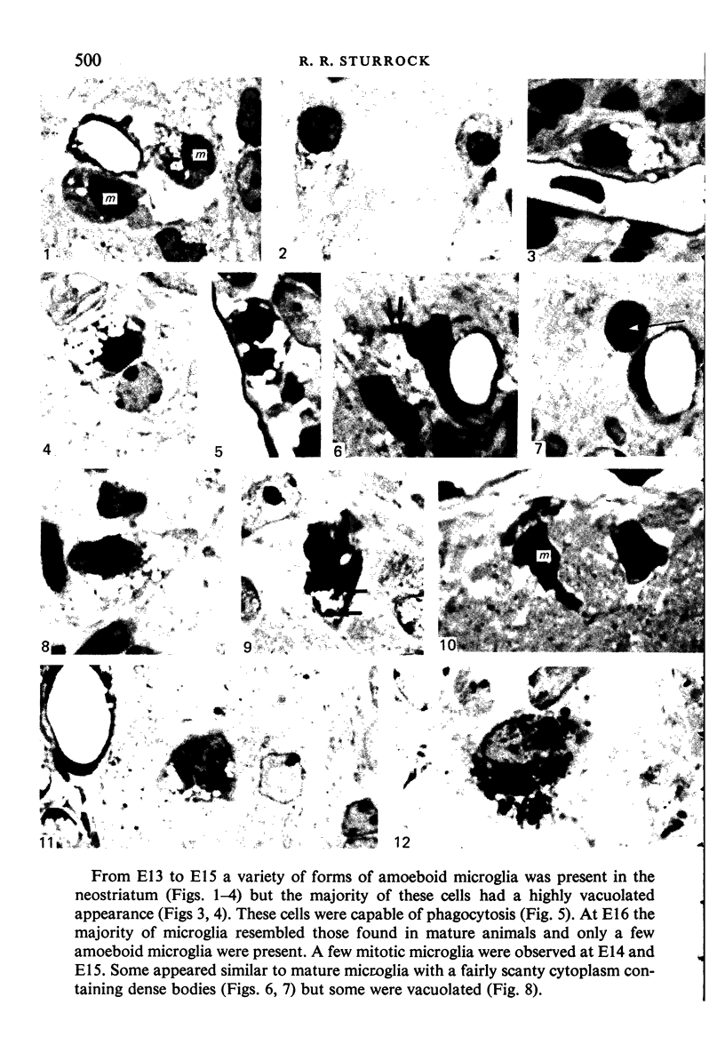
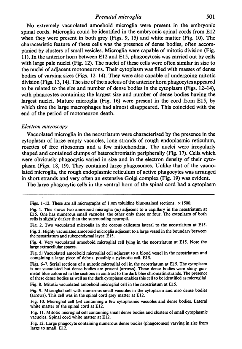
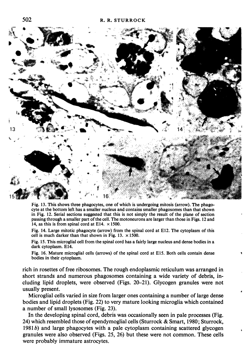
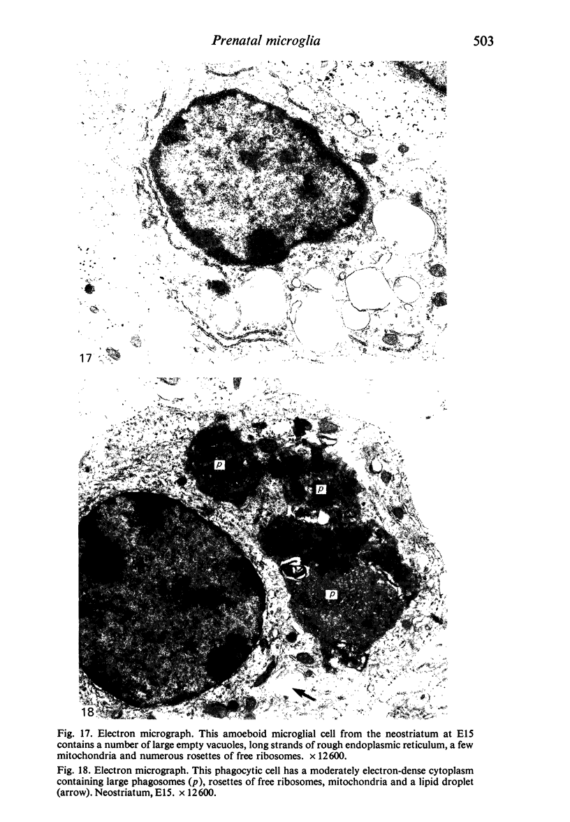
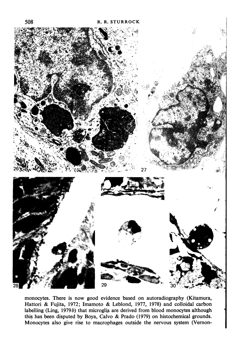
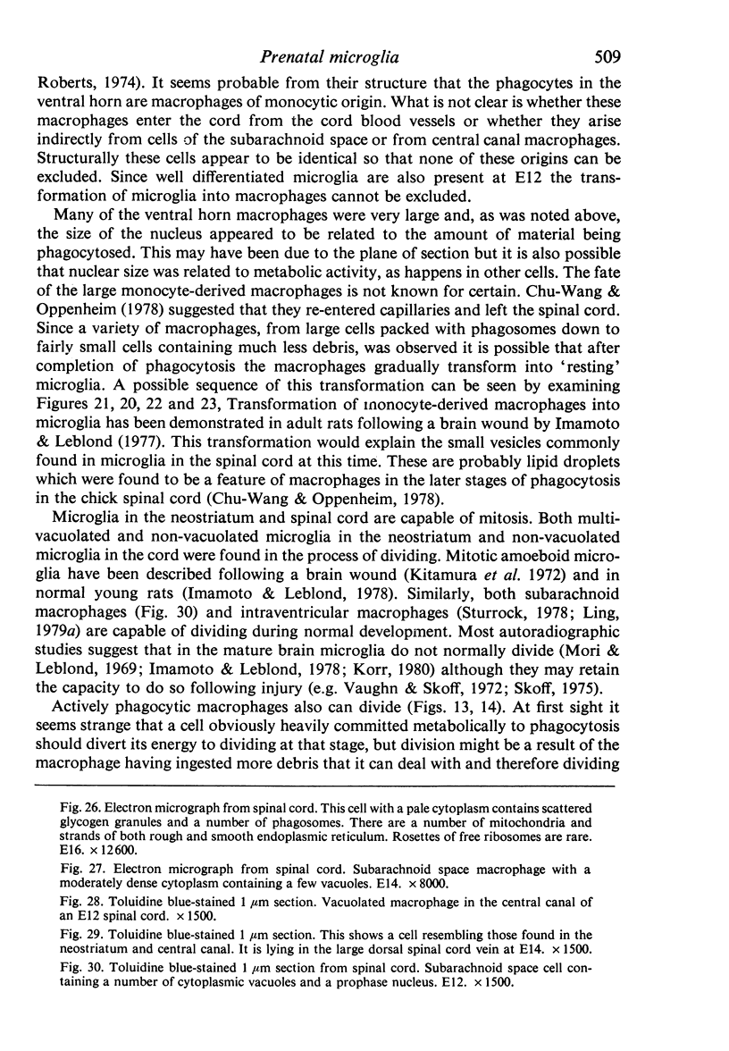
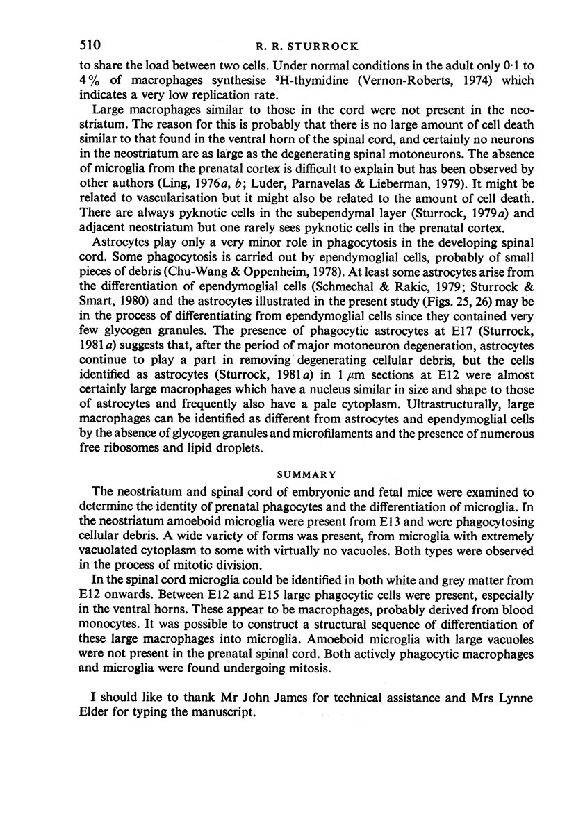
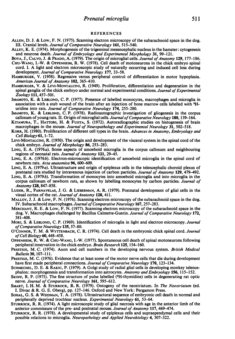
Images in this article
Selected References
These references are in PubMed. This may not be the complete list of references from this article.
- Alley K. E. Morphogenesis of the trigeminal mesencephalic nucleus in the hamster: cytogenesis and neurone death. J Embryol Exp Morphol. 1974 Jan;31(1):99–121. [PubMed] [Google Scholar]
- Boya J., Calvo J., Prado A. The origin of microglial cells. J Anat. 1979 Aug;129(Pt 1):177–186. [PMC free article] [PubMed] [Google Scholar]
- Chu-Wang I. W., Oppenheim R. W. Cell death of motoneurons in the chick embryo spinal cord. I. A light and electron microscopic study of naturally occurring and induced cell loss during development. J Comp Neurol. 1978 Jan 1;177(1):33–57. doi: 10.1002/cne.901770105. [DOI] [PubMed] [Google Scholar]
- HAMBURGER V. Regression versus peripheral control of differentiation in motor hypoplasia. Am J Anat. 1958 May;102(3):365–409. doi: 10.1002/aja.1001020303. [DOI] [PubMed] [Google Scholar]
- Imamoto K., Leblond C. P. Presence of labeled monocytes, macrophages and microglia in a stab wound of the brain following an injection of bone marrow cells labeled with 3H-uridine into rats. J Comp Neurol. 1977 Jul 15;174(2):255–279. doi: 10.1002/cne.901740205. [DOI] [PubMed] [Google Scholar]
- Imamoto K., Leblond C. P. Radioautographic investigation of gliogenesis in the corpus callosum of young rats. II. Origin of microglial cells. J Comp Neurol. 1978 Jul 1;180(1):139–163. doi: 10.1002/cne.901800109. [DOI] [PubMed] [Google Scholar]
- Kitamura T., Hattori H., Fujita S. Autoradiographic studies on histogenesis of brain macrophages in the mouse. J Neuropathol Exp Neurol. 1972 Jul;31(3):502–518. doi: 10.1097/00005072-197207000-00008. [DOI] [PubMed] [Google Scholar]
- Korr H. Proliferation of different cell types in the brain. Adv Anat Embryol Cell Biol. 1980;61:1–72. doi: 10.1007/978-3-642-67577-5. [DOI] [PubMed] [Google Scholar]
- Ling E. A. Electron-microscopic identification of amoeboid microglia in the spinal cord of newborn rats. Acta Anat (Basel) 1976;96(4):600–609. doi: 10.1159/000144707. [DOI] [PubMed] [Google Scholar]
- Ling E. A. Some aspects of amoeboid microglia in the corpus callosum and neighbouring regions of neonatal rats. J Anat. 1976 Feb;121(Pt 1):29–45. [PMC free article] [PubMed] [Google Scholar]
- Ling E. A. Transformation of monocytes into amoeboid microglia in the corpus callosum of postnatal rats, as shown by labelling monocytes by carbon particles. J Anat. 1979 Jun;128(Pt 4):847–858. [PMC free article] [PubMed] [Google Scholar]
- Ling E. A. Ultrastruct and origin of epiplexus cells in the telencephalic choroid plexus of postnatal rats studied by intravenous injection of carbon particles. J Anat. 1979 Oct;129(Pt 3):479–492. [PMC free article] [PubMed] [Google Scholar]
- Malloy J. J., Low F. N. Scanning electron microscopy of the subarachnoid space in the dog. IV. Subarachnoid macrophages. J Comp Neurol. 1976 Jun 1;167(3):257–283. doi: 10.1002/cne.901670302. [DOI] [PubMed] [Google Scholar]
- Merchant R. E., Low F. N. Scanning electron microscopy of the subarachnoid space in the dog. V. Macrophages challenged by bacillus Calmette-Guerin. J Comp Neurol. 1977 Apr 1;172(3):381–407. doi: 10.1002/cne.901720302. [DOI] [PubMed] [Google Scholar]
- Mori S., Leblond C. P. Identification of microglia in light and electron microscopy. J Comp Neurol. 1969 Jan;135(1):57–80. doi: 10.1002/cne.901350104. [DOI] [PubMed] [Google Scholar]
- O'Connor T. M., Wyttenbach C. R. Cell death in the embryonic chick spinal cord. J Cell Biol. 1974 Feb;60(2):448–459. doi: 10.1083/jcb.60.2.448. [DOI] [PMC free article] [PubMed] [Google Scholar]
- Oppenheim R. W., Chu-wang I. Spontaneous cell death of spinal motoneurons following peripheral innervation in the chick embryo. Brain Res. 1977 Apr 8;125(1):154–160. doi: 10.1016/0006-8993(77)90367-5. [DOI] [PubMed] [Google Scholar]
- Prestige M. C. Axon and cell numbers in the developing nervous system. Br Med Bull. 1974 May;30(2):107–111. doi: 10.1093/oxfordjournals.bmb.a071178. [DOI] [PubMed] [Google Scholar]
- Prestige M. C. Evidence that at least some of the motor nerve cells that die during development have first made peripheral connections. J Comp Neurol. 1976 Nov 1;170(1):123–133. doi: 10.1002/cne.901700109. [DOI] [PubMed] [Google Scholar]
- Schmechel D. E., Rakic P. A Golgi study of radial glial cells in developing monkey telencephalon: morphogenesis and transformation into astrocytes. Anat Embryol (Berl) 1979 Jun 5;156(2):115–152. doi: 10.1007/BF00300010. [DOI] [PubMed] [Google Scholar]
- Skoff R. P. The fine structure of pulse labeled (3-H-thymidine cells) in degenerating rat optic nerve. J Comp Neurol. 1975 Jun 15;161(4):595–611. doi: 10.1002/cne.901610408. [DOI] [PubMed] [Google Scholar]
- Sohal G. S., Weidman T. A. Ultrastructural sequence of embryonic cell death in normal and peripherally deprived trachlear nucleus. Exp Neurol. 1978 Aug;61(1):53–64. doi: 10.1016/0014-4886(78)90180-2. [DOI] [PubMed] [Google Scholar]
- Sturrock R. R. A developmental study of epiplexus cells and supraependymal cells and their possible relationship to microglia. Neuropathol Appl Neurobiol. 1978 Sep-Oct;4(5):307–322. doi: 10.1111/j.1365-2990.1978.tb01345.x. [DOI] [PubMed] [Google Scholar]
- Sturrock R. R. A developmental study of the mouse neostriatum. J Anat. 1980 Mar;130(Pt 2):243–261. [PMC free article] [PubMed] [Google Scholar]
- Sturrock R. R. A light microscope study of glial necrosis with age in the anterior limb of the anterior commissure of the pre and postnatal mouse. J Anat. 1974 Jul;117(Pt 3):469–474. [PMC free article] [PubMed] [Google Scholar]
- Sturrock R. R. A quantitative and morphological study of vascularisation of the developing mouse spinal cord. J Anat. 1981 Mar;132(Pt 2):203–221. [PMC free article] [PubMed] [Google Scholar]
- Sturrock R. R. A quantitative lifespan study of changes in cell number, cell division and cell death in various regions of the mouse forebrain. Neuropathol Appl Neurobiol. 1979 Nov-Dec;5(6):433–456. doi: 10.1111/j.1365-2990.1979.tb00642.x. [DOI] [PubMed] [Google Scholar]
- Sturrock R. R. A semithin light microscopic, transmission electron microscopic and scanning electron microscopic study of macrophages in the lateral ventricle of mice from embryonic to adult life. J Anat. 1979 Aug;129(Pt 1):31–44. [PMC free article] [PubMed] [Google Scholar]
- Sturrock R. R. An electron microscopic study of the development of the ependyma of the central canal of the mouse spinal cord. J Anat. 1981 Jan;132(Pt 1):119–136. [PMC free article] [PubMed] [Google Scholar]
- Sturrock R. R., Smart I. H. A morphological study of the mouse subependymal layer from embryonic life to old age. J Anat. 1980 Mar;130(Pt 2):391–415. [PMC free article] [PubMed] [Google Scholar]


