Abstract
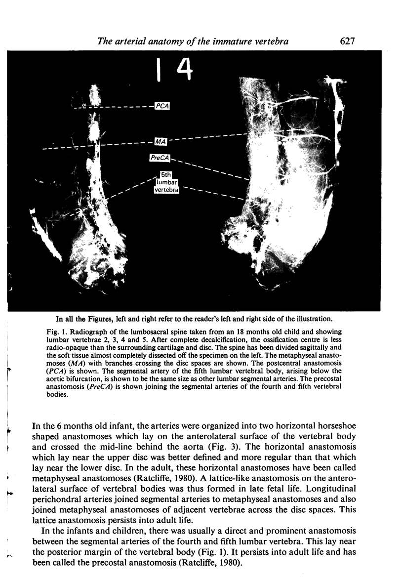
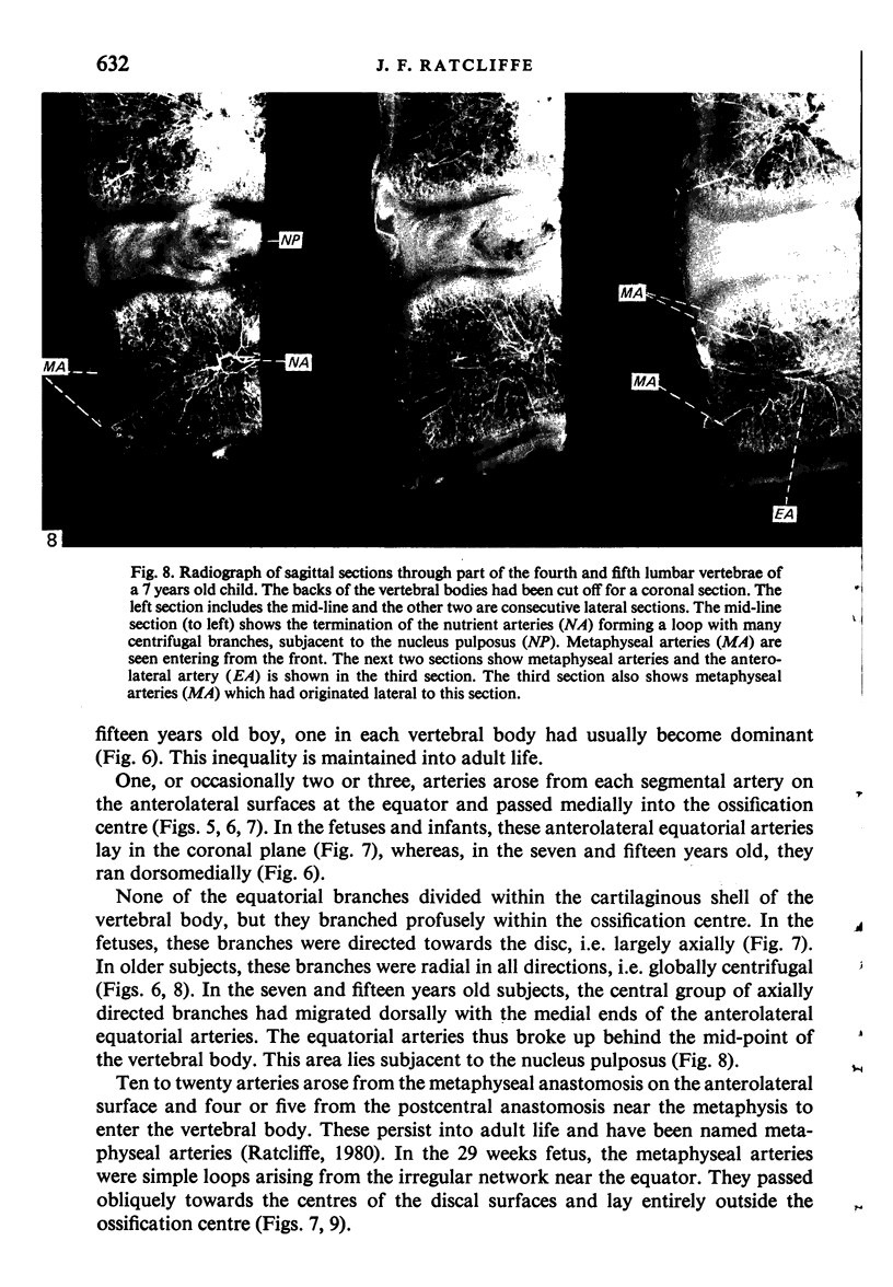
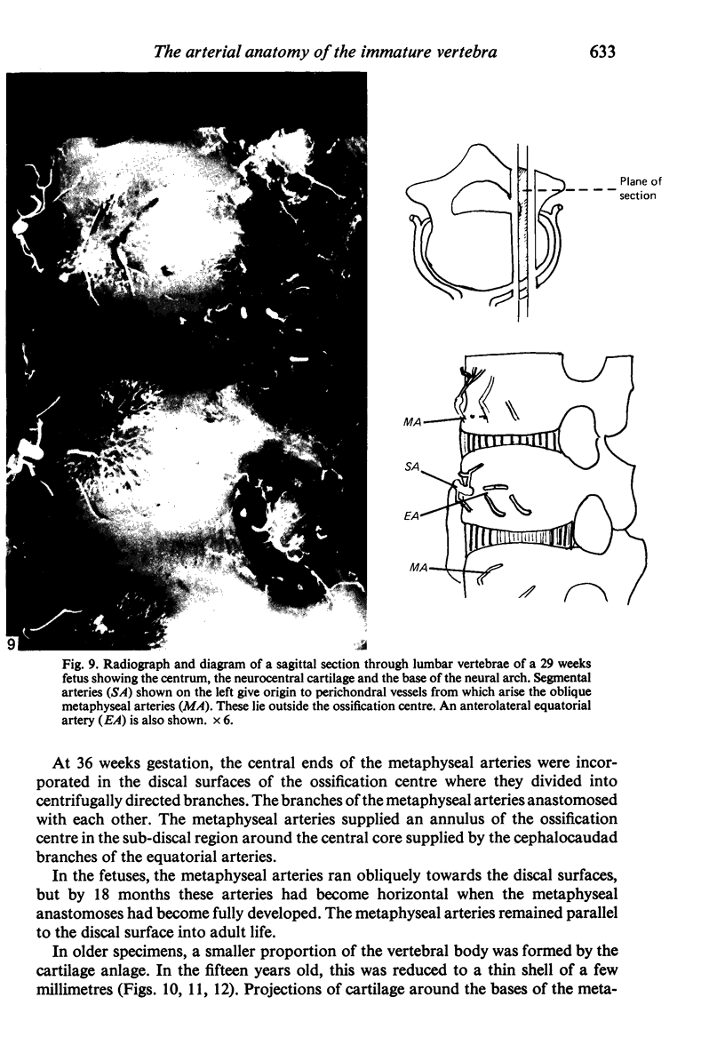
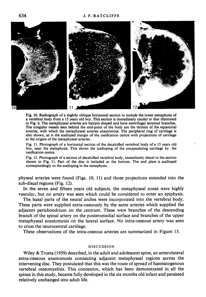
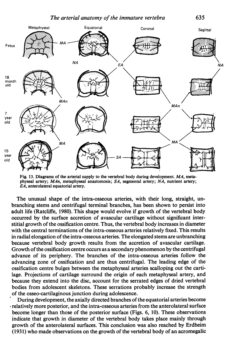
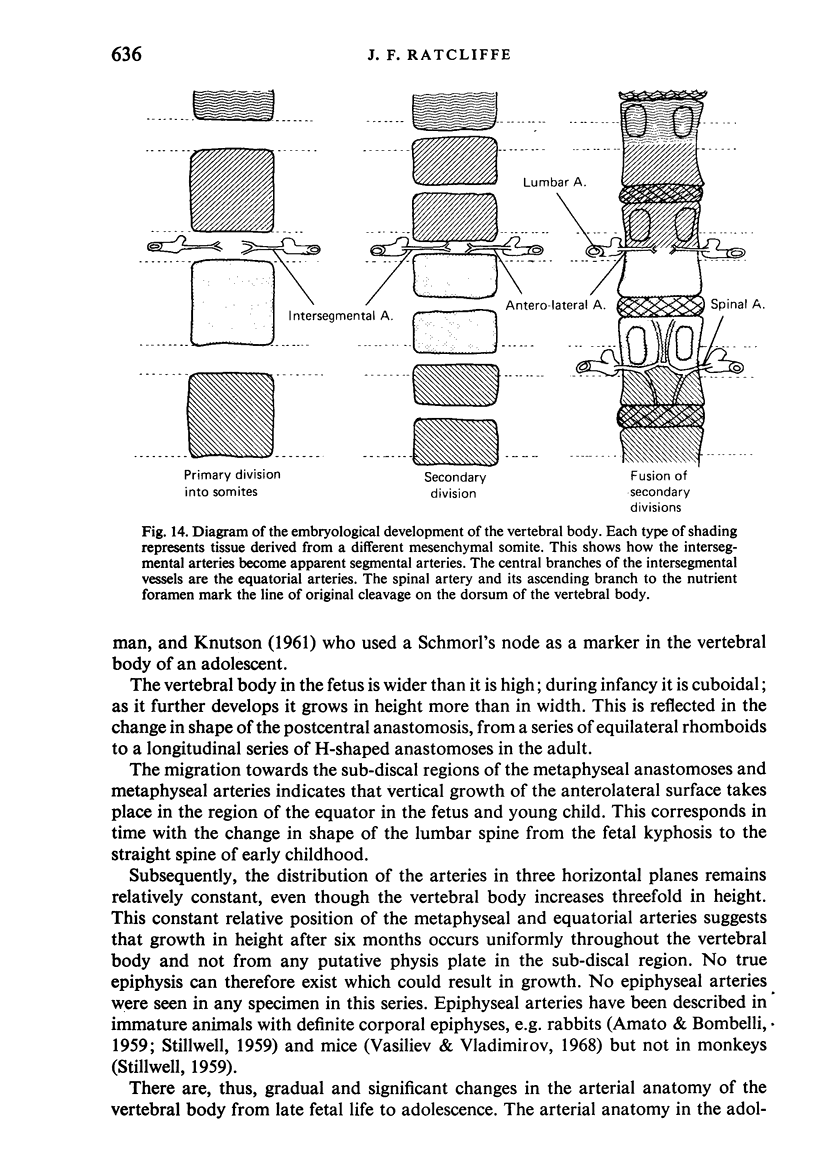
The arterial anatomy of 60 lumbar and lower dorsal vertebral bodies from eight subjects aged between 29 weeks gestation and 15 years was studied. The arteries had been injected with a suspension of barium sulphate and the vertebrae decalcified, sectioned and radiographed. In the specimen of 29 weeks gestation, the equatorial arteries were present. Precursors of the metaphyseal arteries lay obliquely over and completely outside the ossification centre. These precursors originated from an irregular network of perichondral arteries near the equator. By six months of age, the perichondral arteries had migrated discally and had become well organized metaphyseal anastomoses while the metaphyseal arteries had become horizontal. Also by six months, the extra-osseous longitudinal anastomoses had developed into the adult pattern. In the 36 weeks fetus, the ends of the unbranching metaphyseal arteries were incorporated into the ossification centre. This central relationship was maintained into adult life, but, as the ossification centre expanded, the branches of the intra-osseous arteries followed the zone of ossification in a centrifugal manner. In infancy, the metaphyseal arteries were approximately equal in length and the equatorial arteries divided in the middle of the vertebral body; by the age of 15 years, the metaphyseal arteries arising from the anterolateral surfaces were longer than those which arose from the posterior surface, and the equatorial arteries divided behind the mid-point. From these arterial observations, a number of deductions concerning the mode of growth of the vertebral body have been drawn. Preterminal coils and typical peripheral arteries, frequent features in the adult vertebral body, were not seen in any of these specimens. There was no evidence of any epiphyseal growth plate, nor of epiphyseal arteries in these specimens.
Full text
PDF




Images in this article
Selected References
These references are in PubMed. This may not be the complete list of references from this article.
- AMATO V. P., BOMBELLI R. The normal vascular supply of the vertebral column in the growing rabbit. J Bone Joint Surg Br. 1959 Nov;41-B:782–795. doi: 10.1302/0301-620X.41B4.782. [DOI] [PubMed] [Google Scholar]
- Guida G., Cigala F., Riccio V. The vascularization of the vertebral body in the human fetus at term. Clin Orthop Relat Res. 1969 Jul-Aug;65:229–234. [PubMed] [Google Scholar]
- Ratcliffe J. F. Microarteriography of the cadaveric human lumbar spine. Evaluation of a new technique of injection in the anastomotic arterial system. Acta Radiol Diagn (Stockh) 1978;19(4):656–668. doi: 10.1177/028418517801900411. [DOI] [PubMed] [Google Scholar]
- Ratcliffe J. F. The arterial anatomy of the adult human lumbar vertebral body: a microarteriographic study. J Anat. 1980 Aug;131(Pt 1):57–79. [PMC free article] [PubMed] [Google Scholar]
- STILWELL D. L., Jr The vascular supply of vertebral structures. Gross anatomy: rabbit and monkey. Anat Rec. 1959 Nov;135:169–183. doi: 10.1002/ar.1091350303. [DOI] [PubMed] [Google Scholar]
- Vasilev V., Vladimirov B. Uber die Blutversorgung der Zwischenwirbelscheiben. C R Acad Bulg Sci. 1968;21(10):1137–1139. [PubMed] [Google Scholar]
- WILEY A. M., TRUETA J. The vascular anatomy of the spine and its relationship to pyogenic vertebral osteomyelitis. J Bone Joint Surg Br. 1959 Nov;41-B:796–809. doi: 10.1302/0301-620X.41B4.796. [DOI] [PubMed] [Google Scholar]


