Abstract
Chinese herbal medicine has offered an enormous source for developing novel bio-soft materials. In this research, the natural polysaccharide isolated from the Chinese herbal medicine Dendrobium was employed as the secondary building block to fabricate a “hybrid” hydrogel with synthetic poly (vinyl alcohol) (PVA) polymers. Thanks to the presence of mannose units that contain cis-diol motifs on the chain of the Dendrobium polysaccharides, efficient crosslinking with the borax is allowed and reversible covalent borate ester bonds are formed. Eventually, highly dynamic and double-networked hydrogels were successfully prepared by the integration of Dendrobium polysaccharides and PVA. Interestingly, the introduction of polysaccharides has given rise to more robust and dynamic hydrogel networks, leading to enhanced thermal stability, mechanical strength, and tensile capacity (>1000%) as well as the rapid self-healing ability (<5 s) of the “hybrid” hydrogels compared with the PVA/borax single networked hydrogel. Moreover, the polysaccharides/PVA double network hydrogel showed selective antibacterial activity towards S. aureus. The reported polysaccharides/PVA double networked hydrogel would provide a scaffold to hybridize bioactive natural polysaccharides and synthetic polymers for developing robust but dynamic multiple networked hydrogels that are tailorable for biomedical applications.
Keywords: polyvinyl alcohol, natural polysaccharides, double network, hydrogel, herbal medicine
1. Introduction
Hydrogels are three-dimensional materials capable of holding large amounts of water within their network structure [1,2,3,4,5,6]. Known for their high porosity, excellent water retention, and mechanical properties, hydrogels are considered promising artificial biomaterials and have found widespread application, particularly in biomedical fields including wound dressings, drug delivery, tissue engineering, etc. [7,8,9,10]. Currently, the hydrogel’s properties such as mechanical strength, biocompatibility, and biodegradability, to name a few, can be engineered by careful selection of building blocks, crosslinking agents, and adjustment of component ratios, which further allowed tailoring of the hydrogel functionality and performance adaptable to the application scene. Other innovative approaches, including the development of nano-composite hydrogels [11,12], double network hydrogels [13,14], and multi-interaction hydrogels [15], have also been explored to modulate the mechanical properties and functionalities of the hydrogels.
Polyvinyl alcohol (PVA) is a water-soluble polymer that is frequently used for preparing hydrogels due to its excellent solubility, low toxicity, good biocompatibility, and biodegradability [16,17,18,19,20]. Most intriguingly, the abundant hydroxyl groups on the PVA chain have allowed cross-linking with borax to form reversible borate esters and dynamic hydrogels, imparting self-healing properties for fabricating versatile soft materials. However, the mechanical strength of such single PVA-borax networked hydrogels often proved to be weak, and efforts to improve the mechanical properties are usually needed [20,21]. Recent research has demonstrated the incorporation of a polysaccharide block into the PVA-borax hydrogel system to build multiple crosslinking networks that can overcome these limitations. For instance, Zhong et al. developed a resilient and multifunctional composite PVA-borax-based hydrogel by introducing dopamine-grafted oxidized carboxymethyl cellulose and cellulose nanofibers [22]. Ma et al. constructed a robust PVA hydrogel by blending okra polysaccharides with silver nanowires in a unique sandwich structure suitable for strain-sensing devices [23]. Zhang et al. combined Aloe polysaccharides, honey, and PVA with borax, and a freeze-thaw method was used to create a hydrogel with good mechanical strength and excellent biocompatibility [24]. Apparently, more building blocks besides polysaccharides were often necessary to either reinforce the network or endow the hydrogels with desired functions. Therefore, developing simpler hybridized PVA and polysaccharides-based hydrogels with appropriate mechanical properties is highly demanded.
Dendrobium has long been used as a unique herb for disease treatment in Chinese herbal medicine, it is regarded as the “top one in nine immortal herbs” in China. Polysaccharides have been elucidated as a main bioactive constituent in Dendrobium, and they possess diversified bioactivities, including immunomodulatory [25,26], hepatoprotective [27,28], anti-inflammatory [29,30], anti-oxidant [31], anti-tumor [32], and hypoglycemic activities [33]. The Dendrobium polysaccharides primarily consist of glucose and mannose. Hence, the abundance of hydroxyl groups, particularly the cis-diol structure, in mannose from these polysaccharides would allow successful engagement with the chemistry of borate ester, and dynamic networked hydrogels with biofunctions can be envisioned. Previously, Gao et al. [34] have developed an environment-responsive material for postoperative adjuvant therapy by incorporating Mn2+-pectin microspheres into a Dendrobium polysaccharide hydrogel. The MnP@DOP-Gel material exhibits ROS-responsive MnP release, which induces immunogenic cell death in tumor cells and activates dendritic cells and macrophages to trigger a cascade of antitumor immune responses, thereby fulfilling a dual function. This study broadens the application of Dendrobium polysaccharides in drug delivery, although research into its biomedical applications, such as wound healing, selective antibacterial effects, and tissue engineering, remains limited.
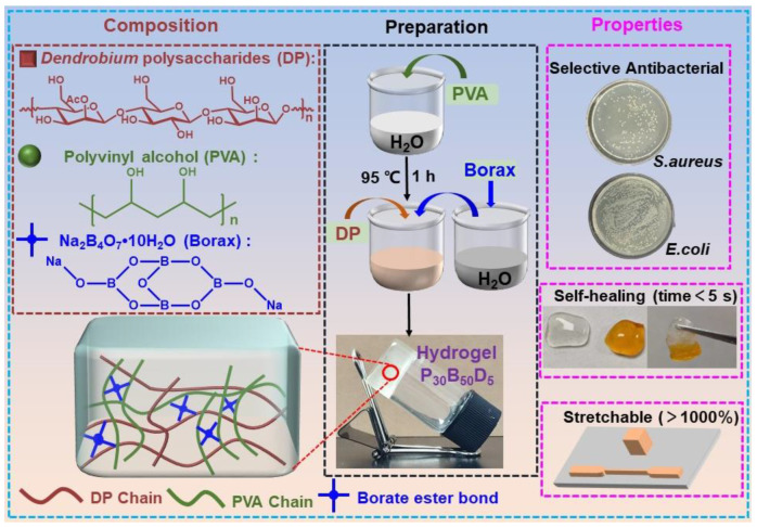
Herein, taking advantage of synthetic PVA polymers and natural Dendrobium polysaccharides (DP), double dynamic cross-linked PVA/DP hydrogels are prepared via the formation of the reversible borate ester bonds (Scheme 1). Such simpler dynamic hydrogels have demonstrated significantly enhanced mechanical strength and rapid self-healing capability. Moreover, the dynamic PVA/DP hydrogels showed outstanding selective antibacterial activity toward S. aureus. We believe the reported hybrid synthetic and natural polymeric hydrogels built by the formation of a double dynamic network will offer scaffolds for developing novel smart bio-soft materials to address the challenges in biomedical fields [35,36].
Scheme 1.
Schematic illustration of the composition, preparation, and properties of double networked PVA/DP hydrogels.
2. Results and Discussion
2.1. Preparation and Characterization of PVA/DP Hydrogels
The PVA/DP hydrogels were prepared by blending PVA (P), DP (D), and borax (B) with different mass ratios in 1 mL of deionized water at 95 °C. The feed amount for all the hydrogel building blocks is listed in Table 1. For brevity, all the hydrogels are labeled as PxByDz. The subscripts X, Y, and Z represent the final concentration of each building block (mg/mL). All the hydrogels were rapidly formed and are assumed to feature a dynamic dual network where PVA and DP are crosslinked with borax through reversible borate ester bonds due to the presence of a cis-diol motif on both polymer chains. For comparison, single networked PVA/borax (P30B10, P30B30, P30B50) hydrogels were also prepared. As shown in Figure S1, the strain dependence of G′ and G″ for hydrogels P30B10, P30B30, and P30B50 was compared, and hydrogel P30B50, exhibiting superior gel strength, was selected for further experimental testing and comparison. Additionally, the G′ and G″ values of the P30B50 hydrogel are higher than those reported in the literature.
Table 1.
Feed amount of hydrogels.
| Samples | PVA (mg) | Borax (mg) | DP (mg) | Water (mL) |
|---|---|---|---|---|
| P30B50 | 60 | 100 | 0 | 2 |
| P30B50D1 | 60 | 100 | 2 | 2 |
| P30B50D5 | 60 | 100 | 10 | 2 |
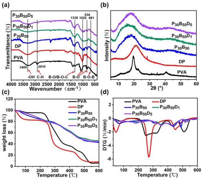
To validate the formation of reversible borate ester in the network, Fourier-transform infrared (FT-IR) spectroscopy was performed. As shown in Figure 1a, the hydrogels P30B50, P30B50D1, and P30B50D5 exhibit asymmetric stretching vibrations at 661, 834, and 1025 cm−1, which are associated with residual borate B-O stretching vibrations and B-O-B bond bending in the borate network [21,36,37]. Additionally, a narrow absorption band at 1338 cm−1 is attributed to the stretching vibrations of B-O and B-O-C groups [38]. All samples show a stretching vibration band at 2910 cm−1, corresponding to saturated C-H bonds, and a broad absorption band around 3400 cm−1, indicating the presence of -OH groups in polysaccharides and hydrogen bonds in the hydrogel, leading to wide absorption peaks. These results confirm the successful fabrication of a three-dimensional crosslinked network in the P30B50, P30B50D1, and P30B50D5 hydrogels through reversible borate ester bonds.
Figure 1.
(a) FT−IR spectra, (b) XRD patterns, (c) TG, (d) DTG curves of P30B50, P30B50D1, and P30B50D5 hydrogels and their compositions. All the samples were freeze-dried before the measurement.
An X-ray diffraction experiment was also performed to confirm the formation of the borate ester-linked dual hydrogel network Figure 1b. The bulk PVA polymer shows its characteristic peaks at 2θ = 19.5°, 22.5°, and 40.6°, reflecting its microcrystalline structures [39,40]. Upon crosslinking with borax, these characteristic peaks disappear. A new broad diffraction peak centered at 18.5° appears for the hydrogel P30B50, suggesting that the material has a predominantly amorphous, low-crystallinity structure. Such changes in diffraction patterns indicate the disruption of the microcrystalline structure of PVA due to the newly formed dynamic borate ester-linked network [18,41,42]. Upon the addition of DP into the system, the diffraction peaks of the hydrogel P30B50D1 and P30B50D5 respectively shift to lower angles at 17.8° and 17.2°. A shoulder at 10.4° also appears for the hydrogel P30B50D5. Notably, the diffraction peak at 21.2° for bulk DP completely vanishes upon cross-linking with borax, suggesting that the molecular structure of hydrogel P30B50D5 may undergo rearrangement, thereby indicating the successful integration of DP into the double-networked hydrogels [43,44].
Having established the formation of the dynamic reversible network for the hydrogel P30B50, P30B50D1, and P30B50D5, we next examined the stability of these crosslinked hydrogels using the thermogravimetric analysis (TGA). As shown in Figure 1c, both the bulk PVA and DP polymer undergoes a rapid 85% loss of mass within 600 °C. However, only a 55% loss of mass was observed for the hydrogels P30B50, P30B50D1, and P30B50D5 within 600 °C, lower than PVA and DP polymers. As depicted in Figure 1d, the DTG curves illustrate the rate of mass loss as temperature changes [45]. The main thermal decomposition temperatures for PVA and DP are observed at 253.7 °C and 270.0 °C, respectively. Furthermore, the main decomposition temperature of hydrogel P30B50D5 reaches 355.4 °C, which is slightly higher than P30B50D1 at 352.3 °C and P3B5 at 350.1 °C. The increase in thermal stability for the P30B50D5 hydrogels can be attributed to the formation of a denser double hydrogel network [46]. Additionally, hydrogels P30B50D0.1 and P30B50D5 exhibit no significant weight loss at the decomposition temperature of ~270.8 °C, which is for individual PVA and DP, suggesting that hydrogels P30B50D1 and P30B50D5 exhibit a synergistic double-network effect, enhancing their thermal stability, further indicating the successful formation of a double-network hydrogel system involving both PVA and DP polymers.
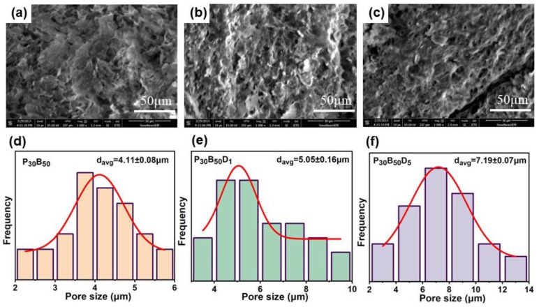
The microstructure of all hydrogels was characterized using scanning electron microscopy (SEM), and the experimental results are shown in Figure 2. All three hydrogels in Figure 2a–c displayed characteristic three-dimensional network structures. With an increase in DP content, oriented microporous structures were formed within the hydrogels. As the mass fraction of DP increased, the micropore size of the hydrogels expanded from 4.11 ± 0.08 μm to 5.05 ± 0.16 μm, and then to 7.19 ± 0.07 μm. As a result, the borate ester bonds between PVA and borax decreased, while the borate ester bonds between DP and borax increased. Meanwhile, the hydrogen bonds between the hydroxyl groups of PVA and the DP crosslinks also increased. Consequently, the stability of the hydrogels increased, resulting in a three-dimensional network with a more uniform pored structure and morphology. The porosity of three hydrogel samples was also assessed using the liquid displacement method. As shown in Figure S2, the porosity of P30B50, P30B50D1, and P30B50D5 hydrogels exceeded 50%, with values of 81.6 ± 4.1%, 64.3 ± 1.5%, and 60.6 ± 2.4%, respectively. Porosity was negatively correlated with DP concentration, meaning higher DP concentrations resulted in lower porosity. This likely resulted from the increased DP content, which made the hydrogel network structure more compact. Newly formed borate ester bonds filled the pores of P30B50, leading to denser three-dimensional structural connections and lower porosity. The porosity results obtained by solution exchange are in contrast to the pore size trend observed by SEM, which may be attributed to the internal three-dimensional interconnected pore structure of the material [47,48].
Figure 2.
(a–c) SEM images of P30B50, P30B50D1, and P30B50D5 hydrogels. (d–f) Pore size of P30B50, P30B50D1, and P30B50D5 hydrogels.
2.2. Rheological Studies
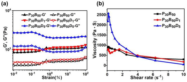
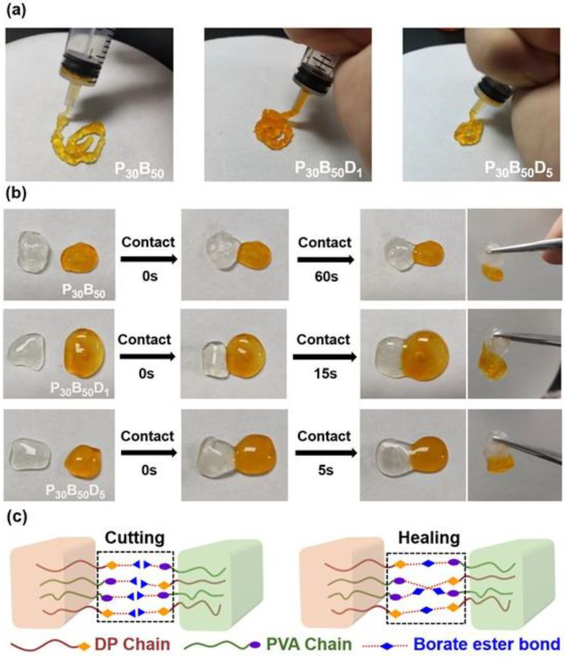
To probe the mechanical properties of the hydrogels, rheological studies were conducted. Initially, a dynamic strain sweep test was carried out to determine the linear viscoelastic range of the hydrogels. As depicted in Figure 3a, the storage modulus (G′) and loss modulus (G″) remained almost constant within the strain range of 0.01–100%, and the G′ is always higher than G″, suggesting that all the hydrogels consistently behave as a viscoelastic solid [49]. Remarkably, hydrogel P30B50D5 possessed a higher G′ than hydrogel P30B50D1 and P30B50, corroborating that a more robust network was formed for P30B50D5 [18]. To ascertain the potential injecting capabilities of such dynamic hydrogels, we measured the viscosity changes at different shear rates for these hydrogels. As shown in Figure 3b, all the hydrogels demonstrated a trend of shear-thinning behavior [50]. In particular, hydrogel P30B50D1 exhibited a prompter response to external shear force, likely due to the synergistic interaction between the double networks, which strengthens intermolecular interactions [51,52]. Such an observation indicates that hydrogel P30B50D5 should have a more dynamic network that would allow excellent injectability. Indeed, hydrogel P30B50D5 can be easily injected by a syringe and keeps the hydrogel state after extrusion Figure 4a [53].
Figure 3.
(a) Strain dependence of G′ and G″ of hydrogel P30B50, P30B50D1, and P30B50D5. (b) Shear rate−dependent viscosity of hydrogel P30B50, P30B50D1, and P30B50D5.
Figure 4.
(a) Injectable properties of P30B50, P30B50D1, and P30B50D5 hydrogels. (b) Self-healing properties of P30B50, P30B50D1, and P30B50D5 hydrogels. (c) Self-healing Mechanism Diagram of P30B50D5 hydrogels.
2.3. Self-Healing Properties
Since the hydrogels P30B50, P30B50D1, and P30B50D5 have been identified as dynamic cross-linked networks, this prompted us to test their capacity for self-healing. The self-healing abilities of all the hydrogels were evaluated by a direct visual method. As shown in Figure 4b, after cutting an intact hydrogel of P30B50, P30B50D1, and P30B50D5 into two halves, they can easily self-heal into a complete one without external aid. A subsequent picking test using a tweezer shows that the healed hydrogel can support its own weight [54,55,56]. Concerning the healing time of the hydrogels, other similar PVA/Borax-based hydrogels showed a typical healing time ranging from 1 to 10 min [56,57], with some reaching 48.22 min [58], and the fastest reported healing time is 8 s [59] (Table S1). Notably, our hydrogel P30B50D5 demonstrated the most rapid healing time within 5 s, which can be ascribed to the formation of a denser double crosslinked network, leading to a more efficient healing process (Video S1 in Supporting Information). The self-healing mechanism of P30B50D5 hydrogels is depicted in Figure 4c. When the hydrogel network undergoes disruption due to external forces, borate ester bonds are cleaved. Once the split hydrogel pieces contact each other, new reversible borate ester bonds are formed at the interface, reestablishing the hydrogel network [55,60].
2.4. Tensile Properties
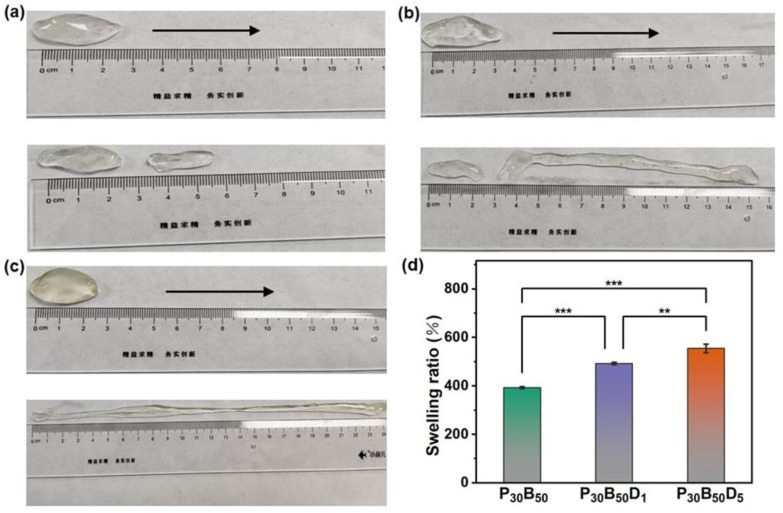
The highly dynamic feature of such double-networked hydrogels prompted us to further explore their tensile performance, which was demonstrated through the direct visual observation method. The hydrogel’s tensile strength was visually evaluated by fixing both ends between the thumb and index finger and stretching it at a constant speed [61]. Figure 5a illustrates that the P30B50 hydrogel, characterized by a single cross-linked network, displays almost no tensile strength and fracture resistance when stretched in one direction. However, with the addition of a small amount of DP, the tensile strength of hydrogel P30B50D1 increases significantly to 600%, as shown in Figure 5b. It further reaches over 1000% with the addition of more DP for the hydrogel P30B50D5, as shown in Figure 5c. As shown in Table S2, most similar PVA/Borax-based hydrogels demonstrated a relatively lower tensile capability, with elongation values of 200% [56], 203.3% [62], and 275% [63]. The best-performing hydrogels reach elongation values of 792% [64] and 975% [58]. In contrast, the polysaccharide-based hydrogel developed in our study exhibits an exceptional elongation value of 1000%, indicating excellent stretchability. These initial tensile test results indicate that the introduction of DP into the P30B50 hydrogel to build a secondary network can significantly boost the overall tensile strength of the hydrogel. This tensile strength enhancement is attributed to the densified but more dynamic crosslinks within the network, which subsequently improves the hydrogel’s elasticity without compromising its structural integrity [21].
Figure 5.
Tensile properties of (a) P30B50, (b) P30B50D1, and (c) P30B50D5 hydrogels. The arrow denotes the stretching direction. (d) Swelling ratio of P30B50, P30B50D1, and P30B50D5 hydrogels in PBS. Data are reported as means ± SD, n = 3 (** p < 0.01, *** p < 0.001).
2.5. Swelling Ratio
Hydrogels are characterized by their inherent ability to swell, increasing in volume and mass upon water absorption. Herein, to evaluate the swelling capability of the hydrogel in the aqueous solution, phosphate-buffered saline (PBS) solution (10 mM, pH 7.4) was selected for the swelling test in light of its future potential for biomedical applications, e.g., wound dressings [21]. Figure 5d illustrates the swelling behavior of different hydrogels immersed in a PBS solution at room temperature. The swelling ratio of the P30B50D5 hydrogel (554.9%) is significantly higher than that of the P30B50D1 hydrogel (492.4%) and the P30B50 hydrogel (392.9%). This enhanced swelling behavior can be attributed to the introduction of more hydrophilic natural polysaccharide blocks into the hydrogel system, which remarkably improves hydrogel hydrophilicity. Furthermore, an increase in the DP content leads to a rise in both the quantity and size of the pores within the hydrogel network, resulting in greater pore expansion and improved water absorption capacity, and eventually boosts the swelling ratio of the P30B50D5 hydrogel. Considering the application of hydrogels in the biomedical field, having an appropriate swelling ability in an aqueous environment is very important. For example, many similar PVA/Borax-based hydrogels with swelling ratios from 32% [65] to 396% [66] and up to 600% [56] have demonstrated potential for wound dressing applications, as shown in Table S3. Hence, the P30B50D5 hydrogel with the highest swelling ratio together with the best tensile properties further guarantees its potential as a wound dressing material.
2.6. Water Retention Properties and In Vitro Degradation Behavior
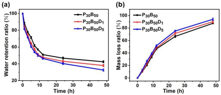
Figure 6a presents the water retention rates of the hydrogels. All hydrogel samples showed rapid water loss during the first 10 h. Notably, while the P30B50D5 hydrogel retained less water than the P30B50 hydrogel, it still maintained a sufficient moisture environment. The water retention rates exhibited an opposite trend to the swelling behavior, but shared the same underlying mechanism related to pore size. An ideal hydrogel should degrade effectively after completing its biofunctions, e.g., for wound healing [67]. Hence, the in vitro degradation behavior of the hydrogels was studied in PBS solution at pH 7.4. Figure 6b shows that the mass of all three hydrogels decreased significantly with a longer immersion time. The degradation of hydrogels would result from the physical dissolution of matrix components during hydration and the chemical cleavage of the dual-network structure [68,69].
Figure 6.
(a) Water retention properties of hydrogel P30B50, P30B50D1, and P30B50D5. (b) In vitro degradation behavior of hydrogel P30B50, P30B50D1, and P30B50D5. Data are reported as means ± SD, n = 3.
2.7. Antibacterial Activity
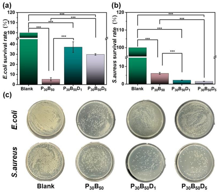
To explore the application potential of such dynamic double networked and particularly natural polysaccharides-integrated hydrogels, we initially evaluate the antibacterial activity of hydrogels P30B50, P30B50D1, and P30B50D5, since hydrogels are now widely applied as wound dressings to prevent wound infections [24,70]. To this end, two representative bacterial strains, the Gram-negative E. coli and Gram-positive S. aureus, were selected. The surface antibacterial activity test was employed to evaluate the antibacterial efficacy of these hydrogels [55,71]. As shown in Figure 7, after 24 h of exposure, the survival rates of E. coli treated with P30B50, P30B50D1, and P30B50D1 hydrogels were 5.27%, 37.11%, and 29.81%, respectively, as shown in Figure 7a. The survival rates of S. aureus treated with P30B50, P30B50D1, and P30B50D5 hydrogels were only 6.12%, 2.23%, and 1.57%, respectively, as shown in Figure 7b. These results indicate that the P30B50 hydrogel exhibits stronger antibacterial properties against E. coli compared with the P30B50D1 and P30B50D5 hydrogels. He et al. [55] reported that treatment with a PBO1 hydrogel can reduce the survival rates of E. coli and S. aureus to 2% and 0.6%, respectively. Similarly, Yi et al. [60] reported survival rates of 15.9% for E. coli and 14.0% for S. aureus after treatment with the PB-EPL/TA@BC hydrogel. The P30B50D5 hydrogel prepared in this study does not demonstrate significant synergistic antibacterial effects against E. coli and S. aureus. However, the P30B50D1 and P30B50D5 hydrogels exhibit stronger antibacterial properties against S. aureus, suggesting a selectivity over different bacterial strains for the polysaccharide-integrated hydrogels [72,73,74]. The cell wall of S. aureus contains a thick peptidoglycan layer, making it susceptible to damage by specific antimicrobial agents. It has been reported that the active components in Dendrobium polysaccharide-based hydrogels may interact with the peptidoglycan layer of the cell wall of S. aureus to inhibit its synthesis or disrupt the cell membrane, thereby hindering bacterial growth [75,76]. In contrast, E. coli has a more complex cell wall structure, including an outer membrane and a lipopolysaccharide layer, which distinguishes it from S. aureus. Such an outer membrane may act as a barrier, preventing the entry of hydrophilic or macromolecular substances into the cell of E. coli [77,78]. As a result, the Dendrobium polysaccharide-based hydrogels may have difficulties penetrating the outer membrane of E. coli, leading to reduced antimicrobial activity. Although PVA/DP hydrogels demonstrate selective antibacterial activity, further investigation of their composition/bioactivity relationship is necessary to better understand their structural characteristics and the molecular mechanisms responsible for their antibacterial effects.
Figure 7.
Antibacterial activity of the P30B50, P30B50D1, and P30B50D5 hydrogels. (a–c) Quantitative statistics of the bacterial killing ratio of the hydrogels against E. coli and S. aureus in the surface antibacterial activity test. Data are reported as means ± SD, n = 3 (*** p < 0.001).
3. Conclusions
In summary, we have successfully prepared novel and simpler double-networked hydrogels by crosslinking Dendrobium polysaccharide and synthetic PVA polymers with borax. Compared with the single-networked PVA/borax hydrogel, double-networked PVA/DP hydrogels demonstrated enhanced thermostability, mechanical strength, and tensile capacity, as well as rapid self-healing ability due to the introduction of a robust but more dynamic hydrogel network. Intriguingly, such PVA/DP hydrogels showed selective antibacterial efficacy against S. aureus over E. coli. Our research highlights a novel and simpler strategy for developing double-networked hydrogels by hybridizing herbal medicine-derived polysaccharides with synthetic polymers. Considering the diversified structures and functions of the herbal-medicine-derived polysaccharides, it is possible to fabricate enormous dynamic double networked bio-soft materials with fewer building blocks while tailoring properties and performance.
4. Materials and Methods
4.1. Materials
Dendrobium polysaccharide (DP, ≥99.5% purity, Mw = 230 KDa, the molar ratio of mannose, glucose, and galactose = 37.8:21.9:1) was purchased from BoRui Saccharide Biotech Co., Ltd. (Yangzhou, China). Its structure has been characterized in previous literature (fraction DHPW1) [79]. Poly (vinyl alcohol) (PVA, 99.8 mol%, Mw~88,000, BR), borax (sodium tetraborate decahydrate, Na2B4O7•10H2O, 99.99% purity), and phosphate buffer solution (PBS, pH = 7.2–7.4) were purchased from Titan Scientific Co., Ltd. (Shanghai, China). All reagents were analytically pure and purchased from Titan Scientific Co., Ltd. (Shanghai, China). Deionized water purchased from Watsons
4.2. Preparation of Hydrogels
The typical procedure for the hydrogel P30B50D5: polyvinyl alcohol (PVA) powder (60 mg) was added to deionized water (1 mL) at 95 °C and stirred for 1 h until it was dissolved completely. Dendrobium polysaccharide (DP) (10 mg) was added to the PVA solution and stirred continuously at 60 °C for an additional hour to obtain a homogeneous mixture. Separately, borax powder (100 mg) was dissolved in 1 mL of deionized water at room temperature with magnetic stirring, and this borax solution was then added to the above polymeric mixture for cross-linking. In approximately 15 s, a homogeneous hydrogel was formed. A similar procedure was followed to prepare all other hydrogels except for the P30B50 hydrogel, which does not contain DP.
4.3. Fourier-Transform Infrared Spectroscopy (FT-IR)
The FT-IR spectra of the hydrogel samples were recorded on a Fourier-transform infrared (FT-IR) spectrometer (Bruker Tensor II, Erlangen, Germany). The recorded wavelength range is between 400–4000 cm−1. Each sample was scanned 32 times.
4.4. Powder X-Ray Diffractometer (PXRD)
The crystalline structures of the hydrogels were analyzed on an XRD diffractometer (Rigaku D/max-IIIA, Osaka, Japan) with Cu Kα radiation (λ = 0.154 nm), and the spectra were recorded from 5–60° (2θ) with a scanning speed of 5°/min.
4.5. Thermogravimetric Analysis (TGA)
The thermal stability of the hydrogels was analyzed using a Netzsch TG 209 F3 instrument (NETZSCH company, Selb, Germany). A sample with a mass of 5~10 mg was placed in an alumina crucible and heated to 600 °C with a heating rate of 5 °C /min and a nitrogen flow rate of 20 mL/min.
4.6. Scanning Electron Microscopy (SEM)
The microstructure of the hydrogel was investigated using a scanning electron microscope (SEM, JSM7100F, JEOL, Tokyo, Japan). Prior to observation, all samples were freeze-dried and sputtered with gold. For the analysis of pore size distribution, Nano Measurer 1.2 software was used. A minimum of 30 pores was selected for each measurement to ensure statistical reliability.
4.7. Porosity of Hydrogel
The porosity of the PVA/DP hydrogels was determined using the solvent displacement method. Initially, the hydrogels were freeze-dried and weighed (Md). The freeze-dried samples were then immersed in anhydrous ethanol until complete saturation and weighed again (Mf). The volume of ethanol consumed (Vs) was recorded during this process, and the procedure was repeated three times for each test. Additionally, the volume change of ethanol and the ethanol density (ρ) were recorded, and the porosity of the hydrogels was calculated using the appropriate equation [47,48]:
| Porosity ratio (%) = [(Mf − Md)/(ρ × Vs)] × 100 |
Data are reported as means ± SD, n = 3 (** p < 0.01, *** p < 0.001).
4.8. Rheological Property
The rheological behavior of the hydrogel was investigated by a rheometer (TA DHR-2, Waters-TA Instruments, New Castle, DE, USA). Rheological tests were conducted over a strain range of 0.01–200% at an angular frequency of 10 rad/s to assess changes in both the storage modulus and the loss modulus.
4.9. Macroscopic Self-Healing Test of Hydrogels
Two hydrogel samples, each with a diameter of 10 mm, were prepared in advance. To visually demonstrate the self-healing capability, one of the hydrogels was dyed with methyl orange, and then sliced into two pieces. The two pieces of the hydrogel were brought to contact and placed at 25 °C for self-healing [60]. To evaluate the strength of the repaired hydrogel, a tweezer was used to lift one corner of the sample to test if it could sustain its own weight. Photographs were taken at different stages of the healing process to document its progression.
4.10. Tensile Property
The hydrogel’s tensile strength was visually evaluated by fixing both ends between the thumb and index finger and stretching it at a constant speed [80,81].
4.11. Swelling Behavior
The swelling behavior of the hydrogels was studied by the gravimetric method [21]. The hydrogel was dried to a constant weight to obtain its dry weight (Ms). Subsequently, the dried hydrogel samples were immersed in a phosphate-buffered saline (PBS) solution (10 mM, pH 7.4) until equilibrium was reached. Finally, excess surface water was absorbed using filter paper, and the swollen hydrogel was weighed (Me). The swelling ratio of the hydrogel was calculated using the following formula:
| Swelling ratio (%) = [(Me − Ms)/Ms] × 100 |
Data are reported as means ± SD, n = 3 (** p < 0.01, *** p < 0.001).
4.12. Water Retention Properties
First, the three hydrogel samples were dried in a freeze-dryer until all moisture was removed(The model of the freeze dryer is ZLGJ-10, Tianjin Kenuo Instrument Equipment Co., Ltd., Tianjin, China). The dried hydrogel samples were then immersed in 10 mM PBS solution at pH 7.4 until swelling equilibrium was reached. Excess surface moisture was removed using filter paper, and the initial weight (M0) was recorded [21]. The samples were placed at room temperature for 48 h and weighed at different time points (Mt). The water retention ratio of the hydrogels was calculated using the following formula:
| Water retention ratio (%) = (Mt/M0) × 100 |
Data are reported as means ± SD, n = 3 (** p < 0.01, *** p < 0.001).
4.13. In Vitro Degradation Behavior
The in vitro degradation behavior of the three hydrogels in PBS solution at pH 7.4 was investigated using the gravimetric method [21]. A certain mass of hydrogel (denoted as M1) was immersed and incubated in PBS solution at pH 7.4 at 37 °C. At regular intervals, the hydrogel was removed, and surface moisture was blotted with filter paper before weighing. The mass of the hydrogel was recorded as M2 after each removal. The mass loss ratio of the hydrogel was calculated using the following formula:
| Mass loss ratio (%) = [(M1 − M2)/M1] × 100 |
Data are reported as means ± SD, n = 3 (** p < 0.01, *** p < 0.001).
4.14. Antibacterial Test
100 µL of either E. coli or S. aureus stock culture were added to 10 mL of Mueller-Hinton (MH) liquid medium. The mixture was incubated at 37 °C while shaking for 24 h, after which the bacterial suspension was stored at 4 °C as the stock solution for E. coli or S. aureus. This stock solution was diluted to 107 CFU/mL and 106 CFU/mL using sterile PBS to prepare working bacterial solutions. The samples were heated in a water bath at 80 °C for 1 h, and then they were allowed to cool for 3 h. Each sample was subsequently cut into eight pieces, each weighing 100 mg, and placed in a laminar flow hood. The samples were exposed to UV light for 60 min. Using the surface drop method, 100 mg of each sample was placed into wells of a 24-well plate. Onto the surface of each sample, 10 µL of the 107 CFU/mL bacterial working solution was added. The plate was sealed and incubated at 37 °C for 2–3 h. After incubation, the samples were washed with 1 mL of Mueller-Hinton (MH) liquid medium. Next, 100 µL of the wash was transferred to agar plates, with three replicates prepared for each sample. The plates were incubated for 24 h, and the colonies were photographed for documentation and counted to determine the antibacterial rate. The final results were expressed as the percentage of bacterial inhibition [55]. Data are reported as means ± SD, n = 3 (** p < 0.01, *** p < 0.001)
Supplementary Materials
The following supporting information can be downloaded at: https://www.mdpi.com/article/10.3390/gels10120821/s1, Figure S1: Strain dependence of G′ and G″ of hydrogel P30B10, P30B30, and P30B50; Figure S2. Porosity of hydrogel P30B50, P30B50D1, and P30B50D5; Table S1: Comparison of self-healing time of similar PVA-based hydrogels [56,57,58,59]; Table S2: Comparison of tensile properties of similar PVA-based hydrogels [56,58,62,63,64]; Table S3: Comparison of the swelling ratio of similar PVA-based hydrogels [56,65,66]. Video S1: Illustration of self-healing property of the hydrogel P30B50D5.
Author Contributions
W.L.: Investigation, Writing and Editing. C.Y.: Investigation. D.W.: Investigation. G.D.: Conceptualization, Writing, Reviewing and Editing, Supervision, Funding acquisition. Y.J.: Investigation. Q.L.: Conceptualization, Writing, Reviewing and Editing, Supervision, Funding acquisition. All authors have read and agreed to the published version of the manuscript.
Institutional Review Board Statement
Not applicable.
Informed Consent Statement
Not applicable.
Data Availability Statement
The original contributions presented in the study are included in the article and Supplementary Material, further inquiries can be directed to the corresponding authors.
Conflicts of Interest
The authors declare no conflicts of interest.
Funding Statement
This work was supported by the National Natural Science Foundation of China (Grants 21901067) and Starting Grant from the Ministry of Human Resource and Social Security of China, and Zhejiang Provincial Natural Science Foundation of China (LY19B040005).
Footnotes
Disclaimer/Publisher’s Note: The statements, opinions and data contained in all publications are solely those of the individual author(s) and contributor(s) and not of MDPI and/or the editor(s). MDPI and/or the editor(s) disclaim responsibility for any injury to people or property resulting from any ideas, methods, instructions or products referred to in the content.
References
- 1.Yang Q., Peng J., Xiao H., Xu X., Qian Z. Polysaccharide hydrogels: Functionalization, construction and served as scaffold for tissue engineering. Carbohydr. Polym. 2021;278:118952. doi: 10.1016/j.carbpol.2021.118952. [DOI] [PubMed] [Google Scholar]
- 2.Arif Z.U., Khalid M.Y., Noroozi R., Hossain M., Shi H.H., Tariq A., Ramakrishna S., Umer R. Additive manufacturing of sustainable biomaterials for biomedical applications. Asian J. Pharm. Sci. 2023;18:100812. doi: 10.1016/j.ajps.2023.100812. [DOI] [PMC free article] [PubMed] [Google Scholar]
- 3.Yan Y., Zhang X., Xu X., Zhou H., Wang H., Yang Y., Liu Y., Ye J. Development and characterization of temperature-sensitive hydrogels embedded with sodium alginate-chitosan microspheres. Acta Mater. Medica. 2024;3:226–238. doi: 10.15212/AMM-2024-0017. [DOI] [Google Scholar]
- 4.Li X., Gong J.P. Design principles for strong and tough hydrogels. Nat. Rev. Mater. 2024;9:380–398. doi: 10.1038/s41578-024-00672-3. [DOI] [Google Scholar]
- 5.Du X., Zhou J., Shi J., Xu B. Supramolecular Hydrogelators and Hydrogels: From Soft Matter to Molecular Biomaterials. Chem. Rev. 2015;115:13165–13307. doi: 10.1021/acs.chemrev.5b00299. [DOI] [PMC free article] [PubMed] [Google Scholar]
- 6.Sharma S., Bhende M., Goel A. A review: Polysaccharide-based hydrogels and their biomedical applications. Polym. Bull. 2024;81:8573–8594. doi: 10.1007/s00289-023-05130-8. [DOI] [Google Scholar]
- 7.Seliktar D. Designing Cell-Compatible Hydrogels for Biomedical Applications. Science. 2012;336:1124. doi: 10.1126/science.1214804. [DOI] [PubMed] [Google Scholar]
- 8.Liao Z., Liu T., Yao Z., Hu T., Ji X., Yao B. Harnessing stimuli-responsive biomaterials for advanced biomedical applications. Exploration. 2024. 20230133. in press . [DOI]
- 9.Baig M.M.F.A., Wong L.K., Zia A.W., Wu H. Development of biomedical hydrogels for rheumatoid arthritis treatment. Asian J. Pharm. Sci. 2024;19:100887. doi: 10.1016/j.ajps.2024.100887. [DOI] [PMC free article] [PubMed] [Google Scholar]
- 10.Dai H., Fan Q., Wang C. Recent applications of immunomodulatory biomaterials for disease immunotherapy. Exploration. 2022;2:20210157. doi: 10.1002/EXP.20210157. [DOI] [PMC free article] [PubMed] [Google Scholar]
- 11.Sun X., Yao F., Li J. Nanocomposite hydrogel-based strain and pressure sensors: A review. J. Mater. Chem. A. 2020;8:18605. doi: 10.1039/D0TA06965E. [DOI] [Google Scholar]
- 12.Cha G.D., Lee W.H., Sunwoo S., Kang D., Kang T., Cho K.W., Kim M., Park O.K., Jung D., Lee J., et al. Multifunctional Injectable Hydrogel for In Vivo Diagnostic and Therapeutic Applications. ACS Nano. 2022;16:554–567. doi: 10.1021/acsnano.1c07649. [DOI] [PubMed] [Google Scholar]
- 13.Gong J.P. Why are double network hydrogels so tough? Soft Matter. 2010;6:2583. doi: 10.1039/b924290b. [DOI] [Google Scholar]
- 14.Sun J., Zhao X., Illeperuma W.R.K., Chaudhuri O., Oh K.H., Mooney D.J., Vlassak J.J., Suo Z. Highly stretchable and tough hydrogels. Nature. 2012;489:133–136. doi: 10.1038/nature11409. [DOI] [PMC free article] [PubMed] [Google Scholar]
- 15.Li C., Wang C., Keplinger C., Zuo J., Jin L., Sun Y., Zheng P., Cao Y., Lissel F., Linder C., et al. A highly stretchable autonomous self-healing elastomer. Nat. Chem. 2016;8:618–624. doi: 10.1038/nchem.2492. [DOI] [PubMed] [Google Scholar]
- 16.Yuan L., Ren L., Tian X., Huang Z., Xiao Y., Wei S., Wang Z. Investigation on polyvinyl-alcohol-based rapidly gelling hydrogels for containment of hazardous chemicals. RSC Adv. 2016;6:71425–71430. doi: 10.1039/C6RA14032G. [DOI] [Google Scholar]
- 17.Seidi F., Jin Y., Han J., Saeb M.R., Akbari A., Hosseini S.H., Shabanian M., Xiao H. Self-healing Polyol/Borax Hydrogels: Fabrications, Properties and Applications. Chem. Rec. 2020;6:71425–71430. doi: 10.1002/tcr.202000060. [DOI] [Google Scholar]
- 18.Wang C., Shen Z., Hu P., Wang T., Zhang X., Liang L., Bai J., Qiu L., Lai X., Yang X., et al. Facile fabrication and characterization of high-performance Borax-PVA hydrogel. J. Sol-Gel Sci. Technol. 2021;101:103–113. doi: 10.1007/s10971-021-05584-0. [DOI] [Google Scholar]
- 19.Xu J., Chunlei L., Qiang H. Modulation of swelling of PVA hydrogel by polymer and crosslinking agent concentration. Polym. Bull. 2022;80:1303–1320. doi: 10.1007/s00289-022-04116-2. [DOI] [Google Scholar]
- 20.Park J., Kim T.Y., Kim Y., An S., Kim K.S., Kang M., Kim S.A., Kim J., Lee J., Cho S., et al. A Mechanically Resilient and Tissue-Conformable Hydrogel with Hemostatic and Antibacterial Capabilities for Wound Care. Adv. Sci. 2023;10:2303651. doi: 10.1002/advs.202303651. [DOI] [PMC free article] [PubMed] [Google Scholar]
- 21.Zhang X., Mu Y., Zhao L., Hong Y., Shen L. Self-healing, antioxidant, and antibacterial Bletilla striata polysaccharide-tannic acid dual dynamic crosslinked hydrogels for tissue adhesion and rapid hemostasis. Int. J. Biol. Macromol. 2024;270:132182. doi: 10.1016/j.ijbiomac.2024.132182. [DOI] [PubMed] [Google Scholar]
- 22.Zhong Y., Seidi F., Li C., Wan Z., Jin Y., Song J., Xiao H. Antimicrobial/Biocompatible Hydrogels Dual-Reinforced by Cellulose as Ultrastretchable and Rapid Self-Healing Wound Dressing. Biomacromolecules. 2021;22:1654–1663. doi: 10.1021/acs.biomac.1c00086. [DOI] [PubMed] [Google Scholar]
- 23.Ma Y., Liu K., Lao L., Li X., Zhang Z., Lu S., Li Y., Li Z. A stretchable, self-healing, okra polysaccharide-based hydrogel for fast-response and ultra-sensitive strain sensors. Int. J. Biol. Macromol. 2022;205:491–499. doi: 10.1016/j.ijbiomac.2022.02.065. [DOI] [PubMed] [Google Scholar]
- 24.Zhang Q., Zhang M., Wang T., Chen X., Li Q., Zhao X. Preparation of aloe polysaccharide/honey/PVA composite hydrogel: Antibacterial activity and promoting wound healing. Int. J. Biol. Macromol. 2022;211:249–258. doi: 10.1016/j.ijbiomac.2022.05.072. [DOI] [PubMed] [Google Scholar]
- 25.Xie S., Liu B., Ye H., Li Q., Pan L., Zha X., Liu J., Duan J., Luo J. Dendrobium huoshanense polysaccharide regionally regulates intestinal mucosal barriers function and intestinal microbiota in mice. Carbohydr. Polym. 2018;206:149–162. doi: 10.1016/j.carbpol.2018.11.002. [DOI] [PubMed] [Google Scholar]
- 26.Zha X., Zhao H., Bansal V., Pan L., Wang Z., Luo J. Immunoregulatory activities of Dendrobium huoshanense polysaccharides in mouse intestine, spleen and liver. Int. J. Biol. Macromol. 2013;64:377–382. doi: 10.1016/j.ijbiomac.2013.12.032. [DOI] [PMC free article] [PubMed] [Google Scholar]
- 27.Wang X., Luo J., Chen R., Zha X., Wang H. The effects of daily supplementation of Dendrobium huoshanense polysaccharide on ethanol-induced subacute liver injury in mice by proteomic analysis. Food Funct. 2014;5:2020–2035. doi: 10.1039/C3FO60629E. [DOI] [PubMed] [Google Scholar]
- 28.Wang X., Luo J., Chen R., Zha X., Pan L. Dendrobium huoshanense polysaccharide prevents ethanol-induced liver injury in mice by metabolomic analysis. Int. J. Biol. Macromol. 2015;78:354–362. doi: 10.1016/j.ijbiomac.2015.04.024. [DOI] [PubMed] [Google Scholar]
- 29.Shang Z., Qin D., Li Q., Zha X., Pan L., Peng D., Luo J. Dendrobium huoshanense stem polysaccharide ameliorates rheumatoid arthritis in mice via inhibition of inflammatory signaling pathways. Carbohydr. Polym. 2021;258:117657. doi: 10.1016/j.carbpol.2021.117657. [DOI] [PubMed] [Google Scholar]
- 30.Yu J., Zhao J., Xie H., Cai M., Yao L., Li J., Han L., Chen W., Yu N., Peng D. Dendrobium huoshanense polysaccharides ameliorate ulcerative colitis by improving intestinal mucosal barrier and regulating gut microbiota. J. Funct. Foods. 2022;96:105231. doi: 10.1016/j.jff.2022.105231. [DOI] [Google Scholar]
- 31.Zhu H., Yi X., Jia S., Liu C., Han Z., Han B., Jiang G., Ding Z., Wang R., Lv G. Optimization of Three Extraction Methods and Their Effect on the Structure and Antioxidant Activity of Polysaccharides in Dendrobium huoshanense. Molecules. 2023;28:8019. doi: 10.3390/molecules28248019. [DOI] [PMC free article] [PubMed] [Google Scholar]
- 32.Liu B., Shang Z., Li Q., Zha X., Wu D., Yu N., Han L., Peng D., Luo J. Structural features and anti-gastric cancer activity of polysaccharides from stem, root, leaf and flower of cultivated Dendrobium huoshanense. Int. J. Biol. Macromol. 2020;143:651–664. doi: 10.1016/j.ijbiomac.2019.12.041. [DOI] [PubMed] [Google Scholar]
- 33.Wang H., Li Q., Yu N., Chen W., Zha X., Wu D., Pan L., Duan J., Luo J. Dendrobium huoshanense polysaccharide regulates hepatic glucose homeostasis and pancreatic β-cell function in type 2 diabetic mice. Carbohydr. Polym. 2019;211:39–48. doi: 10.1016/j.carbpol.2019.01.101. [DOI] [PubMed] [Google Scholar]
- 34.Gao N., Huang Y., Jing S., Zhang M., Liu E., Qiu L., Huang J., Muhitdinov B., Huang Y. Environment-responsive dendrobium polysaccharide hydrogel embedding manganese microsphere as a post-operative adjuvant to boost cascaded immune cycle against melanoma. Theranostics. 2024;14:3810–3826. doi: 10.7150/thno.94354. [DOI] [PMC free article] [PubMed] [Google Scholar]
- 35.Yang G., Liu Y., Hu Y., Yuan Y., Qin Y., Li Q., Ma S. Bio-soft matter derived from traditional Chinese medicine: Characterizations of hierarchical structure, assembly mechanism, and beyond. J. Pharm. Anal. 2024;14:100943. doi: 10.1016/j.jpha.2024.01.011. [DOI] [PMC free article] [PubMed] [Google Scholar]
- 36.Nishiyabu R., Takahashi Y., Yabuki T., Gommori S., Yamamoto Y., Kitagishi H., Kubo Y. Boronate sol–gel method for one-step fabrication of polyvinyl alcohol hydrogel coatings by simple cast- and dip-coating techniques. RSC Adv. 2019;10:86–94. doi: 10.1039/C9RA08208E. [DOI] [PMC free article] [PubMed] [Google Scholar]
- 37.Song K., Ye W., Gao X., Fang H., Zhang Y., Zhang Q., Li X., Yang S., Wei H., Ding Y. Synergy between dynamic covalent boronic ester and boron–nitrogen coordination: Strategy for self-healing polyurethane elastomers at room temperature with unprecedented mechanical properties. Mater. Horiz. 2020;8:216–223. doi: 10.1039/D0MH01142H. [DOI] [PubMed] [Google Scholar]
- 38.Wang S., Forster M.C., Xue K., Ehlers F., Pang B., Andreas L.B., Vana P., Zhang K. Macroscalar Helices Co-Assembled from Chirality-Transferring Temperature-Responsive Carbohydrate-Based Bolaamphiphiles and 1,4-Benzenediboronic Acid. Angew. Chem. Int. Ed. 2021;60:9712–9718. doi: 10.1002/anie.202100153. [DOI] [PMC free article] [PubMed] [Google Scholar]
- 39.Lee Y.M., Kimt S.H., Kimt S.J. Preparation and characteristics of β-chitin and poly(vinyl alcohol) blend. Polymer. 1996;37:5897–5905. doi: 10.1016/S0032-3861(96)00449-1. [DOI] [Google Scholar]
- 40.Assender H.E., Windle A.H. Crystallinity in poly(vinyl alcohol) 2. Computer modelling of crystal structure over a range of tacticities. Polymer. 1998;39:4303–4312. doi: 10.1016/S0032-3861(97)10297-X. [DOI] [Google Scholar]
- 41.Lin W., Li Q., Zhu T. New Konjac Glucomannan-PVA Composite Membrane for Application in Pervaporation Dehydration of Caprolactam Solution. Chem. Eng. Technol. 2012;35:1069–1076. doi: 10.1002/ceat.201100205. [DOI] [Google Scholar]
- 42.Ahmad S., Manzoor K., Purwar R., Ikram S. Morphological and Swelling Potential Evaluation of Moringa oleifera Gum/Poly(vinyl alcohol) Hydrogels as a Superabsorbent. ACS Omega. 2020;29:17955–17961. doi: 10.1021/acsomega.0c01023. [DOI] [PMC free article] [PubMed] [Google Scholar]
- 43.Cheng H., Keerthika Devi R., Huang K.Y., Ganesan M., Ravi S.K., Lin C.C. Highly Biocompatible Antibacterial Hydrogel for Wearable Sensing of Macro and Microscale Human Body Motions. Small. 2024;20:2401201. doi: 10.1002/smll.202401201. [DOI] [PubMed] [Google Scholar]
- 44.Yang L., Ma X., Guo N., Zhang Y. Konjac Glucomannan/Poly(vinyl alcohol)/Na + Rectorite Nanocomposite Films: Structure, Characteristic and Drug Delivery Behaviour. J. Inorg. Organomet. Polym. Mater. 2013;23:1459–1467. doi: 10.1007/s10904-013-9950-6. [DOI] [Google Scholar]
- 45.Lei H., Zhao J., Ma X., Li H., Fan D. Antibacterial Dual Network Hydrogels for Sensing and Human Health Monitoring. Adv. Healthc. Mater. 2021;10:2101089. doi: 10.1002/adhm.202101089. [DOI] [PubMed] [Google Scholar]
- 46.Guan Y., Shao L., Dong D., Wang F., Zhang Y., Wang Y. Bio-inspired natural polyphenol cross-linking poly(vinyl alcohol) films with strong integrated strength and toughness. RSC Adv. 2016;6:69966–69972. doi: 10.1039/C6RA08904F. [DOI] [Google Scholar]
- 47.Zhou Z., Xiao J., Guan S., Geng Z., Zhao R., Gao B. A hydrogen-bonded antibacterial curdlan-tannic acid hydrogel with an antioxidant and hemostatic function for wound healing. Carbohydr. Polym. 2022;285:119235. doi: 10.1016/j.carbpol.2022.119235. [DOI] [PubMed] [Google Scholar]
- 48.Ye H., Cheng J., Yu K. In situ reduction of silver nanoparticles by gelatin to obtain porous silver nanoparticle/chitosan composites with enhanced antimicrobial and wound-healing activity. Int. J. Biol. Macromol. 2018;121:633–642. doi: 10.1016/j.ijbiomac.2018.10.056. [DOI] [PubMed] [Google Scholar]
- 49.Chang Y., Chou Y., Lin Y., Chen W., Chen C., Lin H. Microgel-reinforced PVA hydrogel with self-healing and hyaluronic acid drug-releasing properties. Int. J. Polym. Mater. Polym. Biomat. 2020;70:1224–1235. doi: 10.1080/00914037.2020.1785460. [DOI] [Google Scholar]
- 50.Shan M., Chen X., Zhang X., Zhang S., Zhang L., Chen J., Wang X., Liu X. Injectable Conductive Hydrogel with Self-Healing, Motion Monitoring, and Bacteria Theranostics for Bioelectronic Wound Dressing. Adv. Healthc. Mater. 2024;13:2303876. doi: 10.1002/adhm.202303876. [DOI] [PubMed] [Google Scholar]
- 51.Tu Y., Chen N., Li C., Liu H., Zhu R., Chen S., Xiao Q., Liu J., Ramakrishna S., He L. Advances in injectable self-healing biomedical hydrogels. Acta Biomater. 2019;90:1–20. doi: 10.1016/j.actbio.2019.03.057. [DOI] [PubMed] [Google Scholar]
- 52.Li Y., Gu Y., Qian S., Zheng S., Pang Y., Wang L., Liu B., Liu S., Zhao Q. An injectable, self-healable, and reusable PEDOT:PSS/PVA hydrogel patch electrode for epidermal electronics. Nano Res. 2024;17:5479–5490. doi: 10.1007/s12274-024-6515-7. [DOI] [Google Scholar]
- 53.Bucak C.D., Sahin M.O. Super-flexible, moldable, injectable, self-healing PVA/B/CMC hydrogels synthesis and characterization, as potential water-retaining agent in agriculture. Polym. Bull. 2022;80:6591–6608. doi: 10.1007/s00289-022-04379-9. [DOI] [Google Scholar]
- 54.Yuyu E., Ju Y., Wang Z., Chang Z., Jiang J., Li P., Lei F., Yao X., Wang K. Tunable and Self-Healing Properties of Polysaccharide-Based Hydrogels through Polymer Architecture Modulation. ACS Sustain. Chem. Eng. 2022;10:14053–14063. doi: 10.1021/acssuschemeng.2c05125. [DOI] [Google Scholar]
- 55.He Y., Liu K., Zhang C., Guo S., Chang R., Guan F., Yao M. Facile preparation of PVA hydrogels with adhesive, self-healing, antimicrobial, and on-demand removable capabilities for rapid hemostasis. Biomater. Sci. 2022;10:5620. doi: 10.1039/D2BM00891B. [DOI] [PubMed] [Google Scholar]
- 56.Tu L., Fan Y., Deng Y., Hu L., Sun H., Zheng B., Lu D., Guo C., Zhou L. Production and Anti-Inflammatory Performance of PVA Hydrogels Loaded with Curcumin Encapsulated in Octenyl Succinic Anhydride Modified Schizophyllan as Wound Dressings. Molecules. 2023;28:1321. doi: 10.3390/molecules28031321. [DOI] [PMC free article] [PubMed] [Google Scholar]
- 57.Xue S., Liu G., Lai J., An P., Liu Y., Wu Y., Wang Y., Ye Z., Tang Q., Zhou H. Boron Nitride Nanosheets Strengthened PVA/Borax Hydrogels with Highly Efficient Self-Healing and Rapid pH-Driven Shape Memory Effect. Macromol. Mater. Eng. 2021;306:2100415. doi: 10.1002/mame.202100415. [DOI] [Google Scholar]
- 58.Palungan J., Luthfiyah W., Mustopa A.Z., Nurfatwa M., Rahman L., Yulianty R., Wathoni N., Yoo J., Hasan N. The Formulation and Characterization of Wound Dressing Releasing S-Nitrosoglutathione from Polyvinyl Alcohol/Borax Reinforced Carboxymethyl Chitosan Self-Healing Hydrogel. Pharmaceutics. 2024;16:344. doi: 10.3390/pharmaceutics16030344. [DOI] [PMC free article] [PubMed] [Google Scholar]
- 59.Liu L., Zhang Y., Jiang F., He Q., Lin B. A co-type ductile film with high tensile strength and fast self-healing properties for shaped fruit preservation. J. Mat. Chem. B. 2024;12:3262–3272. doi: 10.1039/D3TB02862C. [DOI] [PubMed] [Google Scholar]
- 60.Yi X., He J., Wei X., Li H., Liu X., Cheng F. A polyphenol and ε-polylysine functionalized bacterial cellulose/PVA multifunctional hydrogel for wound healing. Int. J. Biol. Macromol. 2023;247:125663. doi: 10.1016/j.ijbiomac.2023.125663. [DOI] [PubMed] [Google Scholar]
- 61.Zhang C., Liu K., He Y., Chang R., Guan F., Yao M. A multifunctional hydrogel dressing with high tensile and adhesive strength for infected skin wound healing in joint regions. J. Mat. Chem. B. 2023;11:11135–11149. doi: 10.1039/D3TB01384G. [DOI] [PubMed] [Google Scholar]
- 62.Yu H., Zhao L., Wang L. Double-network PVA/gelatin/borax hydrogels with self-healing, strength, stretchable, stable, and transparent properties. J. Appl. Polym. Sci. 2023;140:e53852. doi: 10.1002/app.53852. [DOI] [Google Scholar]
- 63.Kwon H., Ryu I., Choe G., Yim S. Enhanced Stretchability and Resilience of PVA–Sucrose Composite Hydrogel and Its Application to All-In-One Supercapacitors. ACS Appl. Energ. Mater. 2022;6:537–545. doi: 10.1021/acsaem.2c03565. [DOI] [Google Scholar]
- 64.Xu Y., Sun S., Maimaitiyiming X. High tensile poly(vinyl alcohol)/Carboxymethyl cellulose sodium/Polyacrylamide/Borax dual network hydrogel for lifting heavy weight and multi-functional sensors. Cellulose. 2023;30:11721–11736. doi: 10.1007/s10570-023-05548-3. [DOI] [Google Scholar]
- 65.Tavakoli J., Tang Y. Honey/PVA hybrid wound dressings with controlled release of antibiotics: Structural, physico-mechanical and in-vitro biomedical studies. Biomater. Adv. 2017;77:318–325. doi: 10.1016/j.msec.2017.03.272. [DOI] [PubMed] [Google Scholar]
- 66.Zandraa O., Ngwabebhoh F.A., Patwa R., Nguyen H.T., Motiei M., Saha N., Saha T., Saha P. Development of dual crosslinked mumio-based hydrogel dressing for wound healing application: Physico-chemistry and antimicrobial activity. Int. J. Pharm. 2021;607:120952. doi: 10.1016/j.ijpharm.2021.120952. [DOI] [PubMed] [Google Scholar]
- 67.Li Z., Li B., Li X., Lin Z., Chen L., Chen H., Jin Y., Zhang T., Xia H., Lu Y., et al. Ultrafast in-situ forming halloysite nanotube-doped chitosan/oxidized dextran hydrogels for hemostasis and wound repair. Carbohydr. Polym. 2021;267:118155. doi: 10.1016/j.carbpol.2021.118155. [DOI] [PubMed] [Google Scholar]
- 68.Liu J., Li J., Yu F., Zhao Y., Mo X., Pan J. In situ forming hydrogel of natural polysaccharides through Schiff base reaction for soft tissue adhesive and hemostasis. Int. J. Biol. Macromol. 2020;147:653–666. doi: 10.1016/j.ijbiomac.2020.01.005. [DOI] [PubMed] [Google Scholar]
- 69.Ruixin L., Cheng X., Yingjie L., Hao L., Caihong S., Weihua S., Weining A., Yinghai Y., Xiaoli Q., Yunqiang X., et al. Degradation behavior and compatibility of micro, nanoHA/chitosan scaffolds with interconnected spherical macropores. Int. J. Biol. Macromol. 2017;103:385–394. doi: 10.1016/j.ijbiomac.2017.03.175. [DOI] [PubMed] [Google Scholar]
- 70.Xi Y., Ge J., Wang M., Chen M., Niu W., Cheng W., Xue Y., Lin C., Lei B. Bioactive Anti-inflammatory, Antibacterial, Antioxidative Silicon-Based Nanofibrous Dressing Enables Cutaneous Tumor Photothermo-Chemo Therapy and Infection-Induced Wound Healing. ACS Nano. 2020;14:2904–2916. doi: 10.1021/acsnano.9b07173. [DOI] [PubMed] [Google Scholar]
- 71.Ge W., Cao S., Shen F., Wang Y., Ren J., Wang X. Rapid self-healing, stretchable, moldable, antioxidant and antibacterial tannic acid-cellulose nanofibril composite hydrogels. Carbohydr. Polym. 2019;224:115147. doi: 10.1016/j.carbpol.2019.115147. [DOI] [PubMed] [Google Scholar]
- 72.Mo M., Chen W., Jiang F., Ding Z., Bi Y., Kong F. Effect of Ultrasonic Treatment on Structure, Antibacterial Activity of Sugarcane Leaf Polysaccharides. Chem. Biodivers. 2023;20:e202300006. doi: 10.1002/cbdv.202300006. [DOI] [PubMed] [Google Scholar]
- 73.Han Q., Wu Z., Huang B., Sun L., Ding C., Yuan S., Zhang Z., Chen Y., Hu C., Zhou L., et al. Extraction, antioxidant and antibacterial activities of Broussonetia papyrifera fruits polysaccharides. Int. J. Biol. Macromol. 2016;92:116–124. doi: 10.1016/j.ijbiomac.2016.06.087. [DOI] [PubMed] [Google Scholar]
- 74.Lu Y., Qin L., Mao Y., Lnong X., Wei Q., Su J., Chen S., Wei Z., Wang L., Liao X., et al. Antibacterial activity of a polysaccharide isolated from litchi (Litchi chinensis Sonn.) pericarp against Staphylococcus aureus and the mechanism investigation. Int. J. Biol. Macromol. 2024;279:134788. doi: 10.1016/j.ijbiomac.2024.134788. [DOI] [PubMed] [Google Scholar]
- 75.Reed P., Atilano M.L., Alves R., Hoiczyk E., Sher X., Reichmann N.T., Pereira P.M., Roemer T., Filipe S.R., Pereira-Leal J.B., et al. Staphylococcus aureus Survives with a Minimal Peptidoglycan Synthesis Machine but Sacrifices Virulence and Antibiotic Resistance. PLoS Pathog. 2015;11:e1004891. doi: 10.1371/journal.ppat.1004891. [DOI] [PMC free article] [PubMed] [Google Scholar]
- 76.Nikolic P., Mudgil P. The Cell Wall, Cell Membrane and Virulence Factors of Staphylococcus aureus and Their Role in Antibiotic Resistance. Microorganisms. 2023;11:259. doi: 10.3390/microorganisms11020259. [DOI] [PMC free article] [PubMed] [Google Scholar]
- 77.Hartmann M., Berditsch M., Hawecker J., Ardakani M.F., Gerthsen D., Ulrich A.S. Damage of the bacterial cell envelope by antimicrobial peptides gramicidin S and PGLa as revealed by transmission and scanning electron microscopy. Antimicrob. Agents Chemother. 2010;54:3132–3142. doi: 10.1128/AAC.00124-10. [DOI] [PMC free article] [PubMed] [Google Scholar]
- 78.Hou Y., Tan T., Guo Z., Ji Y., Hu J., Zhang Y. Gram-selective antibacterial peptide hydrogels. Biomater. Sci. 2022;10:3831–3844. doi: 10.1039/D2BM00558A. [DOI] [PubMed] [Google Scholar]
- 79.Li X., Zhang Q., Zhu Y., Li Y., Mei S., Luo H., Wu K. Structural characterization of a mannoglucan polysaccharide from Dendrobium huoshanense and evaluation of its osteogenesis promotion activities. Int. J. Biol. Macromol. 2022;211:441–449. doi: 10.1016/j.ijbiomac.2022.05.036. [DOI] [PubMed] [Google Scholar]
- 80.Zhao L., Ren Z., Liu X., Ling Q., Li Z., Gu H. A Multifunctional, Self-Healing, Self-Adhesive, and Conductive Sodium Alginate/Poly(vinyl alcohol) Composite Hydrogel as a Flexible Strain Sensor. ACS Appl. Mater. Interfaces. 2021;13:11344–11355. doi: 10.1021/acsami.1c01343. [DOI] [PubMed] [Google Scholar]
- 81.Song M., Yu H., Zhu J., Ouyang Z., Abdalkarim S.Y.H., Tam K.C., Li Y. Constructing stimuli-free self-healing, robust and ultrasensitive biocompatible hydrogel sensors with conductive cellulose nanocrystals. Chem. Eng. J. 2020;398:125547. doi: 10.1016/j.cej.2020.125547. [DOI] [Google Scholar]
Associated Data
This section collects any data citations, data availability statements, or supplementary materials included in this article.
Supplementary Materials
Data Availability Statement
The original contributions presented in the study are included in the article and Supplementary Material, further inquiries can be directed to the corresponding authors.


