

Abstract
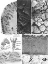
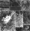
The histological structure of the frog digestive mucosa changes at the oesophagogastric junction. The pseudostratified ciliated mucosal epithelium of oesophageal type changes to a simple mucus-secreting epithelium of gastric type. The glands straighten and the muscularis mucosae develops as a complete layer. The muscularis increases in thickness. Unlike the mammalian stomach, in the frog the surface of the plicae forms convoluted ridges that delimit furrow-shaped pits. Two types of gastric glands are distinguished, fundal and pyloric. The former consist of mucous, oxynticopeptic and endocrine cells. The pyloric glandular cells are mainly of mucus-secreting type with scattered endocrine cells. Scattered endocrine cells of P, D, G, A, EC, and EC-L-like types are found in the glands along the stomach. It is concluded that the mucosal structure of the anuran oesophagogastric junction and stomach is less complicated than that of mammals, including man.
Full text
PDF


Images in this article
Selected References
These references are in PubMed. This may not be the complete list of references from this article.
- Bani G., Formigli L., Cecchi R. Morphological observations on the glands of the oesophagus and stomach of adult Rana esculenta and Bombina variegata. Ital J Anat Embryol. 1992 Apr-Jun;97(2):75–87. [PubMed] [Google Scholar]
- Buchan A. M., Polak J. M., Pearse A. G. Gut hormones in Salamandra salamandra. An immunocytochemical and electron microscopic investigation. Cell Tissue Res. 1980;211(2):331–343. doi: 10.1007/BF00236453. [DOI] [PubMed] [Google Scholar]
- Díaz de Rada O., Sesma P., Vázquez J. J. Endocrine cells of the gastric mucosa of Rana temporaria L. Histol Histopathol. 1987 Apr;2(2):119–128. [PubMed] [Google Scholar]
- El-Salhy M., Grimelius L., Wilander E., Abu-Sinna G., Lundqvist G. Histological and immunohistochemical studies of the endocrine cells of the gastrointestinal mucosa of the toad (Bufo regularis). Histochemistry. 1981;71(1):53–65. doi: 10.1007/BF00592570. [DOI] [PubMed] [Google Scholar]
- Geuze J. J. Light and electron microscope observations on the gastric mucosa of the frog (Rana esculenta). I. Normal structure. Z Zellforsch Mikrosk Anat. 1971;117(1):87–102. doi: 10.1007/BF00331104. [DOI] [PubMed] [Google Scholar]
- Geuze J. J. Light and electron microscope observations on the gastric mucosa of the frog (Rana esculenta). II. Structural alterations during hibernation. Z Zellforsch Mikrosk Anat. 1971;117(1):103–117. doi: 10.1007/BF00331105. [DOI] [PubMed] [Google Scholar]
- Giraud A. S., Yeomans N. D. Fine structure of the gastric mucous and endocrine cells of the toad, Bufo marinus. Cell Tissue Res. 1981;218(3):663–668. doi: 10.1007/BF00210123. [DOI] [PubMed] [Google Scholar]
- Loo S. K., Wong W. C. Histochemical observations on the mucins of the gastrointestinal tract in the toad (Bufo melanostictus). Acta Anat (Basel) 1975;91(1):97–103. doi: 10.1159/000144374. [DOI] [PubMed] [Google Scholar]
- Michelangeli F., Ruiz M. C., Dominguez M. G., Parthe V. Mammalian-like differentiation of gastric cells in the shark Hexanchus griseus. Cell Tissue Res. 1988 Jan;251(1):225–227. doi: 10.1007/BF00215469. [DOI] [PubMed] [Google Scholar]
- Michelangeli F., Sulcas D. M., Ruiz M. C. Ultrastructural studies of endocrine-like cells in the fundic gastric mucosa of the bullfrog, Rana catesbeiana. Cell Tissue Res. 1987 Nov;250(2):413–419. doi: 10.1007/BF00219085. [DOI] [PubMed] [Google Scholar]
- NORRIS J. L. The normal histology of the esophageal and gastric mucosae of the frog. Rana pipiens. J Exp Zool. 1959 Jun;141:155–173. doi: 10.1002/jez.1401410108. [DOI] [PubMed] [Google Scholar]
- Ruiz M. C., Acosta A., Abad M. J., Michelangeli F. Nonparallel secretion of pepsinogen and acid by gastric oxyntopeptic cells of the toad (Bufo marinus). Am J Physiol. 1993 Nov;265(5 Pt 1):G934–G941. doi: 10.1152/ajpgi.1993.265.5.G934. [DOI] [PubMed] [Google Scholar]
- SEDAR A. W. Electron microscopy of the oxyntic cell in the gastric glands of the bullfrog, Rana catesbiana. II. The acid-secreting gastric mucosa. J Biophys Biochem Cytol. 1961 May;10:47–57. doi: 10.1083/jcb.10.1.47. [DOI] [PMC free article] [PubMed] [Google Scholar]
- Suganuma T., Katsuyama T., Tsukahara M., Tatematsu M., Sakakura Y., Murata F. Comparative histochemical study of alimentary tracts with special reference to the mucous neck cells of the stomach. Am J Anat. 1981 Jun;161(2):219–238. doi: 10.1002/aja.1001610206. [DOI] [PubMed] [Google Scholar]