

Abstract
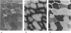
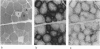
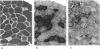
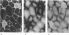
A quantitative histochemical study was made of superficial thigh muscle specimens (semimembranosus and some vastus lateralis) from topi, hartebeest, wildebeest and waterbuck (species listed in order of increasing size). Fibres were largest (up to 120 microns diameter) in waterbuck but smallest (maximum approximately 90 microns) in wildebeest. Type 2B fibres, most of them large, highly glycolytic and weakly aerobic, constituted approximately 75% of the cross-section of topi specimens and approximately 83% of the others, greater area fractions than in other large herbivores. Type 1 fibres, highly aerobic but weakly glycolytic, occupied only 2-3.5% of the area fractions, very low figures, even for these superficial sites. Type 2A fibres occupied > 20% areas in topi, approximately 15% in the other species. In waterbuck they were never more than moderately aerobic, but quite highly glycolytic; elsewhere their characteristic metabolic profiles were close to those of type 1 fibres. The 2B fractions indicate that glycolytic metabolism predominates over lipolytic in all 4 species. Mean enzymic capacities were compared semiquantitatively between species on the basis of wide-area photometric readings. Much the greatest difference was in aerobic (succinate dehydrogenase) capacities: the mean reading in topi was x 2.6 that in waterbuck, but wildebeest capacity came close to that of topi. These latter are the 2 most active species. Readings for the force-generating enzyme, actomyosin ATPase, were slightly weaker in the heavier species. This could be predicted on allometric grounds, but mass considerations appear to be overridden by behavioural differences in relation to metabolism.
Full text
PDF


Images in this article
Selected References
These references are in PubMed. This may not be the complete list of references from this article.
- Andrews F. M., Spurgeon T. L. Histochemical staining characteristics of normal horse skeletal muscle. Am J Vet Res. 1986 Aug;47(8):1843–1852. [PubMed] [Google Scholar]
- Ashmore C. R., Doerr L. Comparative aspects of muscle fiber types in different species. Exp Neurol. 1971 Jun;31(3):408–418. doi: 10.1016/0014-4886(71)90243-3. [DOI] [PubMed] [Google Scholar]
- Brooke M. H., Kaiser K. K. Muscle fiber types: how many and what kind? Arch Neurol. 1970 Oct;23(4):369–379. doi: 10.1001/archneur.1970.00480280083010. [DOI] [PubMed] [Google Scholar]
- Bárány M. ATPase activity of myosin correlated with speed of muscle shortening. J Gen Physiol. 1967 Jul;50(6 Suppl):197–218. doi: 10.1085/jgp.50.6.197. [DOI] [PMC free article] [PubMed] [Google Scholar]
- Emmett B., Hochachka P. W. Scaling of oxidative and glycolytic enzymes in mammals. Respir Physiol. 1981 Sep;45(3):261–272. doi: 10.1016/0034-5687(81)90010-4. [DOI] [PubMed] [Google Scholar]
- Ennion S., Sant'ana Pereira J., Sargeant A. J., Young A., Goldspink G. Characterization of human skeletal muscle fibres according to the myosin heavy chains they express. J Muscle Res Cell Motil. 1995 Feb;16(1):35–43. doi: 10.1007/BF00125308. [DOI] [PubMed] [Google Scholar]
- Goldspink G., Ward P. S. Changes in rodent muscle fibre types during post-natal growth, undernutrition and exercise. J Physiol. 1979 Nov;296:453–469. doi: 10.1113/jphysiol.1979.sp013016. [DOI] [PMC free article] [PubMed] [Google Scholar]
- Gunn H. M. Differences in the histochemical properties of skeletal muscles of different breeds of horses and dogs. J Anat. 1978 Dec;127(Pt 3):615–634. [PMC free article] [PubMed] [Google Scholar]
- Guth L., Samaha F. J. Qualitative differences between actomyosin ATPase of slow and fast mammalian muscle. Exp Neurol. 1969 Sep;25(1):138–152. doi: 10.1016/0014-4886(69)90077-6. [DOI] [PubMed] [Google Scholar]
- Johnson M. A., Polgar J., Weightman D., Appleton D. Data on the distribution of fibre types in thirty-six human muscles. An autopsy study. J Neurol Sci. 1973 Jan;18(1):111–129. doi: 10.1016/0022-510x(73)90023-3. [DOI] [PubMed] [Google Scholar]
- Lexell J., Henriksson-Larsén K., Sjöström M. Distribution of different fibre types in human skeletal muscles. 2. A study of cross-sections of whole m. vastus lateralis. Acta Physiol Scand. 1983 Jan;117(1):115–122. doi: 10.1111/j.1748-1716.1983.tb07185.x. [DOI] [PubMed] [Google Scholar]
- Mabuchi K., Sréter F. A. Actomyosin ATPase. II. Fiber typing by histochemical ATPase reaction. Muscle Nerve. 1980 May-Jun;3(3):233–239. doi: 10.1002/mus.880030308. [DOI] [PubMed] [Google Scholar]
- Martin T. P., Bodine-Fowler S., Roy R. R., Eldred E., Edgerton V. R. Metabolic and fiber size properties of cat tibialis anterior motor units. Am J Physiol. 1988 Jul;255(1 Pt 1):C43–C50. doi: 10.1152/ajpcell.1988.255.1.C43. [DOI] [PubMed] [Google Scholar]
- McMahon T. A. Using body size to understand the structural design of animals: quadrupedal locomotion. J Appl Physiol. 1975 Oct;39(4):619–627. doi: 10.1152/jappl.1975.39.4.619. [DOI] [PubMed] [Google Scholar]
- Meijer A. E. Improved histochemical method for the demonstration of the activity of alpha-glucan phosphorylase. I. The use of glucosyl acceptor dextran. Histochemie. 1968;12(3):244–252. doi: 10.1007/BF00306002. [DOI] [PubMed] [Google Scholar]
- ORNSTEIN L. The distributional error in microspectrophotometry. Lab Invest. 1952;1(2):250–265. [PubMed] [Google Scholar]
- Snow D. H., Guy P. S. Muscle fibre type composition of a number of limb muscles in different types of horse. Res Vet Sci. 1980 Mar;28(2):137–144. [PubMed] [Google Scholar]
- Spurway N. C. Objective characterization of cells in terms of microscopical parameters: an example from muscle histochemistry. Histochem J. 1981 Mar;13(2):269–317. doi: 10.1007/BF01006884. [DOI] [PubMed] [Google Scholar]
- Spurway N. C., Rowlerson A. M. Quantitative analysis of histochemical and immunohistochemical reactions in skeletal muscle fibres of Rana and Xenopus. Histochem J. 1989 Aug;21(8):461–476. doi: 10.1007/BF01845796. [DOI] [PubMed] [Google Scholar]
- Taylor C. R., Maloiy G. M., Weibel E. R., Langman V. A., Kamau J. M., Seeherman H. J., Heglund N. C. Design of the mammalian respiratory system. III Scaling maximum aerobic capacity to body mass: wild and domestic mammals. Respir Physiol. 1981 Apr;44(1):25–37. doi: 10.1016/0034-5687(81)90075-x. [DOI] [PubMed] [Google Scholar]
- White N. A., McGavin M. D., Smith J. E. Age-related changes in percentage of fiber types and mean fiber diameters of the ovine quadriceps muscles. Am J Vet Res. 1978 Aug;39(8):1297–1302. [PubMed] [Google Scholar]
- van den Hoven R., Wensing T., Breukink H. J., Meijer A. E., Kruip T. A. Variation of fiber types in the triceps brachii, longissimus dorsi, gluteus medius, and biceps femoris of horses. Am J Vet Res. 1985 Apr;46(4):939–941. [PubMed] [Google Scholar]