Dear Editor,
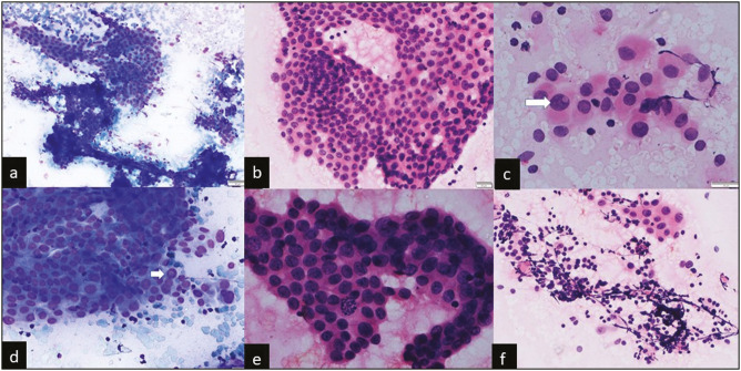
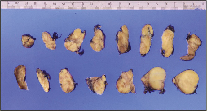
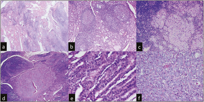
A 32-year-old young woman presented with complaints of swelling in front of her neck for 7 months. On physical examination, there was 5 cm × 4 cm firm, mobile, noncompressible swelling present on the left side of the neck, which was crossing the midline. This swelling was moving with deglutition but not on tongue protrusion. Superiorly, it was extending up to the thyroid notch, inferiorly 1 cm from the suprasternal notch, laterally reaching the anterior border of the left sternocleidomastoid and medially crossing the midline. Her thyroid profile revealed elevated thyroid stimulating hormone—6.43 µIU/mL (range = 0.35–5.50 µIU/mL) and normal total triiodothyronine and thyroxine. Ultrasound neck revealed a well-defined heterogeneously hypoechoic lesion of size 2.8 cm × 2.2 cm in the left lobe and an ill-defined similar lesion measuring 2.3 cm × 1.3 cm in the right lobe with tiny cystic spaces within. Ultrasound-guided fine needle aspiration (FNA) was done from the left lobe since it was well defined larger lesion with the solid component. Smears showed classical features of papillary thyroid carcinoma (PTC) including papillae formation, nuclear crowding, overlapping, grooving, pseudoinclusions, and cellular swirls [Figure 1]. In addition, the background showed an increase in lymphocytes along with lymphocytic infiltration into the follicular cells [Figure 1]. Therefore, the diagnosis of papillary carcinoma thyroid arising in a background of lymphocytic thyroiditis was rendered. The cytological diagnosis led to total thyroidectomy of the patient. Intraoperatively, the surgeon noted a 3 cm × 3 cm hard nodule involving the lower pole of the left lobe of the thyroid. The rest of the thyroid gland was normal. A total thyroidectomy specimen was sent for histopathological examination. Grossly, the thyroid weighed 32 g and measured 8 cm × 5 cm × 3.5 cm. External aspect appeared nodular and irregular however no capsular breech was identified. The cut surface of both lobes showed multiple ill-defined nodules, the largest seen in the left lobe measuring 2.5 cm × 2 cm × 1 cm [Figure 2]. Microscopically, both lobes of the thyroid showed lymphocytic infiltration with germinal centers [Figure 3a and b], and thyroid follicles of varying sizes contained colloid with focal Hurthle cell change [Figure 3c]. Hence, the diagnosis of lymphocytic thyroiditis was made provisionally. Furthermore, it was decided to embed the specimen so that the microscopic focus of PTC is not missed. Moreover, histopathology revealed evidence of microscopic PTC in the left lobe [Figure 3d–f]. Therefore, this was a case of microscopic PTC arising in a background of lobectomy (LT), which was nearly missed on histopathology.
Figure 1.
(a) FNA smears show few papillae and sheets of tumor cells with surrounding lymphocytes (Giemsa, 10×). (b) FNA smears show cellular swirls [hematoxylin and eosin (H&E), 20×]. (c) Tumor cells show pseudoinclusions, nuclear grooving along with Hürthle cell change (H&E, 40×). (d) Tumor cells show pseudoinclusions along with infiltrating lymphocytes at lower and higher power respectively (Giemsa, 20×). (e) Occasional mitosis is seen (H&E, 20×). (f) Smears show numerous lymphocytes surrounding the cells (H&E, 20×)
Figure 2.
Serial gross sections of both the thyroid lobes showing multiple ill-defined nodules
Figure 3.
(a and b) On histopathology, both lobes of the thyroid showed lymphocytic infiltration with germinal centers [hematoxylin and eosin (H&E), 4× and 10×]. (c) Focal Hürthle cell change was also seen (H&E, 20×). (d–f) Microscopic focus of PTC was seen on further sections along with nuclear features of PTC (H&E, 4×, 40×, and 40×)
It can be a real diagnostic war to distinguish reactive nuclear changes associated with LT from PTC arising in a background of LT on FNA cytology (FNAC). The incidence of neoplasia in the setting of LT by FNAC is 4%.[1] FNAC has stood the test of time and has proven its mantle in diagnosing PTC arising in LT with sensitivity >90%.[2] The foci of reactive follicular epithelium in LT usually are adjacent to the inflammatory infiltrate and lack infiltrative edges. The nuclei are mostly round and do not exhibit overlapping and prominent intranuclear inclusions.[1] On the other hand, aspirates of PTC arising in association with LT usually show two types of cell population, first tumor cell fragments devoid of infiltrating lymphocytes with nuclear features of PTC and a second population of reactive follicular epithelium infiltrated by lymphocytes with focal nuclear atypia in isolation showing some but not all features of PTC.[3] A literature search suggests that FNA has an overall accuracy rate of around 95% in the detection of thyroid malignancies.[4] Nevertheless, FNAC has its own limitations and diagnostic pitfalls. These limitations include false negative and false positive results. Histopathological correlation helps in the confirmation.[5] The reported diagnostic pitfalls may occur due to various reasons including those related to specimen adequacy, sampling techniques, the experience of the pathologist interpreting the aspirate, and the overlapping cytological features between some benign and malignant thyroid lesions.
Our case demonstrated the importance of ultrasound-guided FNA in the diagnosis of microscopic PTC, which was nearly missed in histopathology. Cytopathologists must note that the presence of thyroiditis might create confusion in the interpretation of cytological results, so must be reported with caution.[6] Here, we discuss a case of microscopic PTC arising in a background of LT, which was masqueraded by LT features on histopathology. A careful and rigorous search for various cytological features and accurate sampling can help reduce the number of false-positive and false-negative diagnoses. This case taught us that the histopathological samples must be evaluated completely, in the presence of cytological PTC diagnosis; so that microscopic PTC foci are not missed.
Author contributions
This manuscript has been read and approved by all the authors and represents honest work.
Ethical approval
As this is a letter to the editor without identifiers, our institution does not require approval from the Institutional Review Board or its equivalent.
Conflicts of interest
There are no conflicts of interest.
Funding Statement
Nil
REFERENCES
- 1.Makhdoomi R, Mustafa F, Malik R, Bhat S, Alam K, Bashir H, et al. Coexistent papillary carcinoma of thyroid and Hashimoto’s thyroiditis - Diagnosis on fine needle aspiration cytology. Int J Endocrinol Metab. 2013;11:191–4. doi: 10.5812/ijem.7453. [DOI] [PMC free article] [PubMed] [Google Scholar]
- 2.Nguyen GK, Lee MW, Ginsberg J, Wragg T, Bilodeau D. Fine-needle aspiration of the thyroid: An overview. Cytojournal. 2005;2:12. doi: 10.1186/1742-6413-2-12. [DOI] [PMC free article] [PubMed] [Google Scholar]
- 3.Kesmodel SB, Terhune KP, Canter RJ, Mandel SJ, LiVolsi VA, Baloch ZW, et al. The diagnostic dilemma of follicular variant of papillary thyroid carcinoma. Surgery. 2003;134:1005–12. doi: 10.1016/j.surg.2003.07.015. [DOI] [PubMed] [Google Scholar]
- 4.Patel DM, Shah DP, Goswami DH, Gonsai RN, Shah DS, Patel DA. Accuracy of fine needle aspiration cytology in diagnosis of thyroid swelling: Accuracy of FNAC in diagnosis of thyroid swelling. Natl J Integr Res Med. 2012;3:124–9. [Google Scholar]
- 5.Abdullah N, Hajeer M, Abudalu L, Sughayer M. Correlation study of thyroid nodule cytopathology and histopathology at two institutions in Jordan. Cytojournal. 2018;15:24. doi: 10.4103/cytojournal.cytojournal_53_17. [DOI] [PMC free article] [PubMed] [Google Scholar]
- 6.Levy-Blitchtein S, Plasencia-Rebata S, Morales Luna D, Del Valle Mendoza J. Coexistence of papillary thyroid microcarcinoma and mucosa-associated lymphoid tissue lymphoma in a context of Hashimoto’s thyroiditis. Asian Pac J Trop Med. 2016;9:812–4. doi: 10.1016/j.apjtm.2016.06.017. [DOI] [PubMed] [Google Scholar]


