

Abstract
The serous mesothelium of the serosa and mesentery of the small intestine in the mouse were examined by scanning and transmission electron microscopy. The serosa consisted of a single layer of flattened microvilli-bearing cells containing nuclei, caveolae and micropinocytotic vesicles. The observations in this study differed from previous reports on mesothelial surfaces in two respects. A surface layer of amorphous material was present over parts of the serosa. This layer probably represents serous fluid trapped by the mesothelial microvilli but is unaffected by prefixation rinsing in saline or ultrasonic cleaning. The layer is lost following osmication and routine processing for transmission electron microscopy. The possibility that a serous fluid layer may be preserved in this way may be useful in assessing changes in the peritoneum. Stomata were observed in the mesentery but there was no evidence of a connection with the lymphatic system. The presence of mesenteric stomata may explain the difference in permeability reported between parietal peritoneum and mesentery.
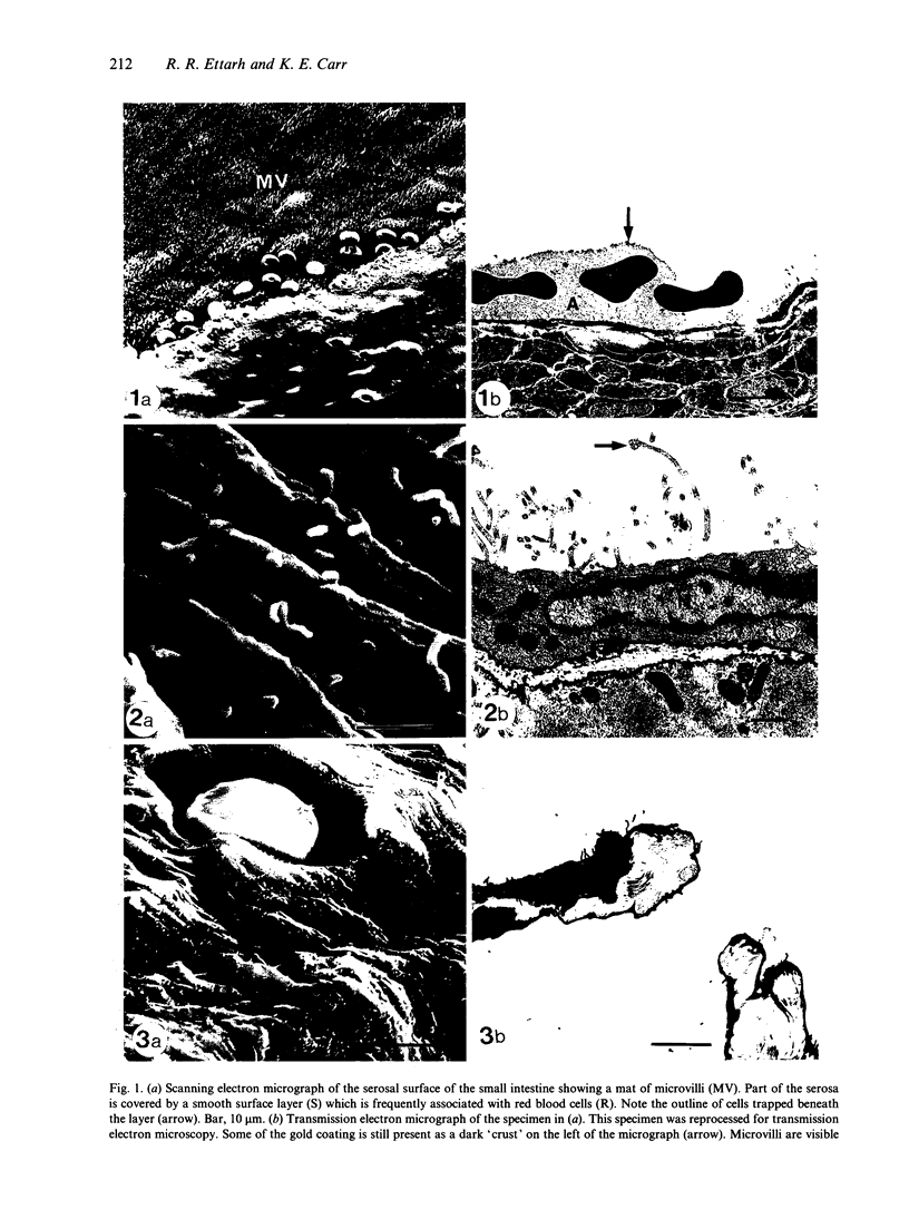
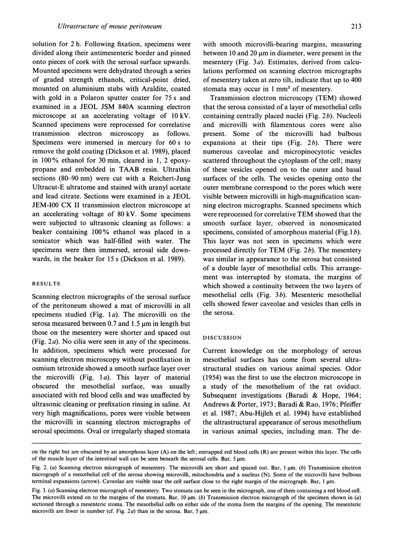
Full text
PDF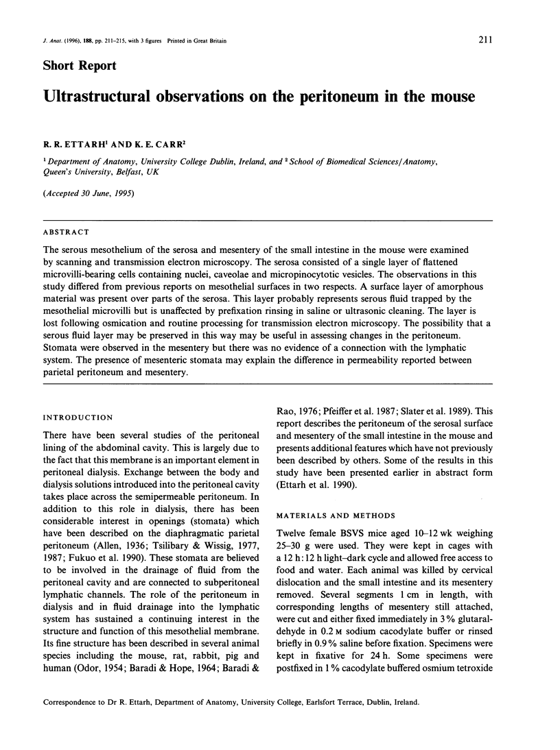
Images in this article

Selected References
These references are in PubMed. This may not be the complete list of references from this article.
- Andrews P. M., Porter K. R. The ultrastructural morphology and possible functional significance of mesothelial microvilli. Anat Rec. 1973 Nov;177(3):409–426. doi: 10.1002/ar.1091770307. [DOI] [PubMed] [Google Scholar]
- BARADI A. F., HOPE J. OBSERVATIONS ON ULTRASTRUCTURE OF RABBIT MESOTHELIUM. Exp Cell Res. 1964 Mar;34:33–44. doi: 10.1016/0014-4827(64)90180-6. [DOI] [PubMed] [Google Scholar]
- Baradi A. F., Rao S. N. A scanning electron microscope study of mouse peritoneal mesothelium. Tissue Cell. 1976;8(1):159–162. doi: 10.1016/0040-8166(76)90027-6. [DOI] [PubMed] [Google Scholar]
- Dickson G. R., McKenna S., McColl K., Carr K. E. Examination of mucosal surfaces by scanning electron microscopy before and after removal of debris. J Microsc. 1989 Jan;153(Pt 1):75–79. doi: 10.1111/j.1365-2818.1989.tb01468.x. [DOI] [PubMed] [Google Scholar]
- Fukuo Y., Shinohara H., Matsuda T. The distribution of lymphatic stomata in the diaphragm of the golden hamster. J Anat. 1990 Apr;169:13–21. [PMC free article] [PubMed] [Google Scholar]
- Leak L. V., Rahil K. Permeability of the diaphragmatic mesothelium: the ultrastructural basis for "stomata". Am J Anat. 1978 Apr;151(4):557–593. doi: 10.1002/aja.1001510409. [DOI] [PubMed] [Google Scholar]
- ODOR D. L. Observations of the rat mesothelium with the electron and phase microscopes. Am J Anat. 1954 Nov;95(3):433–465. doi: 10.1002/aja.1000950304. [DOI] [PubMed] [Google Scholar]
- Shinohara H., Nakatani T., Matsuda T. The presence of lymphatic stomata in the ovarian bursa of the golden hamster. Anat Rec. 1985 Sep;213(1):44–52. doi: 10.1002/ar.1092130107. [DOI] [PubMed] [Google Scholar]
- Slater N. J., Raftery A. T., Cope G. H. The ultrastructure of human abdominal mesothelium. J Anat. 1989 Dec;167:47–56. [PMC free article] [PubMed] [Google Scholar]
- Tsilibary E. C., Wissig S. L. Absorption from the peritoneal cavity: SEM study of the mesothelium covering the peritoneal surface of the muscular portion of the diaphragm. Am J Anat. 1977 May;149(1):127–133. doi: 10.1002/aja.1001490111. [DOI] [PubMed] [Google Scholar]
- Tsilibary E. C., Wissig S. L. Light and electron microscope observations of the lymphatic drainage units of the peritoneal cavity of rodents. Am J Anat. 1987 Oct;180(2):195–207. doi: 10.1002/aja.1001800209. [DOI] [PubMed] [Google Scholar]