Abstract
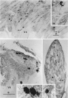
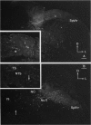
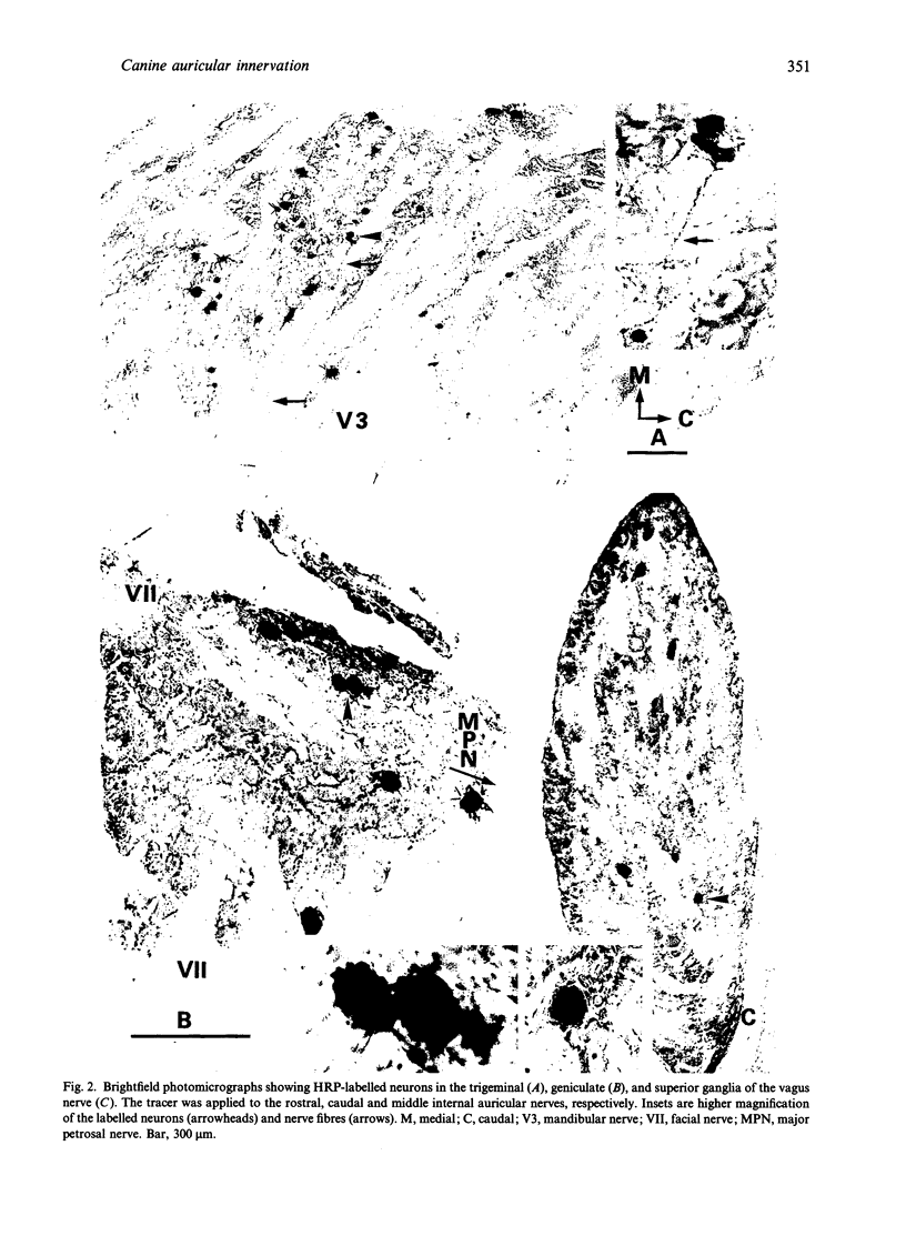
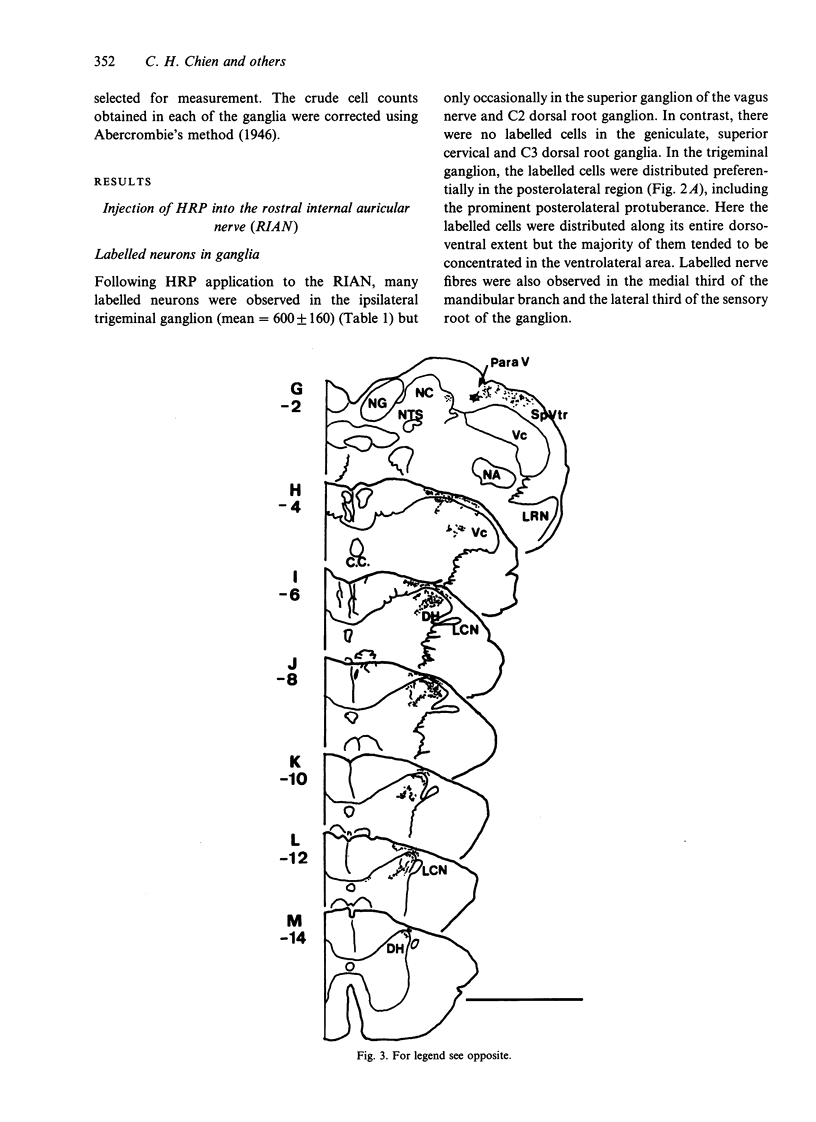
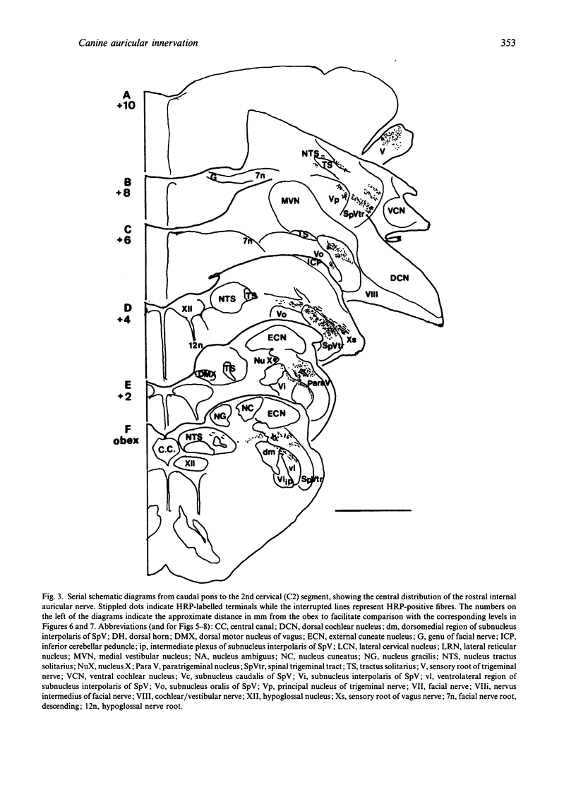
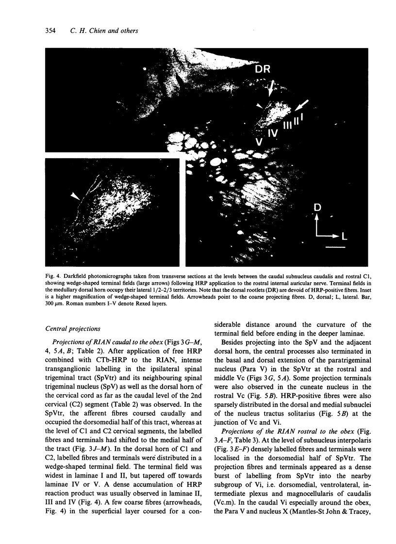
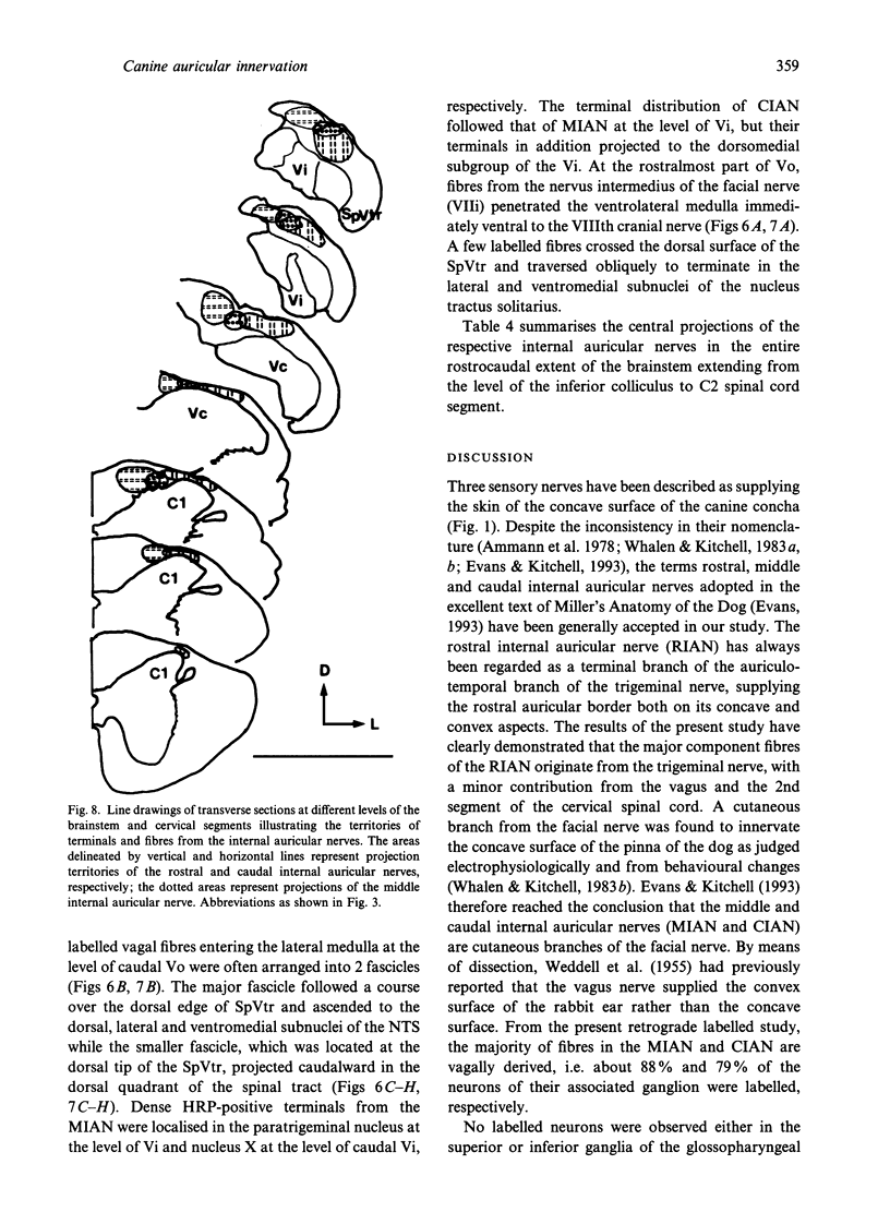
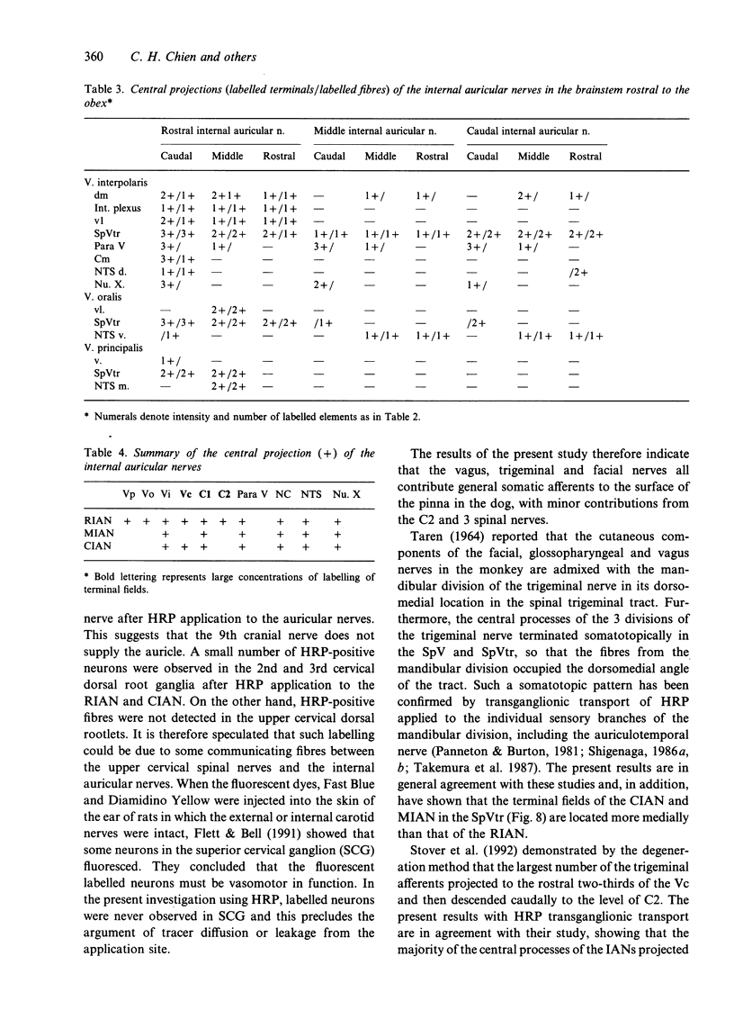
The cranial components and central terminations of the sensory nerves supplying the concave surface of the puppy's pinna, namely, the rostral, middle and caudal internal auricular nerves (RIAN, MIAN and CIAN) were investigated using horseradish peroxidase retrograde and transganglionic labelling techniques. All the 3 internal auricular nerves received contributions from the vagus. The RIAN received additional fibres from the trigeminal nerve while the MIAN and CIAN contained fibres derived from the facial nerves. In the brainstem, collaterals from the descending fibres of the afferents were given off at all levels to the medially located spinal trigeminal nucleus (SpV) which extended rostocaudally from the principal nucleus of the trigeminal nerve, subnuclei oralis, interpolaris and caudalis of the SpV to C1 or C2 cervical segment. The greatest density of central projections was observed in the subnucleus caudalis and C1. In the latter, the terminal field in the dorsal horn was roughly wedge-shaped, tapering off medially from lamina I towards lamina V. A somatotopic organisation was observed in the spinal trigeminal tract (SpVtr) and spinal trigeminal nucleus (SpV) in which the projection fibres and terminal fields of the RIAN were located lateral to those of the MIAN and CIAN. Some nontrigeminal nuclei, e.g. paratrigeminal and cuneate nuclei, nucleus X and the nucleus of the solitary tract were also labelled following HRP application to the internal auricular nerves. The localisation of the central projections of the internal auricular nerves as well as the 2nd order neurons in some specific nuclei to which the afferent fibres project is consistent with the concept of a brainstem somatovisceral link.
Full text
PDF
Images in this article
Selected References
These references are in PubMed. This may not be the complete list of references from this article.
- Abrahams V. C., Richmond F. J., Keane J. Projections from C2 and C3 nerves supplying muscles and skin of the cat neck: a study using transganglionic transport of horseradish peroxidase. J Comp Neurol. 1984 Nov 20;230(1):142–154. doi: 10.1002/cne.902300113. [DOI] [PubMed] [Google Scholar]
- Beckstead R. M., Norgren R. An autoradiographic examination of the central distribution of the trigeminal, facial, glossopharyngeal, and vagal nerves in the monkey. J Comp Neurol. 1979 Apr 1;184(3):455–472. doi: 10.1002/cne.901840303. [DOI] [PubMed] [Google Scholar]
- Bereiter D. A., Hathaway C. B., Benetti A. P. Caudal portions of the spinal trigeminal complex are necessary for autonomic responses and display Fos-like immunoreactivity after corneal stimulation in the cat. Brain Res. 1994 Sep 19;657(1-2):73–82. doi: 10.1016/0006-8993(94)90955-5. [DOI] [PubMed] [Google Scholar]
- Campbell S. K., Parker T. D., Welker W. Somatotopic organization of the external cuneate nucleus in albino rats. Brain Res. 1974 Aug 30;77(1):1–23. doi: 10.1016/0006-8993(74)90801-4. [DOI] [PubMed] [Google Scholar]
- Chan-Palay V. The paratrigeminal nucleus. II. Identification and inter-relations of catecholamine axons, indoleamine axons, and substance P immunoreactive cells in the neuropil. J Neurocytol. 1978 Aug;7(4):419–442. doi: 10.1007/BF01173989. [DOI] [PubMed] [Google Scholar]
- Chudler E. H., Foote W. E., Poletti C. E. Responses of cat C1 spinal cord dorsal and ventral horn neurons to noxious and non-noxious stimulation of the head and face. Brain Res. 1991 Aug 2;555(2):181–192. doi: 10.1016/0006-8993(91)90341-r. [DOI] [PubMed] [Google Scholar]
- Contreras R. J., Beckstead R. M., Norgren R. The central projections of the trigeminal, facial, glossopharyngeal and vagus nerves: an autoradiographic study in the rat. J Auton Nerv Syst. 1982 Nov;6(3):303–322. doi: 10.1016/0165-1838(82)90003-0. [DOI] [PubMed] [Google Scholar]
- Craig A. D. Propriospinal input to thoracolumbar sympathetic nuclei from cervical and lumbar lamina I neurons in the cat and the monkey. J Comp Neurol. 1993 May 22;331(4):517–530. doi: 10.1002/cne.903310407. [DOI] [PubMed] [Google Scholar]
- Flett D. L., Bell C. Topography of functional subpopulations of neurons in the superior cervical ganglion of the rat. J Anat. 1991 Aug;177:55–66. [PMC free article] [PubMed] [Google Scholar]
- Jacquin M. F., Rhoades R. W. Cell structure and response properties in the trigeminal subnucleus oralis. Somatosens Mot Res. 1990;7(3):265–288. doi: 10.3109/08990229009144709. [DOI] [PubMed] [Google Scholar]
- Jacquin M. F., Semba K., Egger M. D., Rhoades R. W. Organization of HRP-labeled trigeminal mandibular primary afferent neurons in the rat. J Comp Neurol. 1983 Apr 20;215(4):397–420. doi: 10.1002/cne.902150405. [DOI] [PubMed] [Google Scholar]
- Jacquin M. F., Semba K., Rhoades R. W., Egger M. D. Trigeminal primary afferents project bilaterally to dorsal horn and ipsilaterally to cerebellum, reticular formation, and cuneate, solitary, supratrigeminal and vagal nuclei. Brain Res. 1982 Aug 26;246(2):285–291. doi: 10.1016/0006-8993(82)91177-5. [DOI] [PubMed] [Google Scholar]
- KERR F. W. Facial, vagal and glossopharyngeal nerves in the cat. Afferent connections. Arch Neurol. 1962 Apr;6:264–281. doi: 10.1001/archneur.1962.00450220006003. [DOI] [PubMed] [Google Scholar]
- Mantle-St John L. A., Tracey D. J. Somatosensory nuclei in the brainstem of the rat: independent projections to the thalamus and cerebellum. J Comp Neurol. 1987 Jan 8;255(2):259–271. doi: 10.1002/cne.902550209. [DOI] [PubMed] [Google Scholar]
- Matsushita M., Okado N., Ikeda M., Hosoya Y. Descending projections from the spinal and mesencephalic nuclei of the trigeminal nerve to the spinal cord in the cat. A study with the horseradish peroxidase technique. J Comp Neurol. 1981 Feb 20;196(2):173–187. doi: 10.1002/cne.901960202. [DOI] [PubMed] [Google Scholar]
- McHaffie J. G., Larson M. A., Stein B. E. Response properties of nociceptive and low-threshold neurons in rat trigeminal pars caudalis. J Comp Neurol. 1994 Sep 15;347(3):409–425. doi: 10.1002/cne.903470307. [DOI] [PubMed] [Google Scholar]
- Menétrey D., Basbaum A. I. Spinal and trigeminal projections to the nucleus of the solitary tract: a possible substrate for somatovisceral and viscerovisceral reflex activation. J Comp Neurol. 1987 Jan 15;255(3):439–450. doi: 10.1002/cne.902550310. [DOI] [PubMed] [Google Scholar]
- Mesulam M. M. Tetramethyl benzidine for horseradish peroxidase neurohistochemistry: a non-carcinogenic blue reaction product with superior sensitivity for visualizing neural afferents and efferents. J Histochem Cytochem. 1978 Feb;26(2):106–117. doi: 10.1177/26.2.24068. [DOI] [PubMed] [Google Scholar]
- Panneton W. M., Burton H. Corneal and periocular representation within the trigeminal sensory complex in the cat studied with transganglionic transport of horseradish peroxidase. J Comp Neurol. 1981 Jul 1;199(3):327–344. doi: 10.1002/cne.901990303. [DOI] [PubMed] [Google Scholar]
- Pfaller K., Arvidsson J. Central distribution of trigeminal and upper cervical primary afferents in the rat studied by anterograde transport of horseradish peroxidase conjugated to wheat germ agglutinin. J Comp Neurol. 1988 Feb 1;268(1):91–108. doi: 10.1002/cne.902680110. [DOI] [PubMed] [Google Scholar]
- Rhoton A. L., Jr, O'Leary J. L., Ferguson J. P. The trigeminal, facial, vagal, and glossopharyngeal nerves in the monkey. Afferent connections. Arch Neurol. 1966 May;14(5):530–540. doi: 10.1001/archneur.1966.00470110074010. [DOI] [PubMed] [Google Scholar]
- Ruggiero D. A., Ross C. A., Reis D. J. Projections from the spinal trigeminal nucleus to the entire length of the spinal cord in the rat. Brain Res. 1981 Nov 30;225(2):225–233. doi: 10.1016/0006-8993(81)90832-5. [DOI] [PubMed] [Google Scholar]
- Shigenaga Y., Chen I. C., Suemune S., Nishimori T., Nasution I. D., Yoshida A., Sato H., Okamoto T., Sera M., Hosoi M. Oral and facial representation within the medullary and upper cervical dorsal horns in the cat. J Comp Neurol. 1986 Jan 15;243(3):388–408. doi: 10.1002/cne.902430309. [DOI] [PubMed] [Google Scholar]
- Shigenaga Y., Okamoto T., Nishimori T., Suemune S., Nasution I. D., Chen I. C., Tsuru K., Yoshida A., Tabuchi K., Hosoi M. Oral and facial representation in the trigeminal principal and rostral spinal nuclei of the cat. J Comp Neurol. 1986 Feb 1;244(1):1–18. doi: 10.1002/cne.902440102. [DOI] [PubMed] [Google Scholar]
- Stover J. D., Schwab C. A., Matthews M. A. Selective deafferentation of convergent inputs to trigeminal subnucleus caudalis: effects on calcitonin gene-related peptide distribution. Somatosens Mot Res. 1992;9(2):107–130. doi: 10.3109/08990229209144766. [DOI] [PubMed] [Google Scholar]
- TAREN J. A. THE POSITIONS OF THE CUTANEOUS COMPONENTS OF THE FACIAL, GLOSSOPHARYNGEAL AND VAGAL NERVES IN THE SPINAL TRACT OF V. J Comp Neurol. 1964 Jun;122:389–397. doi: 10.1002/cne.901220308. [DOI] [PubMed] [Google Scholar]
- Takemura M., Sugimoto T., Sakai A. Topographic organization of central terminal region of different sensory branches of the rat mandibular nerve. Exp Neurol. 1987 Jun;96(3):540–557. doi: 10.1016/0014-4886(87)90217-2. [DOI] [PubMed] [Google Scholar]
- Takemura M., Sugimoto T., Shigenaga Y. Difference in central projection of primary afferents innervating facial and intraoral structures in the rat. Exp Neurol. 1991 Mar;111(3):324–331. doi: 10.1016/0014-4886(91)90099-x. [DOI] [PubMed] [Google Scholar]
- Tavares I., Lima D. Descending projections from the caudal medulla oblongata to the superficial or deep dorsal horn of the rat spinal cord. Exp Brain Res. 1994;99(3):455–463. doi: 10.1007/BF00228982. [DOI] [PubMed] [Google Scholar]
- WEDDELL G., PALLIE W., PALMER E. Studies on the innervation of skin. I. The origin, course and number of sensory nerves supplying the rabbit ear. J Anat. 1955 Apr;89(2):162–174. [PMC free article] [PubMed] [Google Scholar]
- Whalen L. R., Kitchell R. L. Electrophysiologic and behavioral studies of the cutaneous nerves of the concave surface of the pinna and the external ear canal of the dog. Am J Vet Res. 1983 Apr;44(4):628–634. [PubMed] [Google Scholar]
- Whalen L. R., Kitchell R. L. Electrophysiologic studies of the cutaneous nerves of the head of the dog. Am J Vet Res. 1983 Apr;44(4):615–627. [PubMed] [Google Scholar]
- Yasui Y., Itoh K., Mizuno N., Nomura S., Takada M., Konishi A., Kudo M. The posteromedial ventral nucleus of the thalamus (VPM) of the cat: direct ascending projections to the cytoarchitectonic subdivisions. J Comp Neurol. 1983 Oct 20;220(2):219–228. doi: 10.1002/cne.902200209. [DOI] [PubMed] [Google Scholar]
- Yonehara N., Sawada T., Matsuura H., Inoki R. Influence of electro-acupuncture on the release of substance P and the potential evoked by tooth pulp stimulation in the trigeminal nucleus caudalis of the rabbit. Neurosci Lett. 1992 Aug 3;142(1):53–56. doi: 10.1016/0304-3940(92)90618-h. [DOI] [PubMed] [Google Scholar]


