Abstract
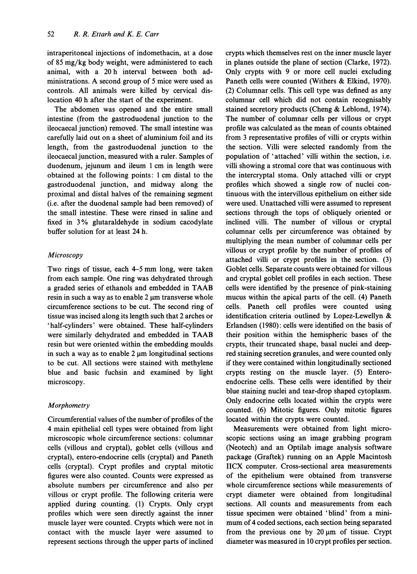
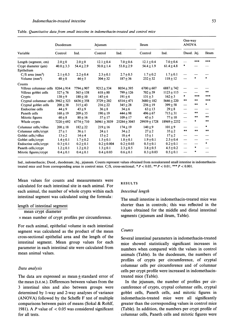
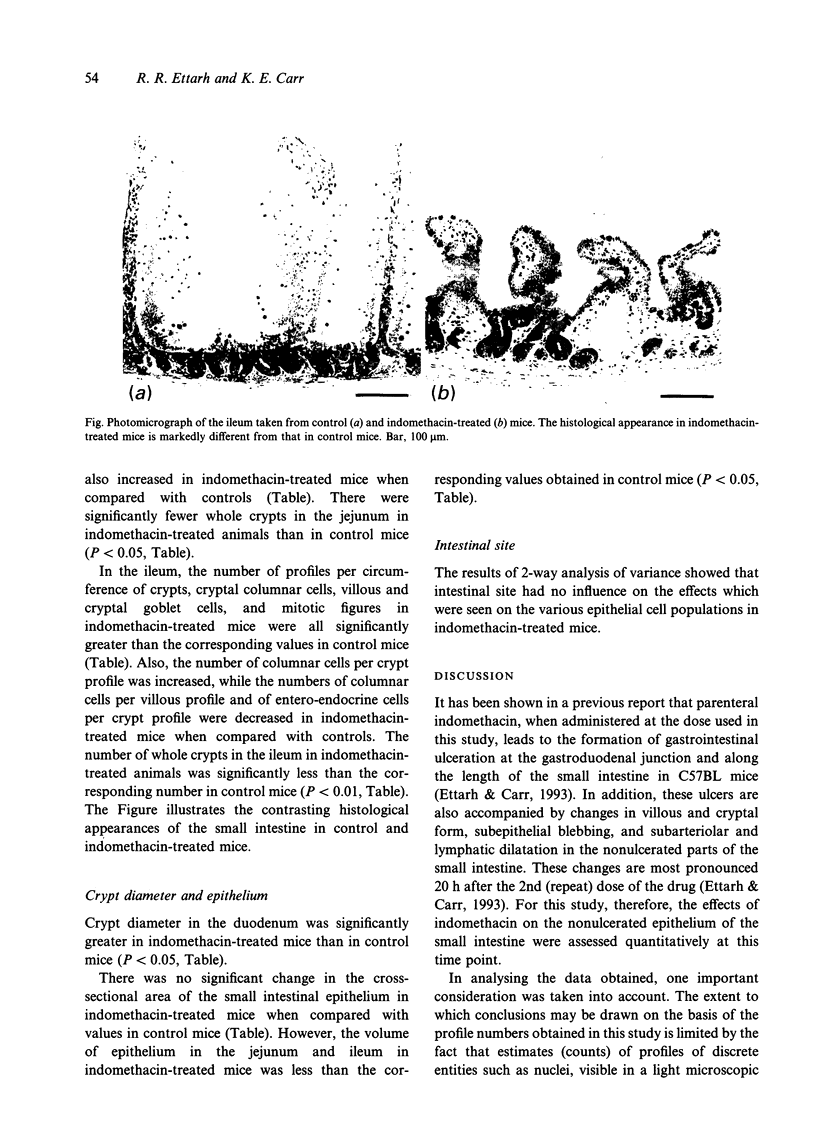
To obtain a clearer understanding of the changes which are induced in the small intestine of the mouse by an ulcerogenic dose of indomethacin, a quantitative analysis of the nonulcerated small intestinal mucosa was performed in mice that were given 2 injections of indomethacin at a dose of 85 mg/kg body weight. At 20 h after the administration of the drug, values were obtained for epithelial volume, whole crypt number, and for the number of profiles of columnar, Paneth, entero-endocrine and goblet cells and cryptal mitotic figures in the small intestine. Comparison of the values obtained from indomethacin-treated mice with those from control mice showed that there were fewer whole crypts and a reduced epithelial volume in the jejunum and ileum in indomethacin-treated mice. The numbers of columnar and Paneth cell profiles and of mitotic figures were significantly greater in the jejunal and ileal crypts in indomethacin-treated mice than in controls. These findings suggest that the administration of high-dose indomethacin in the mouse leads to crypt losses and increased mitotic activity in the nonulcerated parts of the small intestine.
Full text
PDF


Images in this article
Selected References
These references are in PubMed. This may not be the complete list of references from this article.
- Bjarnason I., Macpherson A. The changing gastrointestinal side effect profile of non-steroidal anti-inflammatory drugs. A new approach for the prevention of a new problem. Scand J Gastroenterol Suppl. 1989;163:56–64. doi: 10.3109/00365528909091176. [DOI] [PubMed] [Google Scholar]
- Cheng H., Leblond C. P. Origin, differentiation and renewal of the four main epithelial cell types in the mouse small intestine. I. Columnar cell. Am J Anat. 1974 Dec;141(4):461–479. doi: 10.1002/aja.1001410403. [DOI] [PubMed] [Google Scholar]
- Clarke R. M. The effect of growth and of fasting on the number of villi and crypts in the small intestine of the albino rat. J Anat. 1972 May;112(Pt 1):27–33. [PMC free article] [PubMed] [Google Scholar]
- Ettarh R. R., Carr K. E. Structural and morphometric analysis of murine small intestine after indomethacin administration. Scand J Gastroenterol. 1993 Sep;28(9):795–802. doi: 10.3109/00365529309104012. [DOI] [PubMed] [Google Scholar]
- Gaskin R. J., Gad A., Barros D'sa A. A., Joffe S. N., Baron J. H. Natural history and morphology of secretagogue-induced duodenal ulcers in rats. Gastroenterology. 1975 Oct;69(4):903–910. [PubMed] [Google Scholar]
- Hasan M., Ferguson A. Measurements of intestinal villi non-specific and ulcer-associated duodenitis-correlation between area of microdissected villus and villus epithelial cell count. J Clin Pathol. 1981 Oct;34(10):1181–1186. doi: 10.1136/jcp.34.10.1181. [DOI] [PMC free article] [PubMed] [Google Scholar]
- Hasan M., Sircus W., Ferguson A. Duodenal mucosal architecture in non-specific and ulcer-associated duodenitis. Gut. 1981 Aug;22(8):637–641. doi: 10.1136/gut.22.8.637. [DOI] [PMC free article] [PubMed] [Google Scholar]
- Lopez-Lewellyn J., Erlandsen S. L. Cytodifferentiation of the rat Paneth cell: an immunocytochemical investigation in suckling and weanling animals. Am J Anat. 1980 Jul;158(3):285–297. doi: 10.1002/aja.1001580305. [DOI] [PubMed] [Google Scholar]
- Philp J. R., Buchanan T. J. Quantitative measurement on finite tissue sections. J Anat. 1971 Jan;108(Pt 1):89–97. [PMC free article] [PubMed] [Google Scholar]
- Robert A. Cytoprotection by prostaglandins. Gastroenterology. 1979 Oct;77(4 Pt 1):761–767. [PubMed] [Google Scholar]
- Shorrock C. J., Prescott R. J., Rees W. D. The effects of indomethacin on gastroduodenal morphology and mucosal pH gradient in the healthy human stomach. Gastroenterology. 1990 Aug;99(2):334–339. doi: 10.1016/0016-5085(90)91013-v. [DOI] [PubMed] [Google Scholar]
- Takeuchi K., Johnson L. R. Effect of cell proliferation on healing of gastric and duodenal ulcers in rats. Digestion. 1986;33(2):92–100. doi: 10.1159/000199280. [DOI] [PubMed] [Google Scholar]
- Wallace J. L., Arfors K. E., McKnight G. W. A monoclonal antibody against the CD18 leukocyte adhesion molecule prevents indomethacin-induced gastric damage in the rabbit. Gastroenterology. 1991 Apr;100(4):878–883. doi: 10.1016/0016-5085(91)90259-n. [DOI] [PubMed] [Google Scholar]
- Withers H. R., Elkind M. M. Microcolony survival assay for cells of mouse intestinal mucosa exposed to radiation. Int J Radiat Biol Relat Stud Phys Chem Med. 1970;17(3):261–267. doi: 10.1080/09553007014550291. [DOI] [PubMed] [Google Scholar]


