

Abstract
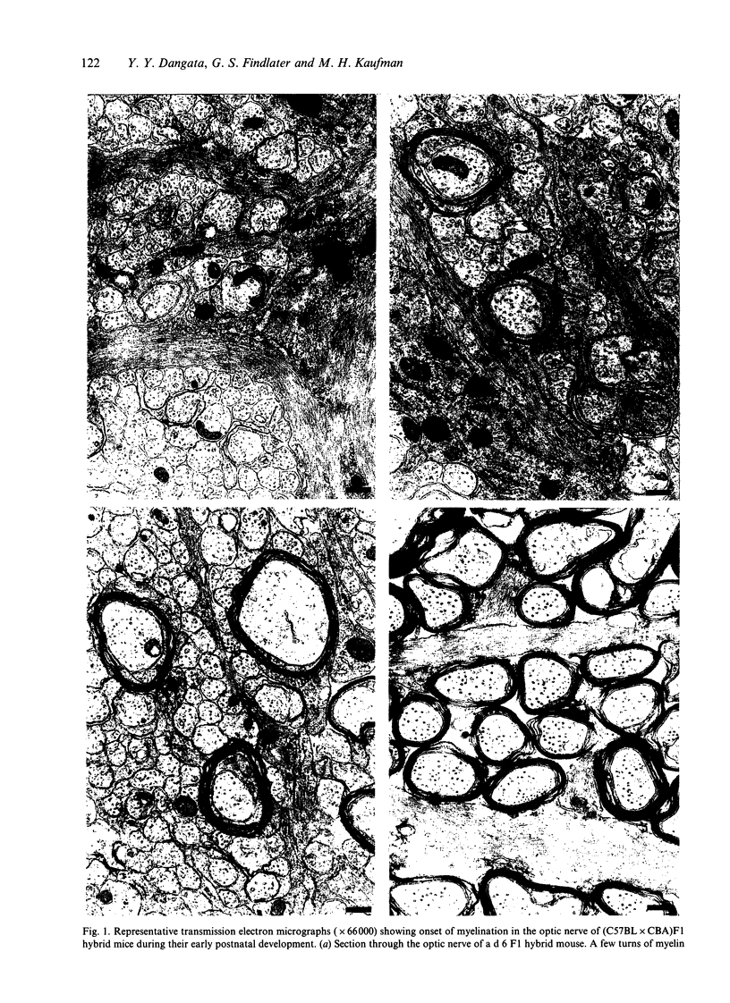
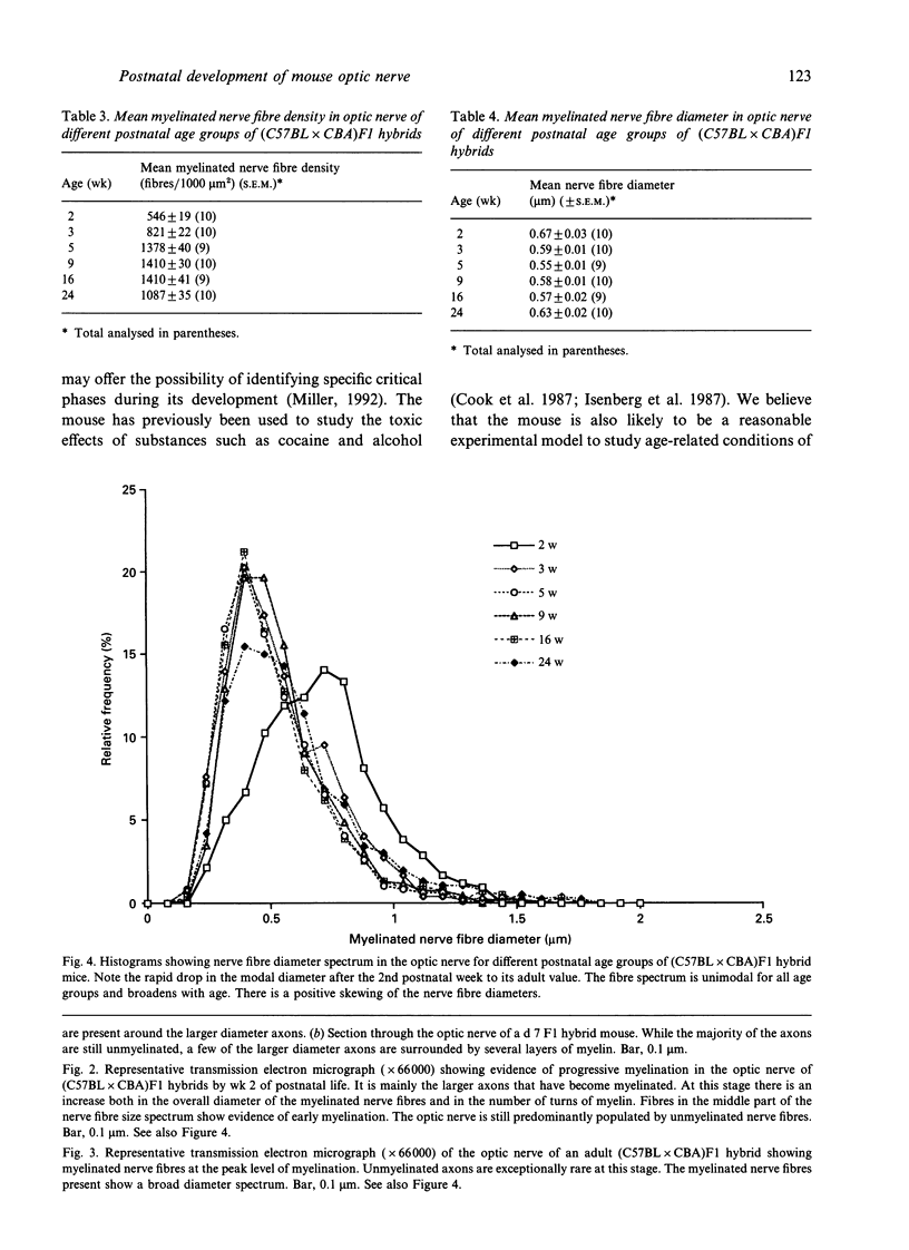
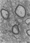
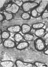
A morphometric analysis of the optic nerve in different age groups of (C57BL x CBA)F1 hybrid mice was carried out. Morphometric parameters examined were mean nerve cross-sectional area (csa), mean myelinated nerve fibre count, mean myelinated nerve fibre density and myelinated nerve fibre size distribution. The findings revealed that the optic nerve continues to develop well into adult life. Growth in calibre was very rapid during the early stage of postnatal life, but progressively slowed with age thereafter. No myelinated nerve fibres were observed before the 5th day of postnatal life. Similarly, once myelination was initiated, it progressed very rapidly during the early stage of postnatal development and, as for the csa, it slowed thereafter. Peak level of myelination within the optic nerve, which corresponded with the age when the maximum number of myelinated nerve fibres, i.e. 94213 +/- 1799 (S.E.M.) was measured, occurred at the 16th week of postnatal life. The earliest myelinated nerve fibres seen were predominantly large in diameter, but with increasing age, fibres of smaller diameter dominated the myelinated nerve fibre spectrum in the nerve. The highest mean myelinated nerve fibre density was observed in mice at the age of peak myelination.
Full text
PDF

Images in this article
Selected References
These references are in PubMed. This may not be the complete list of references from this article.
- Balazsi A. G., Rootman J., Drance S. M., Schulzer M., Douglas G. R. The effect of age on the nerve fiber population of the human optic nerve. Am J Ophthalmol. 1984 Jun;97(6):760–766. doi: 10.1016/0002-9394(84)90509-9. [DOI] [PubMed] [Google Scholar]
- Cook C. S., Nowotny A. Z., Sulik K. K. Fetal alcohol syndrome. Eye malformations in a mouse model. Arch Ophthalmol. 1987 Nov;105(11):1576–1581. doi: 10.1001/archopht.1987.01060110122045. [DOI] [PubMed] [Google Scholar]
- Crespo D., O'Leary D. D., Cowan W. M. Changes in the numbers of optic nerve fibers during late prenatal and postnatal development in the albino rat. Brain Res. 1985 Mar;351(1):129–134. doi: 10.1016/0165-3806(85)90238-x. [DOI] [PubMed] [Google Scholar]
- Dangata Y. Y., Findlater G. S., Dhillon B., Kaufman M. H. Morphometric study of the optic nerve of adult normal mice and mice heterozygous for the Small eye mutation (Sey/+). J Anat. 1994 Dec;185(Pt 3):627–635. [PMC free article] [PubMed] [Google Scholar]
- Dangata Y. Y., Findlater G. S., Kaufman M. H. Morphometric analysis of myelinated fibre composition in the optic nerve of adult C57BL and CBA strain mice and (C57BL x CBA) F1 hybrid: a comparison of interstrain variation. J Anat. 1995 Apr;186(Pt 2):343–348. [PMC free article] [PubMed] [Google Scholar]
- Dolman C. L., McCormick A. Q., Drance S. M. Aging of the optic nerve. Arch Ophthalmol. 1980 Nov;98(11):2053–2058. doi: 10.1001/archopht.1980.01020040905024. [DOI] [PubMed] [Google Scholar]
- Findlater G. S., McDougall R. D., Kaufman M. H. Eyelid development, fusion and subsequent reopening in the mouse. J Anat. 1993 Aug;183(Pt 1):121–129. [PMC free article] [PubMed] [Google Scholar]
- Franz T., Besecke A. The development of the eye in homozygotes of the mouse mutant Extra-toes. Anat Embryol (Berl) 1991;184(4):355–361. doi: 10.1007/BF00957897. [DOI] [PubMed] [Google Scholar]
- GYLLENSTEN L., MALMFORS T. Myelinization of the optic nerve and its dependence on visual function--a quantitative investigation in mice. J Embryol Exp Morphol. 1963 Mar;11:255–266. [PubMed] [Google Scholar]
- Hirose G., Bass N. H. Maturation of oligodendroglia and myelinogenesis in rat optic nerve: a quantitative histochemical study. J Comp Neurol. 1973 Nov 15;152(2):201–209. doi: 10.1002/cne.901520207. [DOI] [PubMed] [Google Scholar]
- Isenberg S. J., Spierer A., Inkelis S. H. Ocular signs of cocaine intoxication in neonates. Am J Ophthalmol. 1987 Feb 15;103(2):211–214. doi: 10.1016/s0002-9394(14)74229-1. [DOI] [PubMed] [Google Scholar]
- Lam K., Sefton A. J., Bennett M. R. Loss of axons from the optic nerve of the rat during early postnatal development. Brain Res. 1982 Mar;255(3):487–491. doi: 10.1016/0165-3806(82)90014-1. [DOI] [PubMed] [Google Scholar]
- Matheson D. F. Some quantitative aspects of myelination of the optic nerve in rat. Brain Res. 1970 Dec 1;24(2):257–269. doi: 10.1016/0006-8993(70)90105-8. [DOI] [PubMed] [Google Scholar]
- Mayhew T. M. An efficient sampling scheme for estimating fibre number from nerve cross sections: the fractionator. J Anat. 1988 Apr;157:127–134. [PMC free article] [PubMed] [Google Scholar]
- Mayhew T. M., Sharma A. K. Sampling schemes for estimating nerve fibre size. I. Methods for nerve trunks of mixed fascicularity. J Anat. 1984 Aug;139(Pt 1):45–58. [PMC free article] [PubMed] [Google Scholar]
- Miller M. T. Ocular teratology. Observations, speculations, questions, principles reaffirmed. Eye (Lond) 1992;6(Pt 2):177–180. doi: 10.1038/eye.1992.35. [DOI] [PubMed] [Google Scholar]
- Mund M. L., Rodrigues M. M., Fine B. S. Light and electron microscopic observations on the pigmented layers of the developing human eye. Am J Ophthalmol. 1972 Feb;73(2):167–182. doi: 10.1016/0002-9394(72)90130-4. [DOI] [PubMed] [Google Scholar]
- Pei Y. F., Rhodin J. A. The prenatal development of the mouse eye. Anat Rec. 1970 Sep;168(1):105–125. doi: 10.1002/ar.1091680109. [DOI] [PubMed] [Google Scholar]
- Perry V. H., Henderson Z., Linden R. Postnatal changes in retinal ganglion cell and optic axon populations in the pigmented rat. J Comp Neurol. 1983 Sep 20;219(3):356–368. doi: 10.1002/cne.902190309. [DOI] [PubMed] [Google Scholar]
- Potts R. A., Dreher B., Bennett M. R. The loss of ganglion cells in the developing retina of the rat. Brain Res. 1982 Mar;255(3):481–486. doi: 10.1016/0165-3806(82)90013-x. [DOI] [PubMed] [Google Scholar]
- Provis J. M., van Driel D., Billson F. A., Russell P. Human fetal optic nerve: overproduction and elimination of retinal axons during development. J Comp Neurol. 1985 Aug 1;238(1):92–100. doi: 10.1002/cne.902380108. [DOI] [PubMed] [Google Scholar]
- REYNOLDS E. S. The use of lead citrate at high pH as an electron-opaque stain in electron microscopy. J Cell Biol. 1963 Apr;17:208–212. doi: 10.1083/jcb.17.1.208. [DOI] [PMC free article] [PubMed] [Google Scholar]
- Repka M. X., Quigley H. A. The effect of age on normal human optic nerve fiber number and diameter. Ophthalmology. 1989 Jan;96(1):26–32. doi: 10.1016/s0161-6420(89)32928-9. [DOI] [PubMed] [Google Scholar]
- Robb R. M., Silver J., Sullivan R. T. Ocular retardation (or) in the mouse. Invest Ophthalmol Vis Sci. 1978 May;17(5):468–473. [PubMed] [Google Scholar]
- SYLVESTER P. E., ARI K. The size and growth of the human optic nerve. J Neurol Neurosurg Psychiatry. 1961 Feb;24:45–49. doi: 10.1136/jnnp.24.1.45. [DOI] [PMC free article] [PubMed] [Google Scholar]
- Skoff R. P., Price D. L., Stocks A. Electron microscopic autoradiographic studies of gliogenesis in rat optic nerve. I. Cell proliferation. J Comp Neurol. 1976 Oct 1;169(3):291–312. doi: 10.1002/cne.901690303. [DOI] [PubMed] [Google Scholar]
- Skoff R. P., Price D. L., Stocks A. Electron microscopic autoradiographic studies of gliogenesis in rat optic nerve. II. Time of origin. J Comp Neurol. 1976 Oct 1;169(3):313–334. doi: 10.1002/cne.901690304. [DOI] [PubMed] [Google Scholar]
- TRUSLOVE G. M. A gene causing ocular retardation in the mouse. J Embryol Exp Morphol. 1962 Dec;10:652–660. [PubMed] [Google Scholar]
- Tennekoon G. I., Cohen S. R., Price D. L., McKhann G. M. Myelinogenesis in optic nerve. A morphological, autoradiographic, and biochemical analysis. J Cell Biol. 1977 Mar;72(3):604–616. doi: 10.1083/jcb.72.3.604. [DOI] [PMC free article] [PubMed] [Google Scholar]
- Theiler K., Varnum D. S. Development of coloboma (Cm/+), a mutation with anterior lens adhesion. Anat Embryol (Berl) 1981;162(1):121–126. doi: 10.1007/BF00318098. [DOI] [PubMed] [Google Scholar]
- Theiler K., Varnum D. S., Stevens L. C. Development of Dickie's small eye: an early lethal mutation in the house mouse. Anat Embryol (Berl) 1980;161(1):115–120. doi: 10.1007/BF00304672. [DOI] [PubMed] [Google Scholar]
- Vaughn J. E. An electron microscopic analysis of gliogenesis in rat optic nerves. Z Zellforsch Mikrosk Anat. 1969;94(3):293–324. doi: 10.1007/BF00319179. [DOI] [PubMed] [Google Scholar]