Abstract
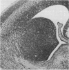
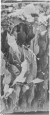
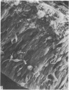
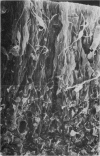
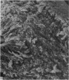
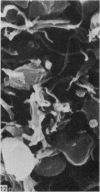
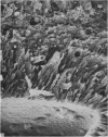
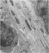
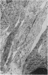
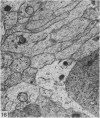
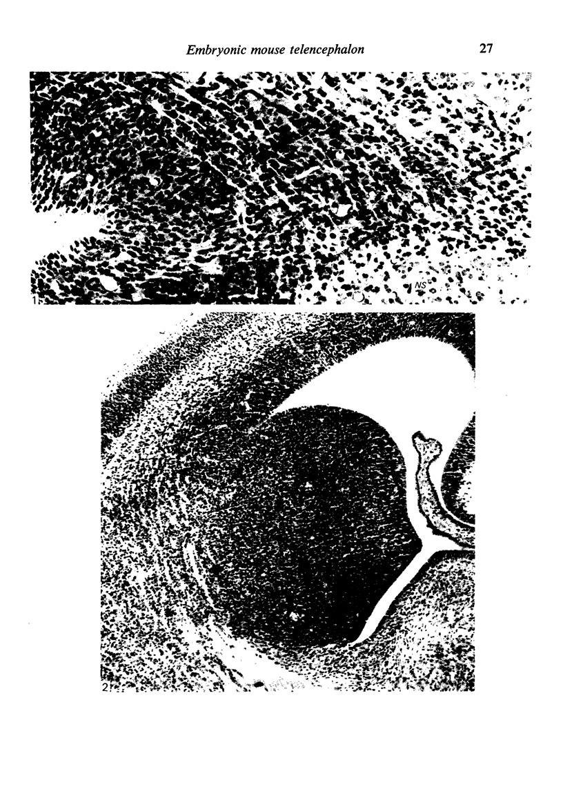
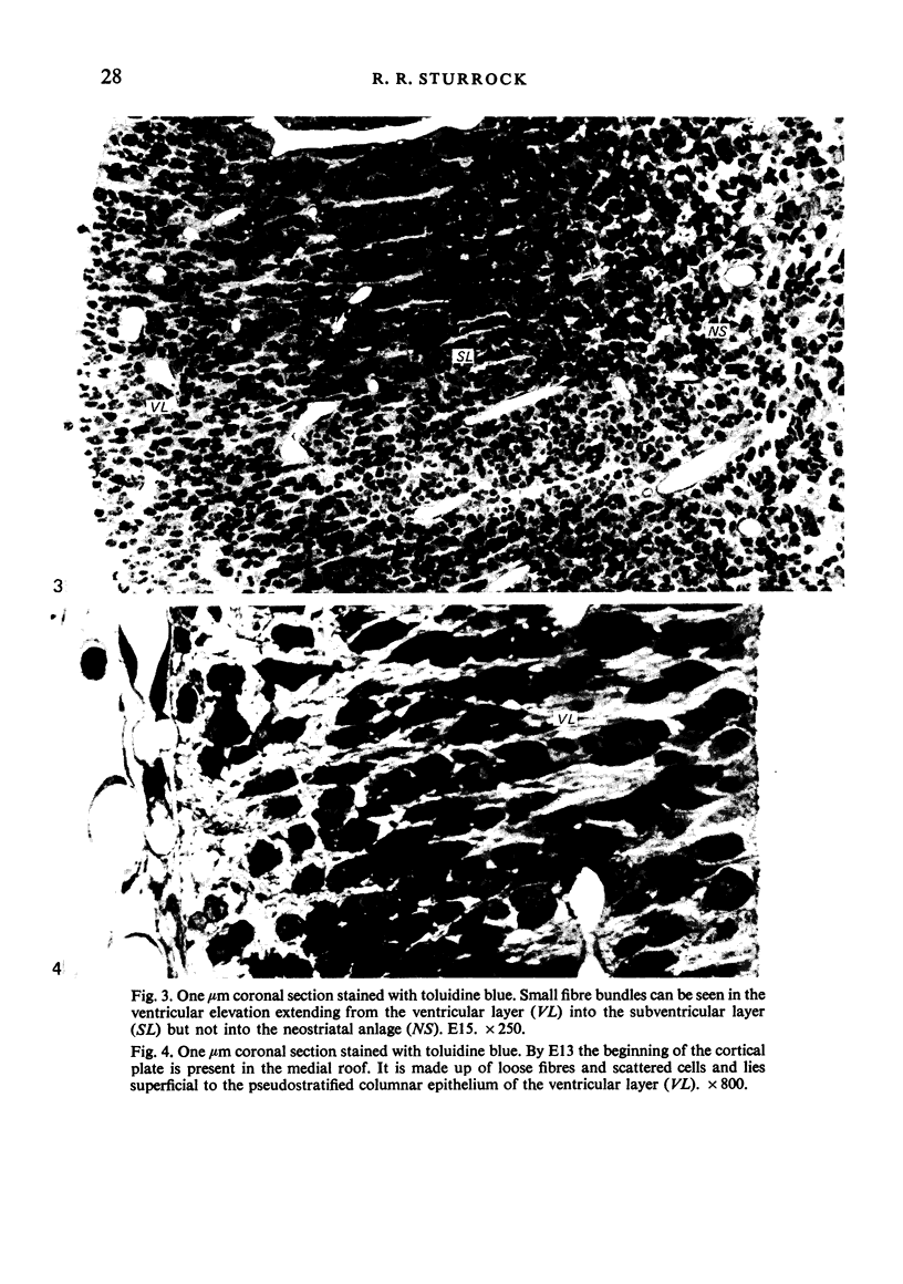
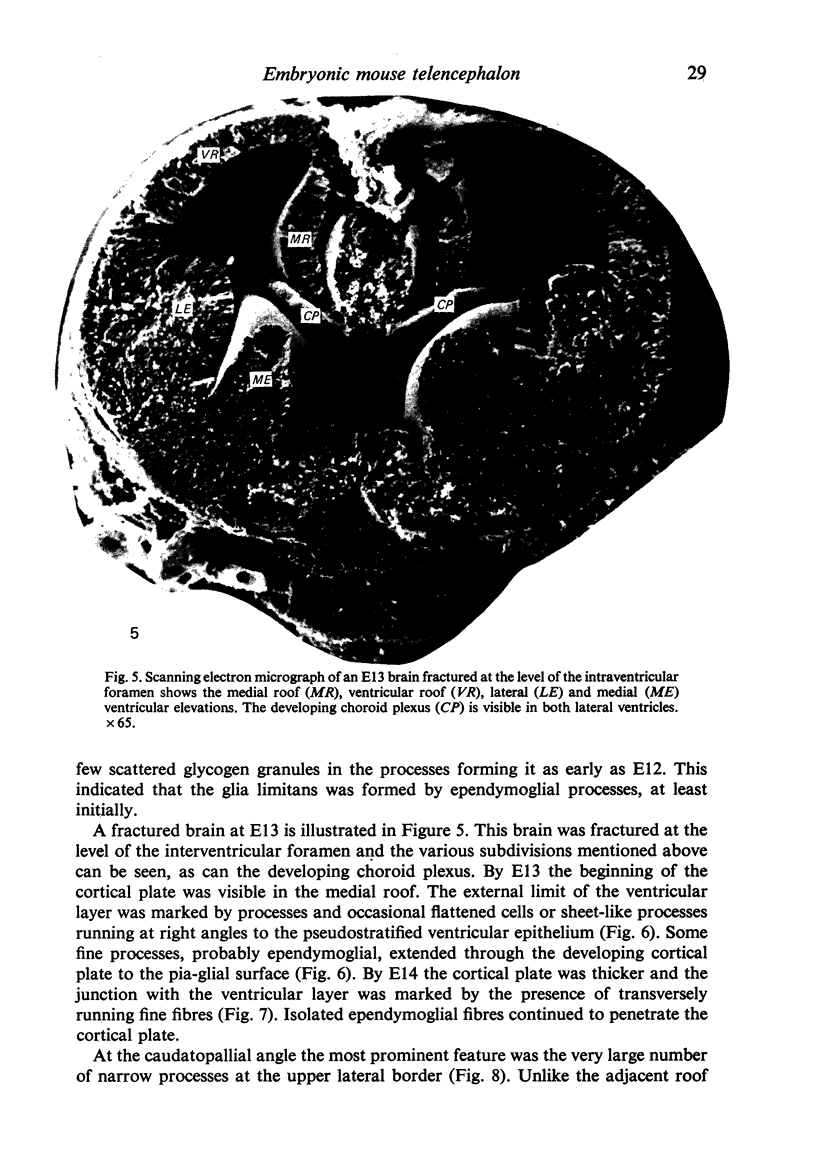
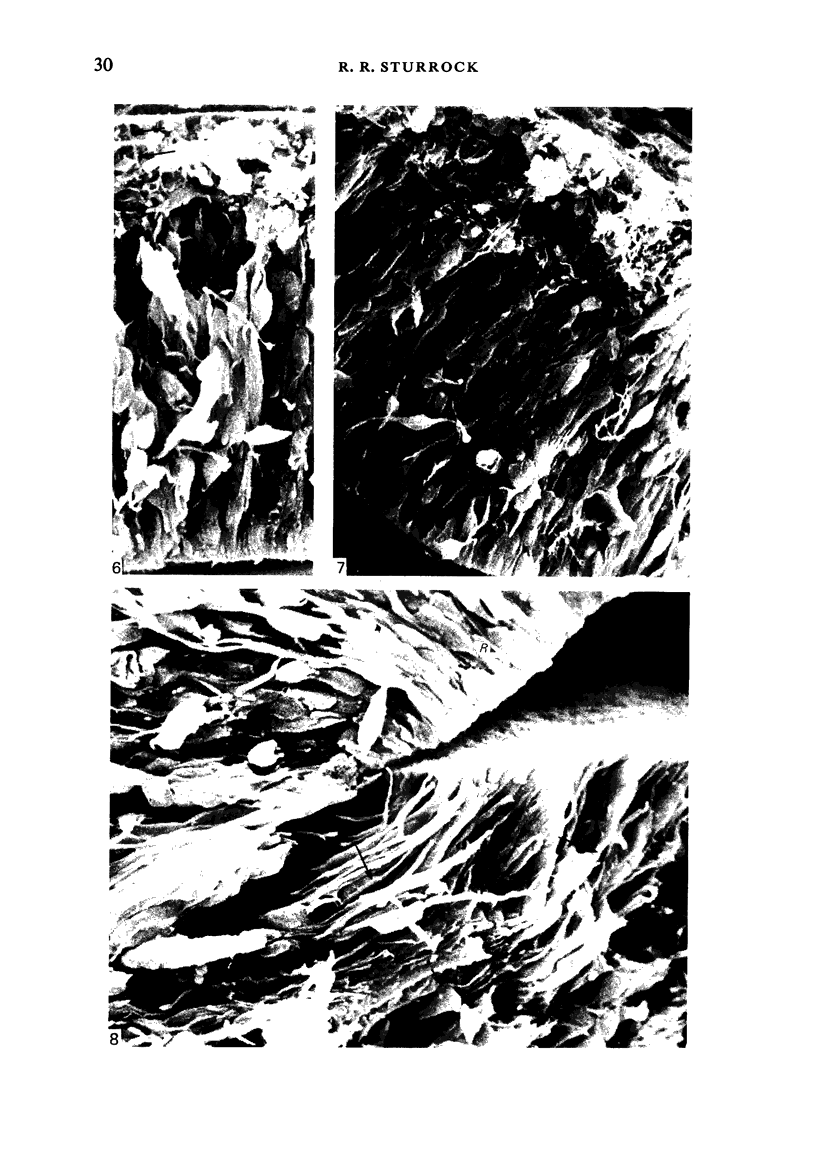
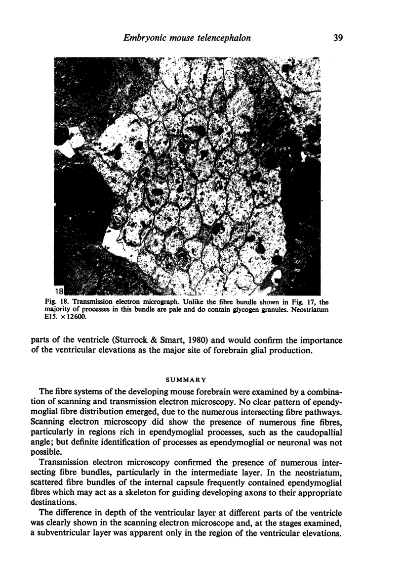
The fibre systems of the developing mouse forebrain were examined by a combination of scanning and transmission electron microscopy. No clear pattern of ependymoglial fibre distribution emerged, due to the numerous intersecting fibre pathways. Scanning electron microscopy did show the presence of numerous fine fibres, particularly in regions rich in ependymoglial processes, such as the caudopallial angle; but definite identification of processes as ependymoglial or neuronal was not possible. Transmission electron microscopy confirmed the presence of numerous intersecting fibre bundles, particularly in the intermediate layer. In the neostriatum, scattered fibre bundles of the internal capsule frequently contained ependymoglial fibres which may act as a skeleton for guiding developing axons to their appropriate destinations. The difference in depth of the ventricular layer at different parts of the ventricle was clearly shown in the scanning electron microscope and, at the stages examined, a subventricular layer was apparent in the region of the ventricular elevations.
Full text
PDF
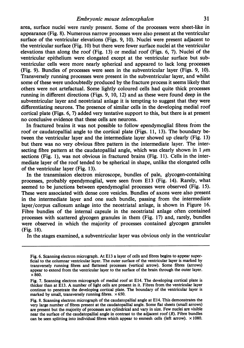
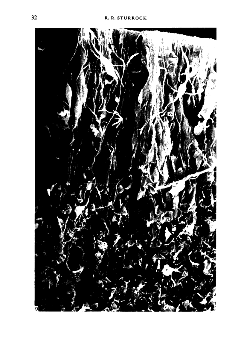
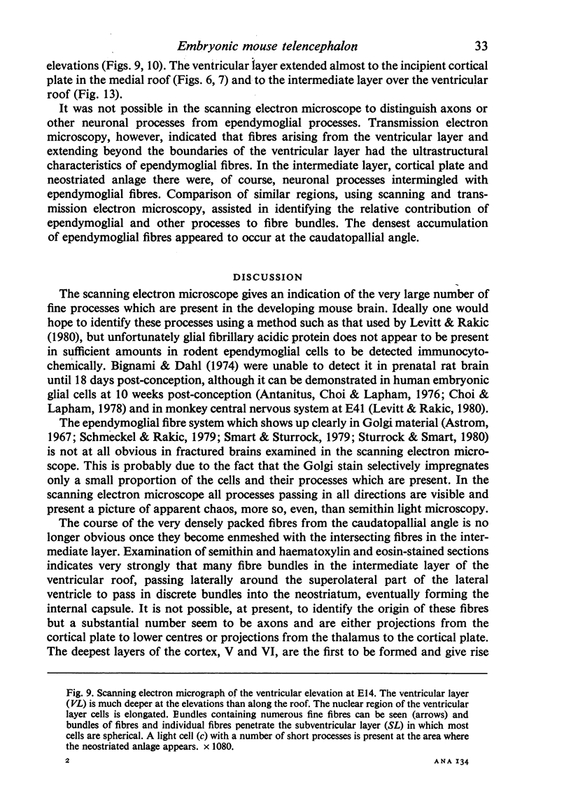
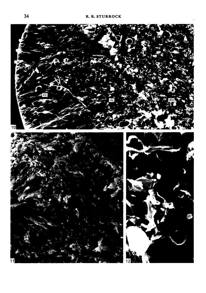
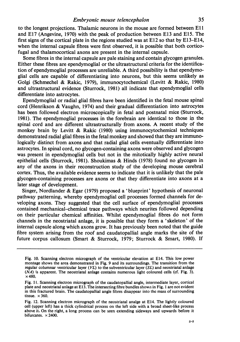
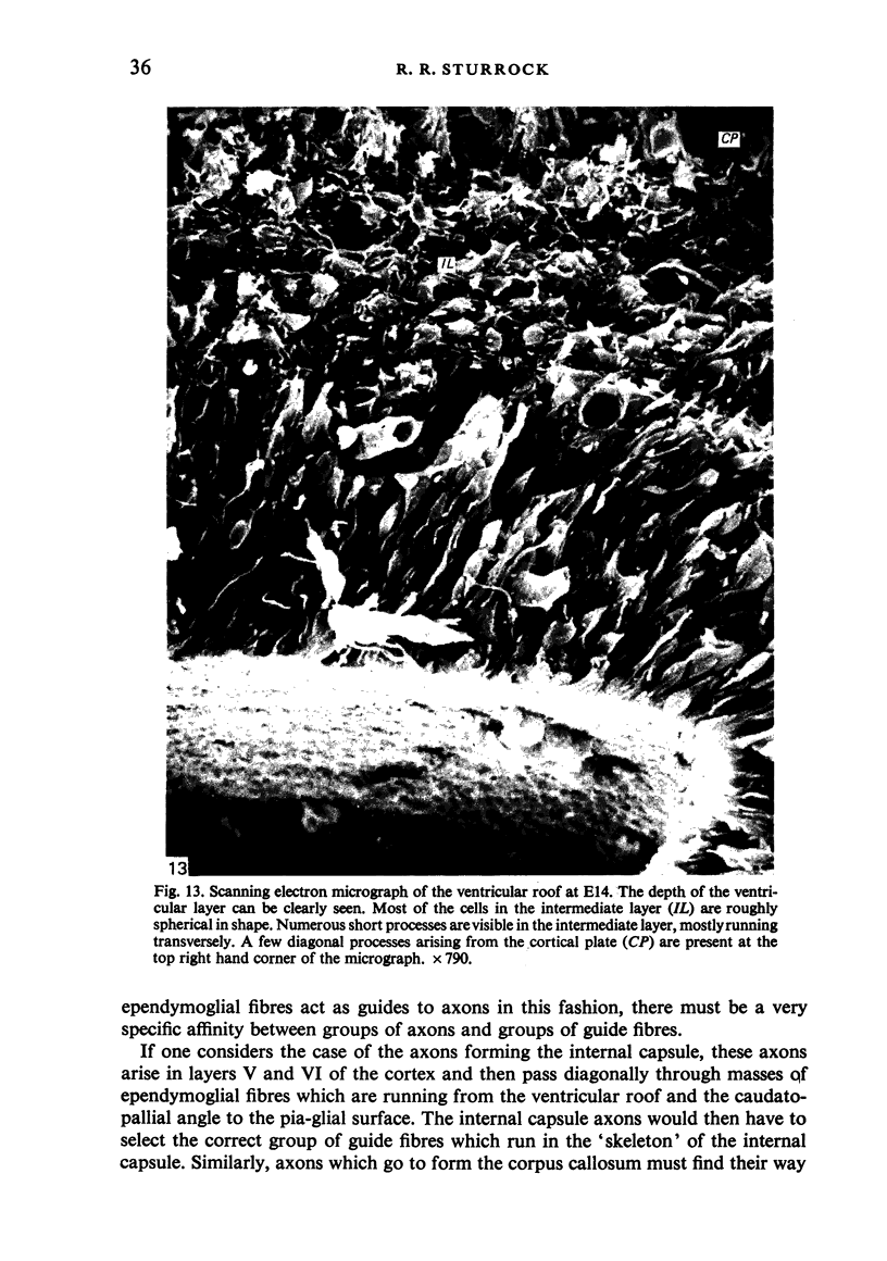
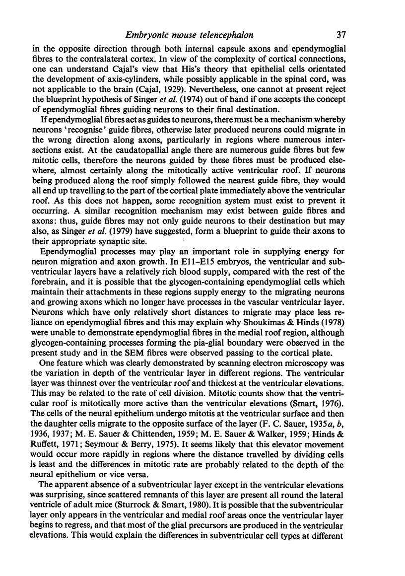
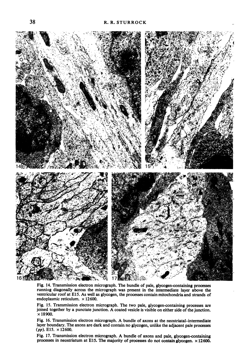
Images in this article
Selected References
These references are in PubMed. This may not be the complete list of references from this article.
- Angevine J. B., Jr Time of neuron origin in the diencephalon of the mouse. An autoradiographic study. J Comp Neurol. 1970 Jun;139(2):129–187. doi: 10.1002/cne.901390202. [DOI] [PubMed] [Google Scholar]
- Antanitus D. S., Choi B. H., Lapham L. W. The demonstration of glial fibrillary acidic protein in the cerebrum of the human fetus by indirect immunofluorescence. Brain Res. 1976 Feb 27;103(3):613–616. doi: 10.1016/0006-8993(76)90464-9. [DOI] [PubMed] [Google Scholar]
- Aström K. E. On the early development of the isocortex in fetal sheep. Prog Brain Res. 1967;26:1–59. [PubMed] [Google Scholar]
- Bignami A., Dahl D. Astrocyte-specific protein and neuroglial differentiation. An immunofluorescence study with antibodies to the glial fibrillary acidic protein. J Comp Neurol. 1974 Jan 1;153(1):27–38. doi: 10.1002/cne.901530104. [DOI] [PubMed] [Google Scholar]
- Choi B. H., Lapham L. W. Radial glia in the human fetal cerebrum: a combined Golgi, immunofluorescent and electron microscopic study. Brain Res. 1978 Jun 16;148(2):295–311. doi: 10.1016/0006-8993(78)90721-7. [DOI] [PubMed] [Google Scholar]
- Henrikson C. K., Vaughn J. E. Fine structural relationships between neurites and radial glial processes in developing mouse spinal cord. J Neurocytol. 1974 Dec;3(6):659–675. doi: 10.1007/BF01097190. [DOI] [PubMed] [Google Scholar]
- Hinds J. W., Ruffett T. L. Cell proliferation in the neural tube: an electron microscopic and golgi analysis in the mouse cerebral vesicle. Z Zellforsch Mikrosk Anat. 1971;115(2):226–264. doi: 10.1007/BF00391127. [DOI] [PubMed] [Google Scholar]
- Levitt P., Rakic P. Immunoperoxidase localization of glial fibrillary acidic protein in radial glial cells and astrocytes of the developing rhesus monkey brain. J Comp Neurol. 1980 Oct 1;193(3):815–840. doi: 10.1002/cne.901930316. [DOI] [PubMed] [Google Scholar]
- Rakic P. Mode of cell migration to the superficial layers of fetal monkey neocortex. J Comp Neurol. 1972 May;145(1):61–83. doi: 10.1002/cne.901450105. [DOI] [PubMed] [Google Scholar]
- SAUER M. E., CHITTENDEN A. C. Deoxyribonucleic acid content of cell nuclei in the neural tube of the chick embryo: evidence for intermitotic migration of nuclei. Exp Cell Res. 1959 Jan;16(1):1–6. doi: 10.1016/0014-4827(59)90189-2. [DOI] [PubMed] [Google Scholar]
- SAUER M. E., WALKER B. E. Radioautographic study of interkinetic nuclear migration in the neural tube. Proc Soc Exp Biol Med. 1959 Jul;101(3):557–560. doi: 10.3181/00379727-101-25014. [DOI] [PubMed] [Google Scholar]
- Schmechel D. E., Rakic P. A Golgi study of radial glial cells in developing monkey telencephalon: morphogenesis and transformation into astrocytes. Anat Embryol (Berl) 1979 Jun 5;156(2):115–152. doi: 10.1007/BF00300010. [DOI] [PubMed] [Google Scholar]
- Seymour R. M., Berry M. Scanning and transmission electron microscope studies of interkinetic nuclear migration in the cerebral vesicles of the rat. J Comp Neurol. 1975 Mar 1;160(1):105–125. doi: 10.1002/cne.901600107. [DOI] [PubMed] [Google Scholar]
- Shoukimas G. M., Hinds J. W. The development of the cerebral cortex in the embryonic mouse: an electron microscopic serial section analysis. J Comp Neurol. 1978 Jun 15;179(4):795–830. doi: 10.1002/cne.901790407. [DOI] [PubMed] [Google Scholar]
- Singer M., Nordlander R. H., Egar M. Axonal guidance during embryogenesis and regeneration in the spinal cord of the newt: the blueprint hypothesis of neuronal pathway patterning. J Comp Neurol. 1979 May 1;185(1):1–21. doi: 10.1002/cne.901850102. [DOI] [PubMed] [Google Scholar]
- Smart I. H. A pilot study of cell production by the ganglionic eminences of the developing mouse brain. J Anat. 1976 Feb;121(Pt 1):71–84. [PMC free article] [PubMed] [Google Scholar]
- Sturrock R. R. A developmental study of the mouse neostriatum. J Anat. 1980 Mar;130(Pt 2):243–261. [PMC free article] [PubMed] [Google Scholar]
- Sturrock R. R. An electron microscopic study of the development of the ependyma of the central canal of the mouse spinal cord. J Anat. 1981 Jan;132(Pt 1):119–136. [PMC free article] [PubMed] [Google Scholar]
- Sturrock R. R., Smart I. H. A morphological study of the mouse subependymal layer from embryonic life to old age. J Anat. 1980 Mar;130(Pt 2):391–415. [PMC free article] [PubMed] [Google Scholar]


