

Abstract
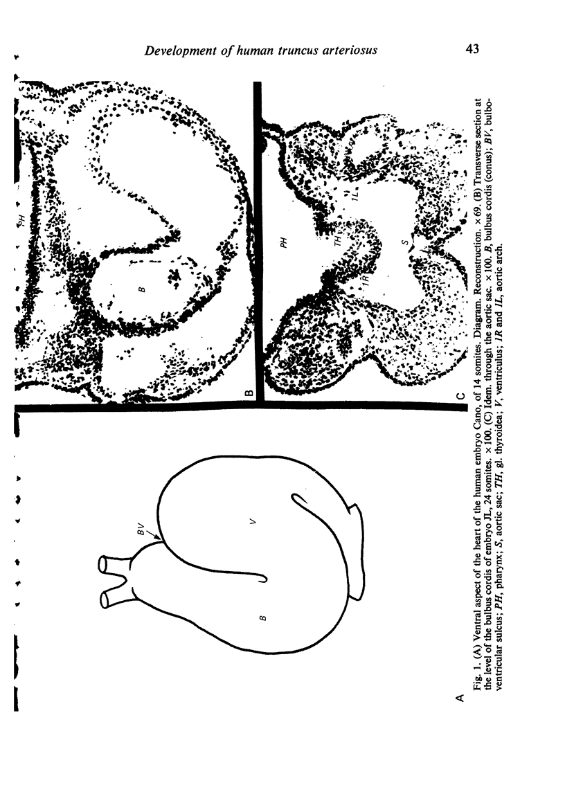
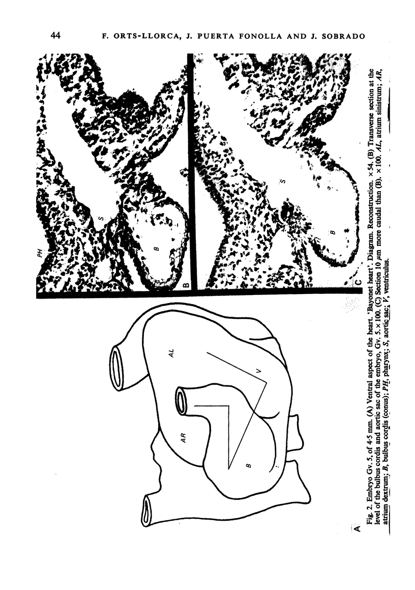
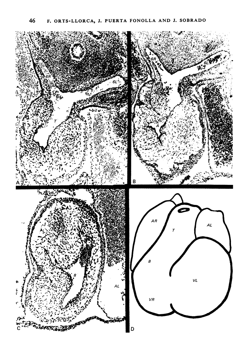
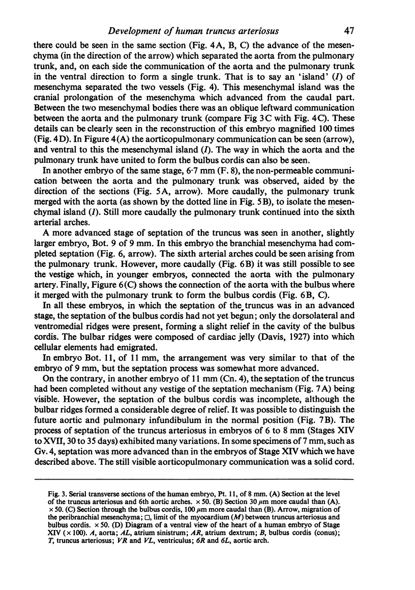
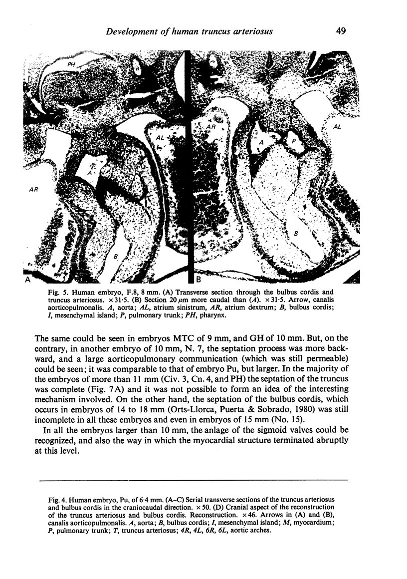
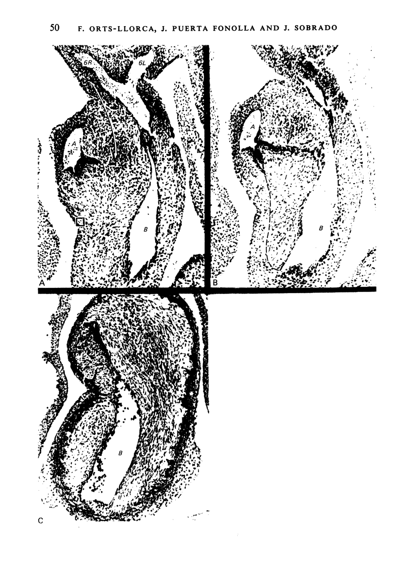
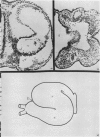
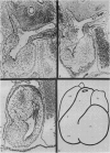
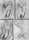
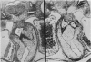
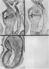
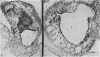
The results of our studies enable us to draw the following conclusions. The truncus appears in the human embryo, between Stages XII and XIII, as a portion of the aortic sac which invaginates into the interior of the pericardial cavity. Therefore it is an arterial portion which is added to the heart. It lengthens progressively. The sigmoid valves form in the angle between the bulbus cordis and the truncus. Septation of the truncus begins when the sixth arterial arches appear in embryos of 6 to 8 mm. The process is very rapid; commencing in embryos of 6 to 7 mm, it is complete in embryos of 10 to 11 mm, that is to say, during only five days. The septation mechanism is extrinsic. The peribranchial mesenchyma which accompanies the aortic sac in its invagination advances principally on the right inferior part and insinuates itself between the fourth and sixth arterial arches, separating the truncus pulmonalis from a portion of the ascending aorta. An aorticopulmonary communication exists for a certain period prior to fusion of the two blocks of mesenchyma; there is a mesenchymal island. On the contrary, in the bulbus cordis septation is effected by the bulbar ridges. Septation of the truncus, which does not exist in the primitive cardiac tube, occurs prior to that of the bulbus cordis. While septation of the truncus has been already completed in embryos of 10 mm, septation of the bulbus cordis is completed only in embryos of 14 to 16 mm. Therefore the bulbus and the truncus are two portions, different in respect of both structure and septation. There is no continuity between the bulbar ridges and the septation of the truncus. They are separated by the sigmoid valves. This makes it possible to observe independent malformations in the bulbus and in the truncus. In the truncus the mesenchyma passes between the two vessels. They do not have a common septum, and it is for this reason that the surgeon can separate them in the mature heart.
Full text
PDF


Images in this article

Selected References
These references are in PubMed. This may not be the complete list of references from this article.
- Asami I. Beitrag zur Entwicklung des Kammerseptums im menschlichen Herzen mit besonderer Berücksichtigung der sogenannten Bulbusdrehung. Z Anat Entwicklungsgesch. 1969;128(1):1–17. [PubMed] [Google Scholar]
- Carr I., Bharati S., Kusnoor V. S., Lev M. Truncus arteriosus communis with intact ventricular septum. Br Heart J. 1979 Jul;42(1):97–102. doi: 10.1136/hrt.42.1.97. [DOI] [PMC free article] [PubMed] [Google Scholar]
- Goor D. A., Dische R., Lillehei C. W. The conotruncus. I. Its normal inversion and conus absorption. Circulation. 1972 Aug;46(2):375–384. doi: 10.1161/01.cir.46.2.375. [DOI] [PubMed] [Google Scholar]
- Hurle J. M. Scanning and light microscope studies of the development of the chick embryo semilunar heart valves. Anat Embryol (Berl) 1979;157(1):69–80. doi: 10.1007/BF00315641. [DOI] [PubMed] [Google Scholar]
- LOS J. A. [Development of the septum sinus venosi cordis. Development of the heart in man, determined from a forgotten structure]. Z Anat Entwicklungsgesch. 1960;122:173–196. [PubMed] [Google Scholar]
- Los J. A. Cardiac septation and development of the aorta, pulmonary trunk, and pulmonary veins: previous work in the light of recent observations. Birth Defects Orig Artic Ser. 1978;14(7):109–138. [PubMed] [Google Scholar]
- Okamoto N., Satow Y., Hidaka N., Akimoto N., Miyabara S. Morphogenesis of congenital heart anomaly--bulboventricular malformations. Jpn Circ J. 1978 Oct;42(10):1105–1120. doi: 10.1253/jcj.42.1105. [DOI] [PubMed] [Google Scholar]
- Pexieder T. Development of the outflow tract of the embryonic heart. Birth Defects Orig Artic Ser. 1978;14(7):29–68. [PubMed] [Google Scholar]
- Pérez-Martinez V., Burgueros M., Quero M., Pérez Leon J., Hafer G. Aorticopulmonary window associated with tetralogy of Fallot. Report of one case and review of the literature. Angiology. 1976 Sep;27(9):526–534. doi: 10.1177/000331977602700906. [DOI] [PubMed] [Google Scholar]
- SHANER R. F. Comparative development of the bulbus and ventricles of the vertebrate heart with special reference to Spitzer's theory of heart malformations. Anat Rec. 1962 Apr;142:519–529. doi: 10.1002/ar.1091420409. [DOI] [PubMed] [Google Scholar]
- Thiene G., Bortolotti U., Gallucci V., Terribile V., Pellegrino P. A. Anatomical study of truncus arteriousus communis with embryological and surgical considerations. Br Heart J. 1976 Nov;38(11):1109–1123. doi: 10.1136/hrt.38.11.1109. [DOI] [PMC free article] [PubMed] [Google Scholar]
- de la Cruz M. V., Sánchez Gómez C., Arteaga M. M., Argüello C. Experimental study of the development of the truncus and the conus in the chick embryo. J Anat. 1977 Jul;123(Pt 3):661–686. [PMC free article] [PubMed] [Google Scholar]