

Abstract
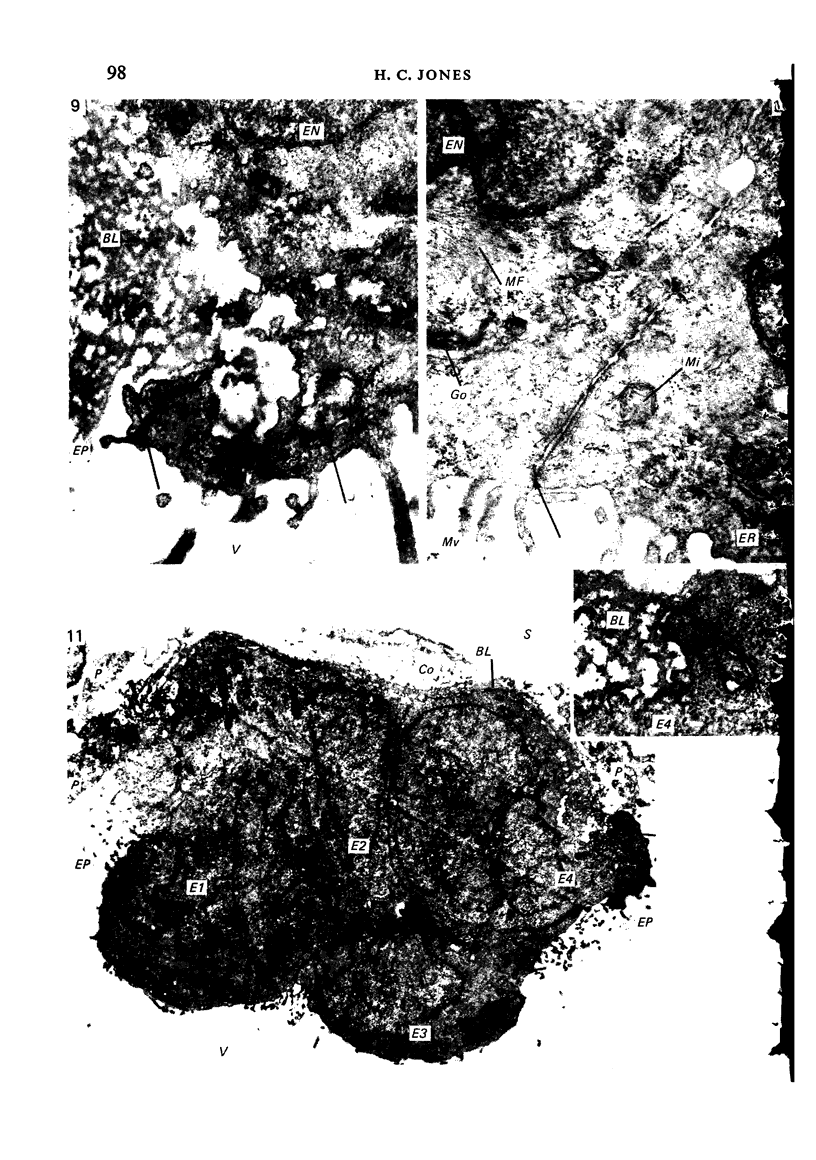
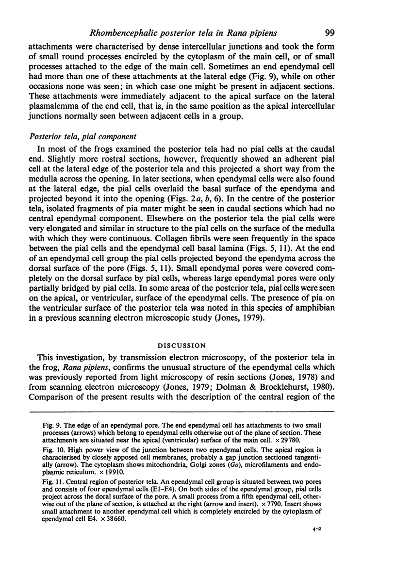
The posterior tela of the rhombencephalon, the overlying arachnoid mater, and the pial surface of the medulla were examined by transmission electron microscopy in the frog, Rana pipiens. The arachnoid mater and the pia mater have, with minor exceptions, similar ultrastructural characteristics to the same structures in mammals. The posterior tela, on the other hand, is an unusual membrane, the ependymal cells of which are modified to form numerous intercellular pores. These cells are pleomorphic (round, columnar or squamous) and have relatively few surface specialisations while their cytoplasm in electron-dense due to numerous microfilaments. Ependymal cells at the edges of pores are characterised by dense coils of basal lamina inserted into indentations of the lateral plasmalemma and, near the apical surface, by small specialised attachments to adjacent ependymal cells which are otherwise in a different plane.
Full text
PDF


Images in this article


Selected References
These references are in PubMed. This may not be the complete list of references from this article.
- BRIGHTMAN M. W. Perivascular spaces in the brains of Necturus maculosus Rafinesque and Mus norwegicus albinus. Anat Rec. 1953 Nov;117(3):427–447. doi: 10.1002/ar.1091170307. [DOI] [PubMed] [Google Scholar]
- Bradley O C. On the Development of the Hind-Brain of the Pig: Part II. J Anat Physiol. 1906 Jan;40(Pt 2):133–151.6. [PMC free article] [PubMed] [Google Scholar]
- Brightman M. W., Reese T. S. Junctions between intimately apposed cell membranes in the vertebrate brain. J Cell Biol. 1969 Mar;40(3):648–677. doi: 10.1083/jcb.40.3.648. [DOI] [PMC free article] [PubMed] [Google Scholar]
- Brocklehurst G. The structure of the rhombencephalic roof in the frog. Acta Neurochir (Wien) 1976;35(1-3):205–214. doi: 10.1007/BF01405948. [DOI] [PubMed] [Google Scholar]
- Dolman G. S., Brocklehurst G. The roof of the hindbrain in Rana pipiens and Rana temporaria. Brain Res. 1980 Feb 24;184(2):506–510. doi: 10.1016/0006-8993(80)90818-5. [DOI] [PubMed] [Google Scholar]
- Jones H. C. Continuity between the ventricular and subarachnoid cerebrospinal fluid in an amphibian, Rana pipiens. Cell Tissue Res. 1978 Dec 14;195(1):153–167. doi: 10.1007/BF00233683. [DOI] [PubMed] [Google Scholar]
- Jones H. C. Fenestration of the epithelium lining the roof of the fourth cerebral ventricle in amphibia. Cell Tissue Res. 1979 Apr 30;198(1):129–136. doi: 10.1007/BF00234840. [DOI] [PubMed] [Google Scholar]
- Klika E. The ultrastructure of meninges in vertebrates. Acta Univ Carol Med (Praha) 1967;13(1):53–71. [PubMed] [Google Scholar]
- Nabeshima S., Reese T. S., Landis D. M., Brightman M. W. Junctions in the meninges and marginal glia. J Comp Neurol. 1975 Nov 15;164(2):127–169. doi: 10.1002/cne.901640202. [DOI] [PubMed] [Google Scholar]
- PEASE D. C., SCHULTZ R. L. Electron microscopy of rat cranial meninges. Am J Anat. 1958 Mar;102(2):301–321. doi: 10.1002/aja.1001020207. [DOI] [PubMed] [Google Scholar]
- Perez-Gomez J., Bindslev N., Orkand P. M., Wright E. M. Electrical properties and structure of the frog arachnoid membrane. J Neurobiol. 1976 May;7(3):259–270. doi: 10.1002/neu.480070309. [DOI] [PubMed] [Google Scholar]
- REYNOLDS E. S. The use of lead citrate at high pH as an electron-opaque stain in electron microscopy. J Cell Biol. 1963 Apr;17:208–212. doi: 10.1083/jcb.17.1.208. [DOI] [PMC free article] [PubMed] [Google Scholar]
- Spurr A. R. A low-viscosity epoxy resin embedding medium for electron microscopy. J Ultrastruct Res. 1969 Jan;26(1):31–43. doi: 10.1016/s0022-5320(69)90033-1. [DOI] [PubMed] [Google Scholar]
- Tornheim P. A., Foltz F. M. Circulation of cerebrospinal fluid in the bullfrog, Rana catesbiana. Anat Rec. 1979 Jul;194(3):389–403. doi: 10.1002/ar.1091940306. [DOI] [PubMed] [Google Scholar]
- Tornheim P. A., Michaels J. E. Fine structure of the rhombencephalic tela of the bullfrog, Rana catesbeiana. Cell Tissue Res. 1979 Nov;202(3):479–491. doi: 10.1007/BF00220439. [DOI] [PubMed] [Google Scholar]