Abstract
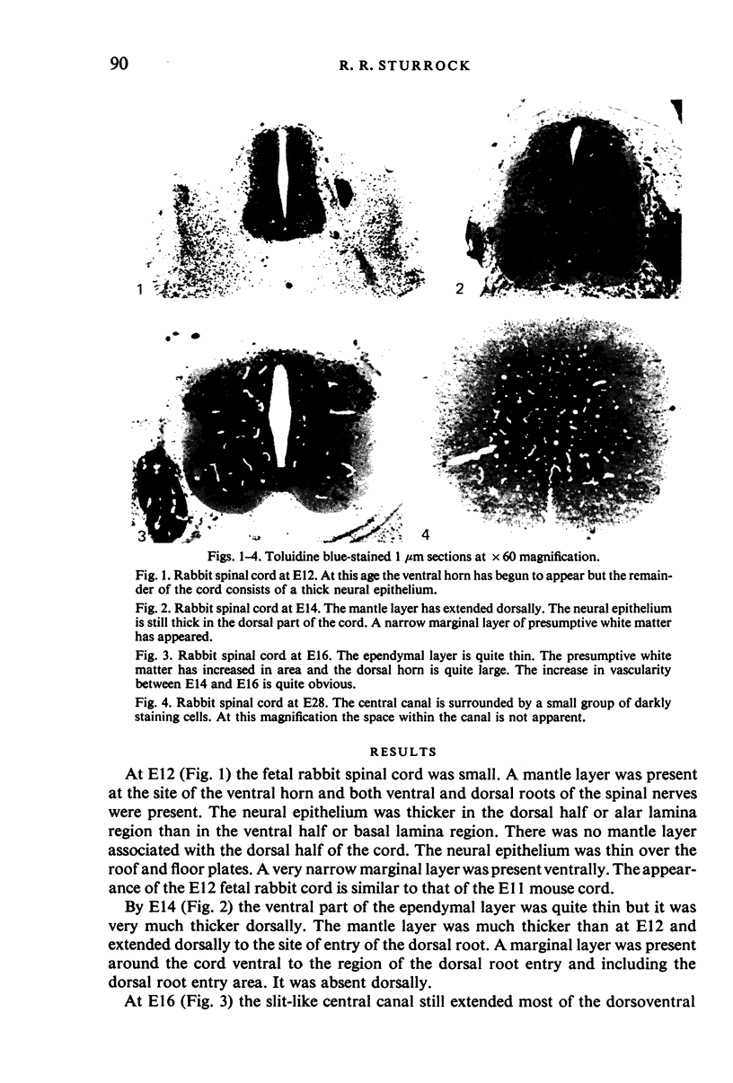
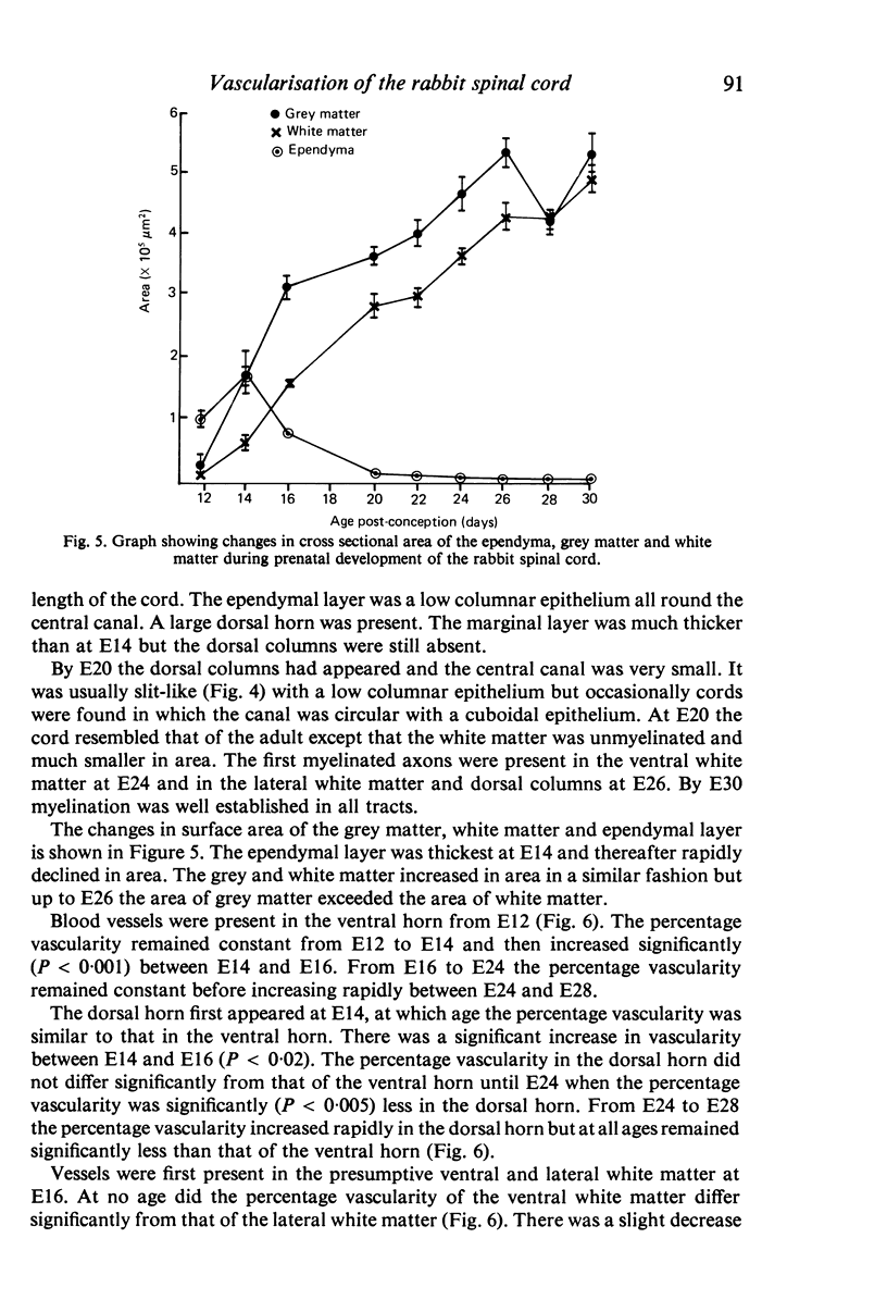
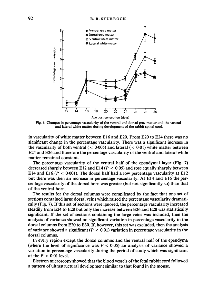
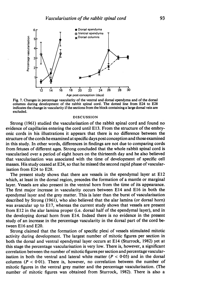
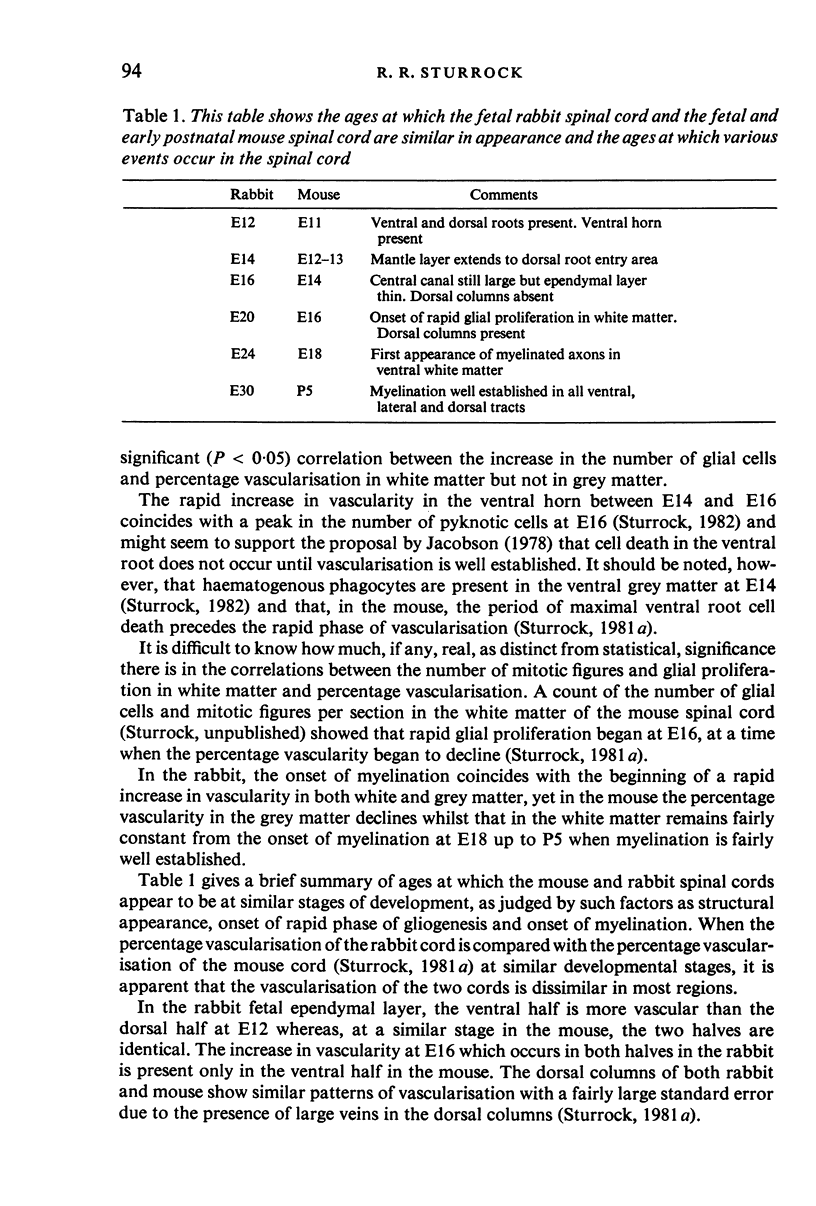
Vascularisation of the fetal rabbit spinal cord was studied by quantitative histology. From E12 to E16 blood vessels were most numerous in the ependymal layer of the cord. Two rapid phases of vascularisation of the grey matter were found. The first, between E14 and E16, was followed by a period when the percentage vascularisation remained fairly constant until the second began at E24. The second rapid phase lasted until E28. In the ventral and lateral white matter the percentage vascularity was fairly constant from E16 until E24. There was a rapid increase in vascularity between E24 and E26. From E26 to E30 the percentage vascularity in the ventral and lateral grey matter was constant. The increase in vascularity from E24 coincided with the onset of myelination in the ventral white matter. The percentage vascularity in the dorsal columns seemed to increase steadily between E24 and E28 but this was somewhat confused due to the presence of large veins in the dorsal columns.
Full text
PDF
Images in this article
Selected References
These references are in PubMed. This may not be the complete list of references from this article.
- Stensaas L. J., Gilson B. C. Ependymal and subependymal cells of the caudato-pallial junction in the lateral ventricle of the neonatal rabbit. Z Zellforsch Mikrosk Anat. 1972;132(3):297–322. doi: 10.1007/BF02450711. [DOI] [PubMed] [Google Scholar]
- Sturrock R. R. A quantitative and morphological study of vascularisation of the developing mouse spinal cord. J Anat. 1981 Mar;132(Pt 2):203–221. [PMC free article] [PubMed] [Google Scholar]
- Sturrock R. R. An electron microscopic study of the development of the ependyma of the central canal of the mouse spinal cord. J Anat. 1981 Jan;132(Pt 1):119–136. [PMC free article] [PubMed] [Google Scholar]
- Sturrock R. R. Gliogenesis in the prenatal rabbit spinal cord. J Anat. 1982 Jun;134(Pt 4):771–793. [PMC free article] [PubMed] [Google Scholar]


