Abstract
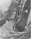
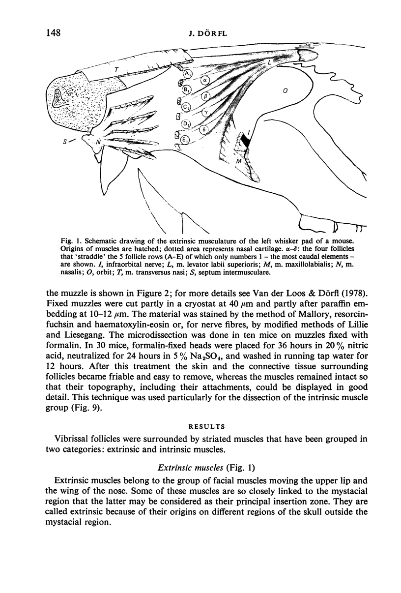
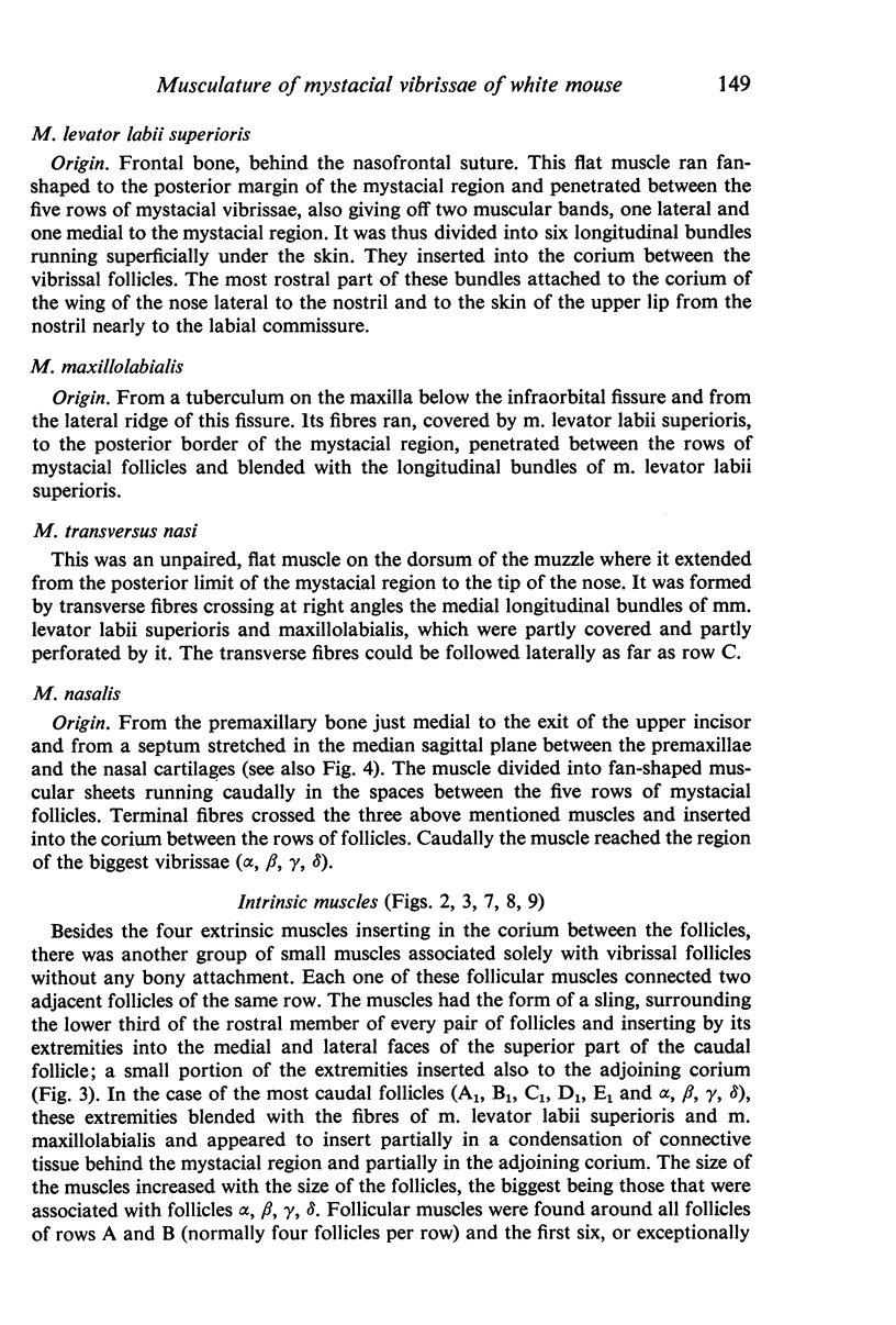
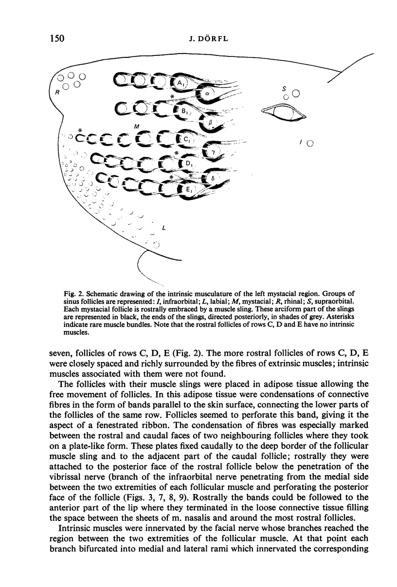
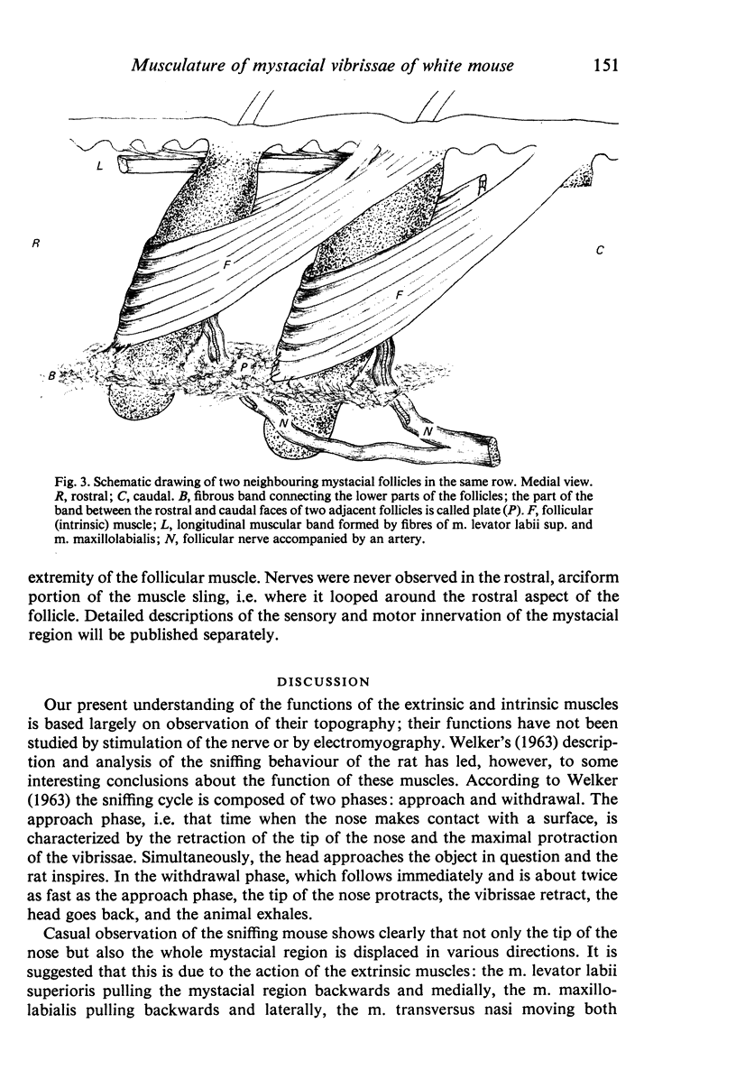
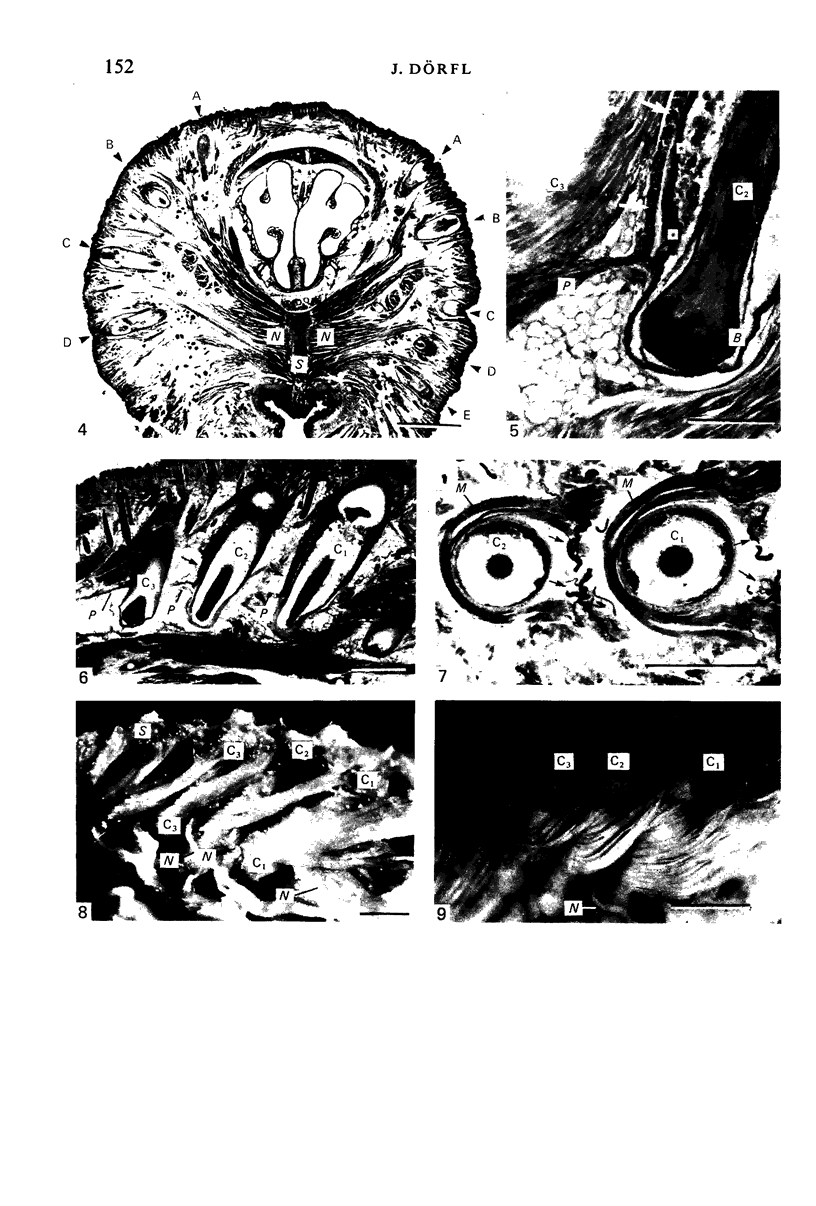
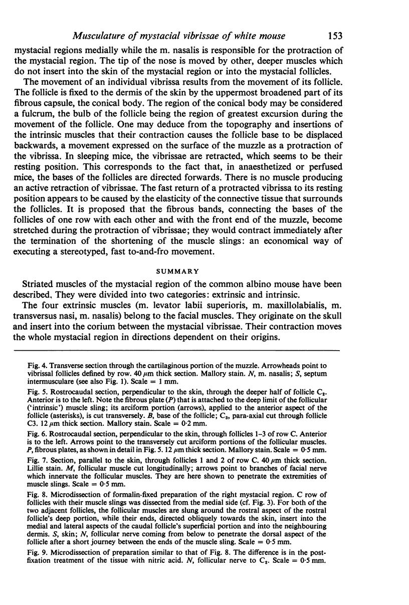
Striated muscles of the mystacial region of the common albino mouse have been described. They were divided into two categories: extrinsic and intrinsic. The four extrinsic muscles (m. levator labii superioris, m. maxillolabialis, m. transversus nasi, m. nasalis) belong to the facial muscles. They originate on the skull and insert into the corium between the mystacial vibrissae. Their contraction moves the whole mystacial region in directions dependent on their origins. Intrinsic (follicular) muscles are associated solely with the vibrissal follicles and have no bony attachment. They were found around follicles alpha, beta, gamma, delta, around all follicles of rows A and B, and around the first six follicles of rows C, D and E. The form of the follicular muscle is a sling connecting two adjacent follicles of the same row. The arc of the sling surrounds the inferior part of the rostral follicle and the two extremities insert to the conical body of the caudal follicle and to the neighbouring corium. They are the protractors of the vibrissae. The inferior parts of the vibrissal follicles of a given row are fixed in a fibrous band which inserts in the anterior part of the muzzle. It is proposed that these bands become stretched during the protraction of vibrissae and contract, by their elasticity, immediately upon the end of the follicular muscles' contraction, executing the fast return of vibrissae to their resting, retracted position.
Full text
PDF
Images in this article
Selected References
These references are in PubMed. This may not be the complete list of references from this article.
- Belford G. R., Killackey H. P. Vibrissae representation in subcortical trigeminal centers of the neonatal rat. J Comp Neurol. 1979 Jan 15;183(2):305–321. doi: 10.1002/cne.901830207. [DOI] [PubMed] [Google Scholar]
- Harris R. M., Woolsey T. A. Morphology of golgi-impregnated neurons in mouse cortical barrels following vibrissae damage at different post-natal ages. Brain Res. 1979 Jan 26;161(1):143–149. doi: 10.1016/0006-8993(79)90201-4. [DOI] [PubMed] [Google Scholar]
- Jeanmonod D., Rice F. L., Van der Loos H. Mouse somatosensory cortex: alterations in the barrelfield following receptor injury at different early postnatal ages. Neuroscience. 1981;6(8):1503–1535. doi: 10.1016/0306-4522(81)90222-0. [DOI] [PubMed] [Google Scholar]
- Shipley M. T. Response characteristics of single units in the rat's trigeminal nuclei to vibrissa displacements. J Neurophysiol. 1974 Jan;37(1):73–90. doi: 10.1152/jn.1974.37.1.73. [DOI] [PubMed] [Google Scholar]
- Simons D. J. Response properties of vibrissa units in rat SI somatosensory neocortex. J Neurophysiol. 1978 May;41(3):798–820. doi: 10.1152/jn.1978.41.3.798. [DOI] [PubMed] [Google Scholar]
- Steffen H., Van der Loos H. Early lesions of mouse vibrissal follicles:: their influence on dendrite orientation in the cortical barrelfield. Exp Brain Res. 1980;40(4):419–431. doi: 10.1007/BF00236150. [DOI] [PubMed] [Google Scholar]
- Van der Loos H., Woolsey T. A. Somatosensory cortex: structural alterations following early injury to sense organs. Science. 1973 Jan 26;179(4071):395–398. doi: 10.1126/science.179.4071.395. [DOI] [PubMed] [Google Scholar]
- Waite P. M. The responses of cells in the rat thalamus to mechanical movements of the whiskers. J Physiol. 1973 Jan;228(2):541–561. doi: 10.1113/jphysiol.1973.sp010099. [DOI] [PMC free article] [PubMed] [Google Scholar]
- White E. L. Ultrastructure and synaptic contacts in barrels of mouse SI cortex. Brain Res. 1976 Mar 26;105(2):229–251. doi: 10.1016/0006-8993(76)90423-6. [DOI] [PubMed] [Google Scholar]
- Wong-Riley M. T., Welt C. Histochemical changes in cytochrome oxidase of cortical barrels after vibrissal removal in neonatal and adult mice. Proc Natl Acad Sci U S A. 1980 Apr;77(4):2333–2337. doi: 10.1073/pnas.77.4.2333. [DOI] [PMC free article] [PubMed] [Google Scholar]
- Woolsey T. A., Van der Loos H. The structural organization of layer IV in the somatosensory region (SI) of mouse cerebral cortex. The description of a cortical field composed of discrete cytoarchitectonic units. Brain Res. 1970 Jan 20;17(2):205–242. doi: 10.1016/0006-8993(70)90079-x. [DOI] [PubMed] [Google Scholar]
- Yohro T. Arrangement and structure of sinus hair muscles in the big-clawed shrew, Sorex unguiculatus. J Morphol. 1977 Aug;153(2):317–331. doi: 10.1002/jmor.1051530210. [DOI] [PubMed] [Google Scholar]


