Abstract
Background
Psoriasis represents a persistent, immune-driven inflammatory condition affecting the skin, characterized by a lack of well-established biologic treatments without adverse events. Consequently, the identification of novel targets and therapeutic agents remains a pressing priority in the field of psoriasis research.
Methods
We collected single-cell RNA sequencing (scRNA-seq) datasets and inferred T cell differentiation trajectories through pseudotime analysis. Bulk transcriptome and scRNA-seq data were integrated to identify differentially expressed genes (DEGs). Machine learning was employed to screen candidate genes. Correlation analysis was used to predict the interactions between cells expressing insulin-induced gene 1 (INSIG1) and other immune cells. Finally, drug docking was performed on INSIG1, and the expression levels of INSIG1 in psoriasis were verified through clinical and in vivo experiments, and further in vivo experiments established the efficacy of tetrandrine in the treatment of psoriasis.
Results
T cells were initially categorized into seven states, with differentially expressed genes in T cells (TDEGs) identified and their functions and signaling pathways. INSIG1 emerged as a characteristic gene for psoriasis and was found to be downregulated in psoriasis and potentially negatively associated with T cells, influencing psoriasis fatty acid metabolism, as inferred from enrichment and immunoinfiltration analyses. In the cellular communication network, cells expressing INSIG1 exhibited close interactions with other immune cells through multiple signaling channels. Furthermore, drug sensitivity showed that tetrandrine stably binds to INSIG1, could be a potential therapeutic agent for psoriasis.
Conclusion
INSIG1 emerges as a specific candidate gene potentially regulating the fatty acid metabolism of patients with psoriasis. In addition, tetrandrine shows promise as a potential treatment for the condition.
Keywords: psoriasis, single-cell RNA sequencing, machine learning, INSIG1, pseudotime analysis
Introduction
Psoriasis, a chronic systemic inflammatory condition, impacts not only the skin and joints but also the nails, posing substantial psychological and social challenges.1 Its treatment poses challenges due to its tendency to relapse and difficulty in achieving a cure. The global incidence of psoriasis is estimated to be between 1% and 3%.2,3 The disease results from a complex interaction of factors, predominantly driven by an abnormal immune response in the skin, which is influenced by genetic predisposition and various environmental triggers.4 Psoriasis not only results in physical disabilities and work-related incapacitations but also markedly diminishes the quality of life for sufferers.5 Additionally, individuals afflicted with psoriasis face an elevated risk of associated health issues including psoriatic arthritis, metabolic disorders, excess body weight, mood disorders,6 cancer, as well as cardiovascular disease,7 contributing to a significant global disease burden.
Over the past few decades, phototherapy and local therapy have been widely used to treat stable psoriatic lesions.8 Recent advancements in understanding the pathobiology of psoriasis have facilitated the creation of highly targeted biologic therapies, demonstrating encouraging outcomes in treatment.9 These biologics offer advantages including swift symptom relief, sustained effectiveness, positive safety profiles, and positive impacts on related health conditions.10,11 Most biologics target cytokines and play a central role in managing psoriasis.12 Their effectiveness is demonstrated by the use of treatments such as tumor necrosis factor (TNF)-α inhibitors,13 interleukin (IL)-23 inhibitors, and IL-17 inhibitors. However, existing biologics are not considered ideal for patients with mild to moderate psoriasis14 and can cause severe side effects such as immune drift, relapse after discontinuation, and high costs. Limited therapeutic options persist due to the lack of biomarkers or validated treatment targets, underscoring the need to discover new therapeutic targets and elucidate the molecular mechanisms driving psoriasis progression.
INSIG1, which encodes an endoplasmic reticulum membrane protein, plays a role in regulating cellular lipid metabolism,15 cholesterol metabolism,16 glucose metabolism,17 and immune and inflammatory responses.18 Research indicates that INSIG1 plays a role in modulating cholesterol homeostasis and the innate antiviral response in macrophages through its interaction with T cell immunoglobulin and mucin domain-containing molecule 4.18 Upon lipopolysaccharide stimulation, macrophages exhibit increased INSIG1 expression, which is implicated in the inflammatory response.19 INSIG1 contributes to the modulation of immune and inflammatory responses, playing a role in the regulation of various inflammatory conditions, including the healing of diabetic wounds,20 atherosclerosis,21 and gout.22 INSIG1 exhibits reduced expression in both autoimmune and cardiometabolic diseases. These functions are potentially valuable for diagnosing and treating psoriasis.
Transcriptome sequencing has played a significant role in deciphering the mechanisms behind a range of skin conditions. The evolution of scRNA-seq techniques has enabled us to uncover cellular heterogeneity on the basis of single cells level, pinpointing differential gene candidates, and constructing a detailed immune profile and cellular genomic map for skin diseases. This approach has opened up novel perspectives for understanding the etiology of these conditions.23
In our study, we systematically delineated the trajectory of T cell differentiation during psoriasis progression and identified TDEGs associated with T cell differentiation using scRNA-seq data. By integrating bulk transcriptomics, machine learning, and scRNA-seq data, INSIG1 was identified as a potential target influencing T cell expression in psoriasis. Additionally, tetrandrine was identified as a promising immunotherapeutic agent for psoriasis, binding stably to INSIG1 as predicted by drug database analyses and molecular docking, thereby offering new clinical treatment options.
Methods
Single-Cell RNA Sequencing Data Processing
Single-cell transcriptome data underwent rigorous quality checks using R software (version 4.2.1), in line with established single-cell data handling protocols. The Seurat package (version 4.0.4), specifically the “Read10X” function, was employed to ingest the count matrix, which was then transformed into “dgCMatrix” format. The merging of individual datasets into a unified structure was facilitated by the “merge” function, with cell label uniqueness ensured by “RenameCells”. Cells deemed of inferior quality were culled, applying criteria such as the exclusion of genes detected in less than three cells and the elimination of cells with gene expression levels below 200. Additionally, we removed cells with a mitochondrial gene percentage greater than 20%. The DoubletFinder method was used to remove potential doublets. Gene expression normalization was executed via the global-scaling “LogNormalize” method, utilizing a scaling factor set at 10,000. For downstream analysis, the “FindVariableFeatures” function identified the top 2000 most variable genes. The “ScaleData” function was then applied, with “vars.to.regress” accounting for UMI and mitochondrial content to minimize extraneous variability. Principal component analysis (PCA) was conducted on the selected variable genes, retaining the initial 30 principal components for subsequent steps. The “Harmony” algorithm was utilized to correct for batch effects across samples. Further refinement of the data’s dimensionality was achieved through t-Distributed Stochastic Neighbor Embedding (t-SNE). Cluster identification was guided by the construction of a shared nearest-neighbor graph, leveraging the Louvain method within the “FindNeighbors” and “FindClusters” functions. The resolution parameter was fine-tuned through a range of 0.1 to 1, with the “clustree” function visualizing cell clustering at various resolutions, identifying an optimal clustering at a resolution of 0.5. Cell annotation was performed based on cell surface markers and literature-referenced genes. Spatial transcriptomic data was predominantly handled through the Semla package, which involved converting Seurat objects to Semla objects using the UpdateSeuratForSemla function. Subsequently, gene expression levels were visualized with the MapFeatures function.
Pseudotime Analysis
Pseudotime analysis was conducted using the “monocle” package (version 2.24.1). The initial object for pseudotime analysis was generated using the “newCellDataSet” function. Following the use of the detectGenes function to remove genes expressed in less than 10% of cells, we proceed with the dispersionTable function to identify genes that are valuable for differentiation directions. Utilizing the expression of these genes, we employ the reduceDimension function to construct a cellular differentiation skeleton and project it into a two-dimensional coordinate system. Subsequently, we use the orderCells function to sort all cells within the differentiation skeleton, with each branch representing distinct differentiation states. To calculate the differentially expressed genes (TDEGs) for each state of T cells, we apply the FindAllMarkers function from the Seurat package, setting the threshold at |log2 fold change| (|log2FC|) > 0.585 and an adjusted P value < 0.05, and these genes are defined as TDEGs.
Enrichment Analysis
Enrichment analysis of differential genes was performed using the “gene set enrichment analysis (GSEA) Base”, “ClusterProfiler”, “org.Hs.eg.db”, “ggplot2”, and “GSVA” packages. Enrichment analysis at single-cell level was conducted using the irGSEA (version 1.1.3) software.
Bulk Transcriptome Data Acquisition and Pre-Processing
All bulk transcriptome data were downloaded from GEO datasets (https://www.ncbi.nlm.nih.gov/geo/). Gene annotation was meticulously performed based on the specific probes associated with each dataset. When encountering genes associated with multiple probes, we calculated the average value across these probes to ensure accuracy. The “normalizeBetweenArrays” function was subsequently applied to refine the expression matrix. As each cohort was handled and analyzed independently, concerns regarding batch effects across different datasets were rendered unnecessary.
Machine Learning
Three distinct machine learning methodologies were deployed: least absolute shrinkage and selection operator (LASSO), support vector machine (SVM), and random forest (RF). LASSO has strong feature selection capabilities, and by applying L1 regularization, it effectively selects the most influential genes while reducing overfitting. Support Vector Machine (SVM) performs exceptionally well in handling high-dimensional data, especially with its built-in feature selection mechanism, which allows it to identify key features in classification tasks. Random Forest, as an ensemble learning method, not only improves prediction accuracy but also selects the most relevant features by calculating the importance scores of genes. All three methods have feature gene selection functions, helping us identify the most valuable features for prediction from a large set of genes, thereby improving the model’s accuracy and interpretability. LASSO was implemented utilizing the “glmnet” package, with the penalty parameter optimized through a 10-fold cross-validation process to counteract overfitting tendencies. For SVM, the analysis relied on the “e1071” (https://CRAN.R-project.org/package=e1071), “kernlab” (https://CRAN.R-project.org/package=kernlab), and “caret” (https://CRAN.R-project.org/package=caret) packages, employing the “rfe” function with the “svmRadial” kernel as the criterion for gene selection, which was based on minimizing cross-validation error. The “randomForest” package (https://cran.r-project.org/doc/Rnews/Rnews_2002-3.pdf#page=18) was applied for RF analysis, constructing forests with a variable number of trees ranging from 0 to 1000, and identifying the optimal number of trees by selecting those with the lowest out-of-bag error. The “randomForest” function was subsequently reapplied to refine the model fit. Gene significance was evaluated through the importance metric, leading to the selection of the top 10 genes as potential biomarkers, as indicated by their importance scores. The performance of these alternative genes was assessed using receiver operating characteristic (ROC) curves, which were generated with the “pROC” package.
Cellular Communication Network
The analysis of cell–cell interaction was conducted using the “CellChat” R package (version 1.0.0). “CellChat” offers an extensive database of cellular communication details, including ligands, receptors, cofactors, and their interactions. This package serves as a flexible and user-friendly toolkit for surmising, dissecting, and visualizing cell–cell communication from scRNA-seq data. Initially, the “createCellChat” function was employed to create a “cellchat” analysis object, utilizing the built-in “CellChatDB.human” database. No specific cell communication methodology was predetermined. Overexpressed genes were identified using the “identifyOverExpressedGenes” function, followed by the identification of receptor–ligand pairs using the “identifyOverExpressedInteractions” function. The “projectData” function was subsequently employed to map these receptor-ligand interactions onto a protein-protein interaction (PPI) network, and the “computeCommunProb” function quantified communication strength. Subsequently, cellular signaling pathways were inferred using the “computeCommunProbPathway” function with default parameters. Visualization was achieved using the “netVisual_bubble” function, maintaining default settings.
Spearman Correlation Analysis of Immune Cell Content and Immune Function
Analysis of immune cell content and functionality was executed utilizing the “gsva” function from the “GSVA” package, configuring the “kcdf” parameter to “Gaussian” to suit the analysis of continuous expression value inputs like log-CPMs, log-RPKMs, or log-TPMs. The method parameter was set to “ssgsea”. Visualization of the outcomes was accomplished using the “ggpubr” package.
Clinical Samples Information
Skin biopsy specimens (4 mm, punch biopsy) were collected from three psoriatic lesions and three healthy volunteers who provided informed consent. This study received approval from the Clinical Research Ethics Committee of the China-Japan Friendship Hospital (ethics approval number: 2023-KY-181).
Animal Study and Ethical Statement
The animal study involved BALB/c mice (male, 6–8 weeks old, weighing 20–25 g) obtained from Vital River Laboratory Animal Technology Co., Ltd. (Beijing, China). The mice were randomly assigned to four groups, that is, the Control group, the Pso model group, the tetrandrine (obtained from MedChem Express (Cat. No.:92 hY-13764; Shanghai, China) intervention group (30mg/kg, gastric lavage),24 and the methotrexate (MTX) as positive control group. An acute murine model of psoriasis-like skin inflammation was induced using imiquimod.25 The Clinical Research Ethics Committee of the China-Japan Friendship Hospital approved all procedures (ethics approval number: 2023-KY-181). The experiments adhered to the guidelines and regulations approved by the Committee.
Quantitative Reverse Transcription Polymerase Chain Reaction (qRT-PCR)
RNA samples were isolated using the RNAsimple Total RNA Kit (TIANGEN) and reverse transcribed using the HiScript III 1st Strand cDNA Synthesis Kit (+gDNA wiper) (Vazyme). qRT-PCR was conducted using NovoStart® SYBR qPCR SuperMix Plus (Novoprotein). The primer sequences are detailed in Table S1. Relative expression of INSIG1 was evaluated using the 2−ΔΔCt method, with GAPDH as the internal reference.
Immunohistochemical Analysis (IHC)
For IHC examination, clinical or in vivo skin tissue samples, prepared as 4-μm-thick skin sections, underwent deparaffinization, hydration, and antigen retrieval processes.
Endogenous peroxidase activity was inhibited, followed by an overnight incubation at 4°C with an anti-INSIG1 antibody (Proteintech, 55282-1-AP). On the subsequent day, the tissue sections were further incubated with a corresponding secondary antibody at ambient temperature for a period of 90 minutes, developed with DAB, and counterstained with hematoxylin. Images were acquired using the Panoramic (3DHISTECH) Digital Slide Scanner and analyzed using Image Pro Plus 6.0 software.
Molecular Docking
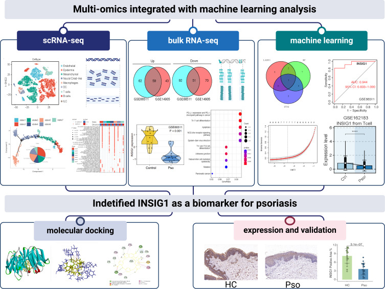
The protein’s crystal structure was sourced from the Protein Data Bank, while the 3D structure of the compound was retrieved from the PubChem compound database. Small molecules were hydrogenated, generating isomers using Discovery Studio software (2019). Protein polyconformation was removed, and molecular docking was performed using CDOCKER (2019). Correlation analysis of molecular docking was conducted using Discovery Studio (2019). Binding energy was calculated through molecular dynamics simulation, with a binding affinity value ≤ −5.0 kcal/mol indicating good binding ability. The flow chart is shown in Figure 1 (Created with BioRender.com).
Figure 1.
Flow chart of this study.
Results
Pseudotime Analysis Elucidated the Diverse States of T Cell Differentiation Within Patients with Psoriasis
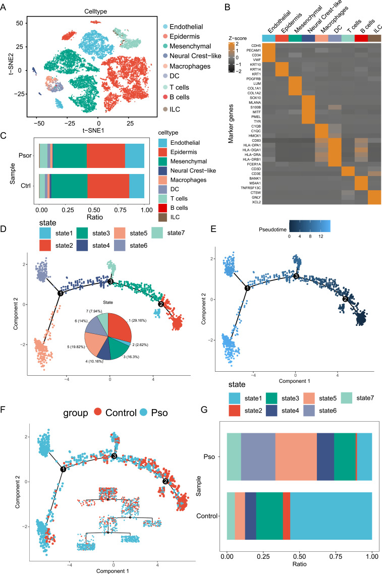
The scRNA-seq data (GSE162183) were obtained for skin tissues from three patients with psoriasis and three healthy individuals, designated as the psoriasis (Pso) group and the Control group, respectively. The data revealed distinct clustering of multiple cell types (Figure 2A), each characterized by their respective marker genes (Figure 2B). Visualization of the distribution of these cells indicated an increase in endothelial and T cells in the Pso group, while epidermal and mesenchymal cells were decreased compared to the Control group (Figure 2C). T cells are recognized for their significant role in the pathology of psoriasis,26 potentially differentiating into various types with either anti-inflammatory or pro-inflammatory functions. These T cells can secrete multiple cytokines, influencing keratinocytes and the immune microenvironment, thereby eliciting an immune-inflammatory response in psoriasis.27,28 However, there has been limited analysis of the T cell differentiation trajectory.
Figure 2.
Individuals afflicted with psoriasis display a spectrum of seven discrete T cell differentiation states. (A) Landscape of different cell clusters and populations in psoriasis. (B) A heatmap depicting the expression levels of genes associated with psoriasis across various cell types. (C) Comparison of cell types and proportions between the Pso and Control groups of psoriasis. (D) Presentation of differentiation trajectories in seven state T cells. (E) Sequence of the pseudotime trajectory of T cells. (F) Differentiation trajectories of T cells in the Pso and Control groups. (G) Proportional distribution of seven state T cells in the Pso and Control groups.
We used the monocle algorithm to analyze T cell differentiation trajectories and visualize their progression in psoriasis. The analysis classified T cells into seven distinct types, each representing a different state in the development of psoriasis (Figure 2D). As depicted in Figure 2E, the fluctuating color saturations along the pseudotime trajectory signify the progression through differentiation phases, where more intense colors denote the initial stages and more subdued tones represent the advanced stages. It was observed that most T cells in the Pso group were at a late stage of differentiation, whereas those in the Control group were predominantly at an early stage (Figure 2F). The cell ratio analysis revealed variations in T cell states between the two groups, specifically a decrease in state one and two T cells and an increase in state five, six, and seven T cells in the Pso group compared to the Control group (Figure 2G). Notably, state six T cells were only present in the psoriasis group. It was speculated that state one and two T cells represent the functional expression of T cells in the skin lesions of healthy individuals, whereas state five, six, and seven T cells may reflect the pathogenesis of psoriasis. Further detailed analysis of these T cell states and their roles in the pathogenesis of psoriasis will enhance understanding of the disease and may lead to new therapeutic options.
Analysis of TDEGs and Functional Enrichment Analysis of Seven State T Cells in Psoriasis
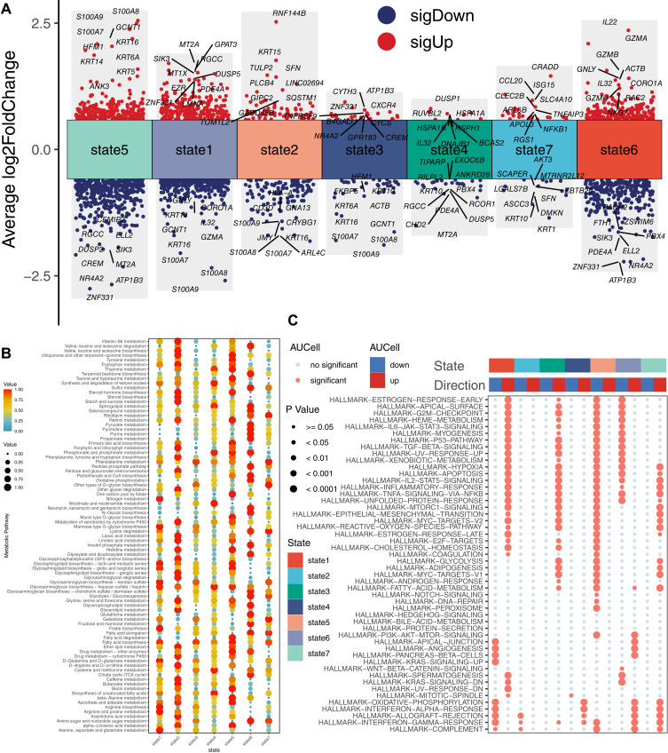
Differential gene expression and functional enrichment analyses provide insights into the roles of various T cell states and their associated biological processes in psoriasis. Initially, TDEGs were visualized using volcano plots (Figure 3A). The analysis indicated that the S100A protein family (S100A7, S100A8, S100A9), Keratin family (KRT5, KRT6A, KRT14, KRT16), GNLY, granzyme family (GZMB, GZMA), CCL20, SCL4A10, and CRADD were upregulated in T cells of states four, five, six, and seven. S100A7 and S100A9 influence psoriatic T helper cell (Th17 and Th22) responses, thereby impacting the progression of psoriasis.29 The upregulation of KRT6, KRT16, and KRT17 alters keratinocyte proliferation, cell adhesion, migration, and inflammatory characteristics, leading to hyperproliferation and innate immune activation of keratinocytes, which are key early barrier alert proteins in psoriasis.30 GZMB, a major marker of CD8 T cells, is highly expressed in psoriasis and may be linked to psoriasis recurrence.31 Additionally, ATP1B3, ZNF331, SIK3, and NR4A2 were found to be downregulated in T cells of states four, five, six, and seven. ATP1B3 is essential for maintaining cellular homeostasis and is associated with immune cell infiltration in various cancers.32 SIK3, a member of the AMP-activated protein kinase family, governs the metabolism of glucose and lipids and functions as an inhibitory regulator to curb the expression of inflammatory genes in macrophages activated by lipopolysaccharide.33 These findings underscore the involvement of these genes in cell growth, differentiation, cycling, apoptosis, and immune-inflammatory responses, providing valuable insights into the pathogenesis of psoriasis.
Figure 3.
Screening and functional analysis of TDEGs in seven state T cells in psoriasis. (A) Upregulated and downregulated TDEGs in the seven state T cells. TDEGs that are upregulated are denoted by red circles, indicating high gene expression levels, whereas downregulated TDEGs are marked by blue circles, signifying low expression levels. (B) Metabolic activity score results for the TDEGs in seven state T cells. (C) Signaling pathways of the TDEGs in seven state T cells through enrichment analysis.
Metabolic activity scores revealed that T cells in seven states exhibit multiple metabolic disorders (Figure 3B), including amino acid metabolism, glucose metabolism, lipid metabolism, and vitamin metabolism. Lipid metabolism, a significant area of research in psoriasis pathogenesis, showed that fatty acid degradation, fatty acid biosynthesis, ether lipid metabolism, and steroid hormone biosynthesis were highly expressed in state five T cells. In state six T cells, sphingolipid metabolism and fatty acid elongation were upregulated, while biosynthesis of unsaturated fatty acids was downregulated. In state seven T cells, fatty acid elongation and fatty acid biosynthesis were highly expressed, whereas fatty acid degradation and ether lipid metabolism were lowly expressed. These metabolic reactions are implicated in psoriasis development and merit further investigation to understand their functions in T cells.
To further extend the analysis, GSEA was used to uncover the potential functions of the seven T cell states (Figure 3C). These pathways were derived from the hallmark gene set (https://www.gsea-msigdb.org/gsea/msigdb/human/genesets.jsp?collection=H). The results indicated that signaling pathways such as G2M checkpoint, IL-6/Janus kinase (JAK)/signal transducer and activator of transcription 3 (STAT3) signaling, tumor protein 53 (P53), transforming growth factor-β (TGF-β), and xenobiotic metabolism were downregulated in states five and six T cells but upregulated in states one and three T cells. Pathways related to hypoxia, apoptosis, IL-2/STAT5 signaling, inflammatory response, and TNF-α/nuclear factor kappa-B (NF-κB) were downregulated in state five and six T cells but upregulated in state one, three, and seven T cells. Glycolysis and fatty acid metabolism were upregulated in state three and seven T cells but downregulated in state five T cells. Therefore, understanding the differential pathways and metabolic activities of the seven T cell states will help elucidate the pathogenesis of psoriasis.
Combined Analysis of Bulk RNA-Seq and scRNA-Seq Data Identified INSIG1 as a Novel Biomarker
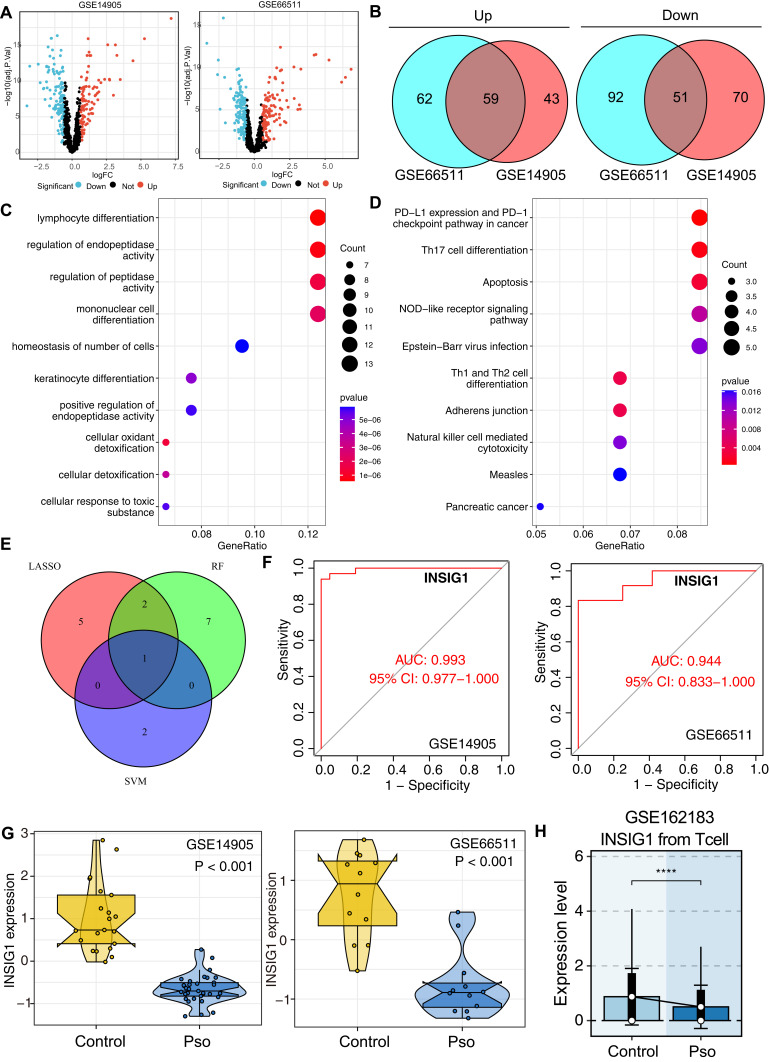
To identify core genes, two RNA-seq datasets (GSE14905 and GSE66511) were analyzed. Initially, within these two cohorts, we conducted differential analysis on TDEGs and screened for DEGs (with |log2FC| > 0.585 and an adjusted P value < 0.05) (Figure 4A). Subsequently, we identified the intersection of genes that were commonly upregulated and commonly downregulated across both datasets, discovering a total of 110 novel DEGs (Figure 4B), including 59 upregulated genes and 51 downregulated genes. Gene Ontology (GO) biological process (BP) enrichment analysis of these 110 novel DEGs revealed enrichment in lymphocyte differentiation, regulation of endopeptidase activity, monocyte differentiation, and keratinocyte differentiation (Figure 4C). Additionally, Kyoto Encyclopedia of Genes and Genomes (KEGG) pathway analysis indicated significant expression of Th17 cell differentiation, apoptosis, nucleotide-binding oligomerization domain (NOD)-like signaling pathway, and Th1 and Th2 cell differentiation in cancer (Figure 4D). The dysfunction of Th1, Th17, and Th22 cells is crucial in the development of psoriasis.27 The NOD-like receptor signaling pathway is a key biological pathway in psoriasis, with NOD2 upregulated in psoriatic epidermis.34 These results further affirm that the identified DEGs are associated with biological processes such as T cell proliferation and differentiation.
Figure 4.
Advanced multi-machine learning analyses identify INSIG1 as an emerging biomarker for psoriasis. (A) DEGs in the GSE14905 and GSE66511 dataset. Red circles represent upregulated genes while blue circles represent downregulated genes. (B) The DEGs that are commonly upregulated and downregulated in the GSE14905 and GSE66511 datasets. (C and D) Results of GO and KEGG enrichment analysis of the new 110 DEGs. (E) Visualization of interactive genes from three machine learning methods. (F) ROC curve reflecting the diagnostic performance of INSIG1 across GSE14905 and GSE66511 datasets and the expression of INSIG1 in the Pso and Control groups of the two datasets. (G) Box plots of differential expression for INSIG1 in the GSE14905 and GSE66511 datasets. (H) Bar charts illustrating the expression levels of INSIG1 in T cells between individuals with psoriasis and those from a healthy cohort within the scRNA-seq dataset of GSE62183.
Machine learning has emerged as an efficient tool for identifying promising biomarkers and we employed it to filter candidate genes from the 110 newly identified DEGs. LASSO regression analysis identified eight genes (Figure S1A), RF analysis identified 10 genes by calculating the importance of each gene (Figure S1B), and SVM analysis identified three genes (Figure S1C). By intersecting the results from LASSO, RF, and SVM analyses, INSIG1 was identified as a common gene (Figure 4E). Furthermore, ROC analysis was conducted to evaluate the diagnostic effectiveness of INSIG1 using external datasets. The results revealed an AUC of 0.993 for GSE14905 and 0.944 for GSE66511 (Figure 4F). INSIG1 was found to be downregulated in patients with psoriasis compared to the Control group (Figure 4G). We conducted an analysis of 12 datasets from psoriasis studies as an external validation cohort. The findings indicated that the expression levels of INSIG1 were markedly lower in individuals with psoriasis than in the Control group across all the cohorts examined (Figure S2A). Additionally, INSIG1 exhibited an AUC value exceeding 0.8 in every cohort (Figure S2B). In the scRNA-seq dataset of GSE162183, we observed a marked reduction in INSIG1 expression within T cells from individuals with psoriasis as compared to Control group (Figure 4H). Additionally, our analysis of the GSE173706 cohort, comprising scRNA-seq data from 14 skin samples affected by psoriasis and 19 healthy skin samples, revealed a similar trend after cell annotation (Figure S3A). Here, INSIG1 expression was notably lower in T cells derived from psoriasis samples than in those from healthy individuals (Figure S3B). Paralleling these findings, our examination of spatial transcriptome data from GSE225475, which included samples from two healthy skins and four psoriasis skins, also indicated a significant downregulation of INSIG1, with these results being consistent across datasets (Figure S3C and D). Notably, within the bulk transcriptome cohort of GSE78097, INSIG1 expression was found to be lowest in the severe psoriasis group (severity of disease were classified by the investigators according to the following six grades: clear, minimal, mild, moderate, severe and very severe)) (Figure S3E), implying a correlation between INSIG1 levels and disease progression of psoriasis. Lastly, we identified the highest expression of INSIG1 in state one T cells (Figure S3F). Given that our previous findings indicated a significant reduction in the proportion of state one T cells in psoriasis patients, the overall decrease in INSIG1 at the bulk transcriptome level is likely attributable to the diminished number of state one T cells. Therefore, modulating T cell differentiation to foster the development of state one T cells could be considered a promising therapeutic approach of psoriasis.
INSIG1 May Affect Fatty Acid Metabolism of Psoriasis and T Cell Function
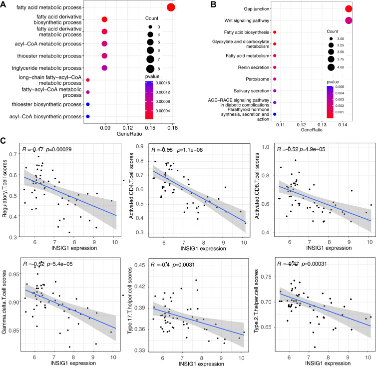
We delved deeper into the potential roles of INSIG1. Initially, within the GSE14905 dataset, we determined the Pearson correlation coefficients for the expression levels of all genes in relation to INSIG1. This analysis allowed us to pinpoint 149 genes that exhibited co-expression with INSIG1, as indicated by correlation coefficients exceeding 0.7. Following this, we conducted an enrichment analysis to further explore the functional implications of these genes. GO BP enrichment analysis revealed that the primary biological processes involved included fatty acid metabolic processes, biosynthetic processes of fatty acid derivatives, and metabolic processes of fatty acid derivatives (Figure 5A). These results further confirmed the close relationship between INSIG1 and lipid metabolism. KEGG pathway analysis implicated the Wnt signaling pathway, fatty acid biosynthesis, and glyoxylate and dicarboxylate metabolism, among others (Figure 5B), providing a basis for subsequent research into the function of INSIG1.
Figure 5.
Functional enrichment and Spearman correlation analysis of INSIG1 co-expressed genes. (A) GO enrichment analysis of INSIG1 co-expressed genes. (B) KEGG enrichment analysis of INSIG1 co-expressed genes. (C) Results of correlation between INSIG1 expression and T cell subtypes via Spearman correlation analysis.
The correlation between INSIG1 expression and T cell subset content was further explored. The results revealed that INSIG1 was negatively correlated with regulatory T cells (Tregs), CD4(+) T cells, CD8(+) T cells, Th17, Th2, and γδ T cells (Figure 5C). Building on these findings, it is hypothesized that INSIG1 may be involved in facilitating the differentiation of T cells into CD4(+) T cells, CD8(+) T cells, or other T cell subtypes.
Interactions Between INSIG1-Expressing T Cells and Immune Cells
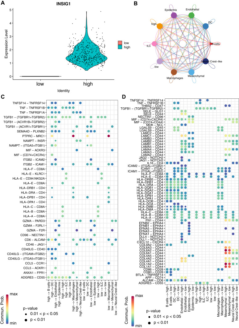
To explore the relationship between INSIG1-expressing T cells and other immune cells, T cells were categorized into those with high and low INSIG1 expression (Figure 6A). A communication network, based on cellular communication analysis, revealed extensive intercellular interactions between both high and low INSIG1-expressing T cells and other immune cells (Figure 6B). This network could be essential in the pathogenesis of psoriasis. Additionally, intercellular ligand-receptor interactions were identified between high and low INSIG1-expressing T cells and other immune cells. As illustrated in Figure 6C, protein tyrosine phosphatase receptor type C (PTPRC) and C1 mannose receptor (MRC1) signaling exhibited strong interactions between macrophages and both high and low INSIG1-expressing T cells, indicating significant communication among them. Previous studies have demonstrated a connection between T cells and macrophages via PTPRC-MRC1 during the development of inflammatory skin diseases.35 Transforming growth factor-beta receptors (TGFBR1 and TGFBR2) showed weaker interactions with high INSIG1-expressing T cells and B cells, dendritic cells, and endothelial cells, and even weaker interactions with low INSIG1-expressing T cells and endothelial cells as well as innate lymphoid cells. TGFBR1 and TGFBR2, as receptors for TGF-β, facilitate the activation of the TGF-β signaling pathway,36 which regulates cell proliferation, differentiation, apoptosis, interactions with the microenvironment, and immune responses in psoriasis.37
Figure 6.
Cellular communication analysis and ligand–receptor interactions of INSIG1 expressing T cells and other cells. (A) Expression level of high and low INSIG1 expressing T cells. (B) Cell communication analysis between high and low INSIG1 expressing T cells and other cells. (C and D) Ligand–receptor interactions between high and low INSIG1 expressing T cells and other cells.
Additionally, when INSIG1-expressing T cells acted as receptors (Figure 6D), interactions involving collagen type VI alpha 2 (COL6A2)-CD44 and collagen type I alpha 2 (COL1A2)-CD44 were noted between INSIG1 high and low T cells and mesenchymal cells. Furthermore, signaling via Jagged-2 (JAG2)-notch receptor (NOTCH2), JAG1-NOTCH2, delta-like protein (DLL) 4-NOTCH2, and DLL1-NOTCH2 was diminished in endothelial cells and INSIG1 high T cells.
Interactions between NOTCH receptors and ligands play a vital role in cell proliferation, differentiation, and survival, and they facilitate the activation and invasion of endothelial cells in psoriasis.38 These findings highlight a significant relationship between INSIG1-expressing T cells and immune cells in the context of psoriasis.
INSIG1 is a Potential Therapeutic Target for Psoriasis, and Tetrandrine Stably Binds with INSIG1
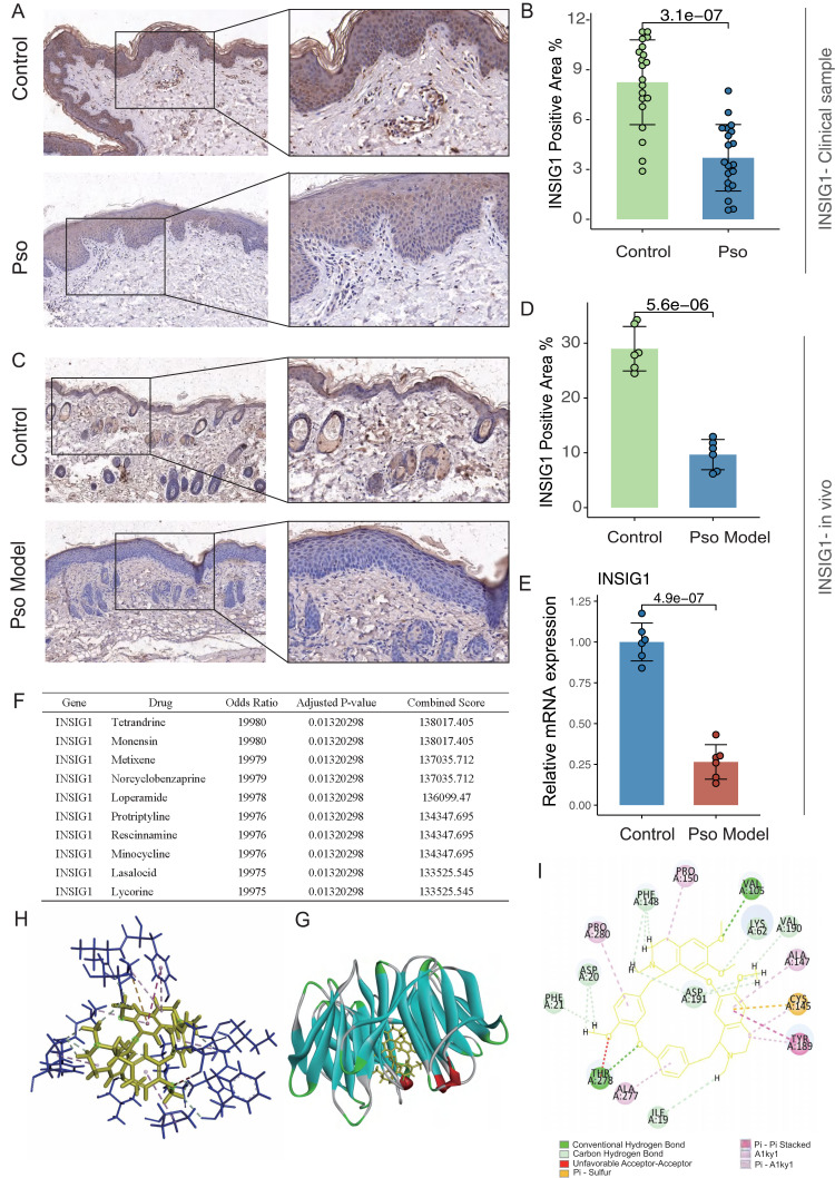
To further verify the expression of INSIG1 in psoriasis tissues, clinical skin samples were collected from patients with psoriasis (Pso) group and from Control group. The IHC results indicated that INSIG1 was lowly expressed in psoriasis lesions (Figure 7A and B). An imiquimod (IMQ)-induced psoriasis-like mouse model was also constructed to analyze the expression of INSIG1 in mouse psoriatic skin tissue through IHC (Figure 7C and D) and qRT-PCR (Figure 7E). Results demonstrated that INSIG1 expression is significantly down-regulated in psoriatic skin (Pso Model) from mice with IMQ-induced psoriasis compared to healthy skin (Control) group. Psoriasis, recognized as a persistent inflammatory and autoimmune condition affecting the skin, is linked to disruptions in lipid metabolism. INSIG1, which manages cholesterol synthesis, the production of fatty acids, and maintains balanced glucose levels across different tissues, also possesses the capacity to modulate inflammatory reactions in autoimmune disorders,18 further confirming the role of INSIG1 in psoriasis.
Figure 7.
Validation of INSIG1 expression in psoriasis through experiments and molecular docking results between INSIG1 and tetrandrine. (A and B) IHC results of INSIG1 expression in the Pso group compared with the Control group in clinical samples (n=20) and (C and D) in in vivo (n=6) experiments (scale bar = 50 μm and 20 μm). (E) mRNA expression level of INSIG1 in the Pso model group compared with the control group in vivo (n=6). (F) Drug sensitivity enrichment analysis of INSIG1. (G and H) Visualization of molecular docking between INSIG1 and tetrandrine. (I) Results of interaction analysis between ligand (tetrandrine) and receptor (INSIG1). Statistical significance: *p < 0.05, **p < 0.01, ***p < 0.001, ****p < 0.0001, compared with the Control group.
To validate the clinical utility of INSIG1, drug sensitivity enrichment analysis was conducted using the Enrichr database, revealing a good binding affinity between tetrandrine and INSIG1 (Figure 7F). Tetrandrine, initially identified as a bis-benzyl-isoquinoline alkaloid, primarily inhibits Th17 cell differentiation and regulates the Th17/Treg balance.39 This compound can promote Treg differentiation and inhibit the expression of IL-22 to suppress the proliferation of keratinocytes, thereby mitigating psoriatic inflammation.40 To substantiate the stability of INSIG1 and tetrandrine and to elucidate their binding dynamics, a series of computational analyses were conducted, encompassing molecular docking, molecular dynamics simulations, and the computation of binding free energies. INSIG1 served as the protein receptor, while tetrandrine acted as the small molecule ligand. Figure 7H and G illustrated the best binding modes of INSIG1 and tetrandrine, with their free binding energy calculated to be −79.6578 kcal/mol. Interaction analysis revealed that tetrandrine’s interaction with INSIG1 consisted of two conventional hydrogen bonds (VAL105, THR278), seven C-H bonds (PHE148, ASP20, PHE21, LYS62, VAL190, ASP191, ILE19), a π-sulfur bond (CYS145), and extensive van der Waals forces (Figure 7I). Docking results illustrated that tetrandrine primarily binds to INSIG1 through hydrogen bonding and van der Waals forces, indicating its potential as a therapeutic drug for psoriasis. Building upon this, we established a murine model of psoriasis and treated it with tetrandrine for a period of one week. The subsequent experimental data revealed that tetrandrine not only markedly alleviated the redness and scaling associated with psoriasis (Figure S4A) but also decreased the psoriasis area and severity index (PASI) score (Figure S4E). Figure S4B and C respectively illustrated the alterations in pathological tissue and body weight of psoriasis mice following tetrandrine treatment.
Additionally, tetrandrine downregulated the peripheral blood levels of pro-inflammatory cytokines, including IL-6, IL-17, IL-23, and TNF-α (Figure S4D), while upregulating INSIG1 expression (Figure S4F). These effects position tetrandrine as a promising candidate for the targeted treatment of psoriasis via INSIG1.
Finally, upon thorough examination and data extraction from public databases, we observed that following biologic therapy, there was a notable restoration and increase in INSIG1 levels among psoriasis patients. These findings underscore the responsiveness of INSIG1 to biologic interventions, hinting at its potential role as a therapeutic target for psoriasis and its possible correlation with disease severity (Figure S4G).
Discussion
Despite notable advancements in managing moderate to severe psoriasis through biologic treatments in recent years, cessation of systemic and biologic interventions could potentially lead to a relapse of the condition and the development of resistance to therapy.10 Some patients are limited in their choice of biologics due to the severity of their diseases and contraindications. As pivotal components of precision medicine, biomarkers are invaluable for clinical practice. They hold the potential to forecast the risks associated with psoriasis comorbidities, expedite the development of innovative therapeutics, refine the accuracy of targeted treatment approaches, and ultimately, elevate the standards of clinical efficacy. Therefore, the exploration of new, effective, and emerging therapeutic biomarkers and targets to regulate immune balance remains imperative.
This research identified elevated T cell expression in psoriasis patients relative to healthy controls through the analysis of publicly available scRNA-seq data. The findings underscore the pivotal role of T cells in the pathogenesis of psoriasis, corroborating earlier research.41 Unlike the previous classification of T cell subpopulations, T cells were classified into seven states by the proposed temporal analysis. States one and two T cells decreased, while states five, six, and seven T cells increased in psoriasis compared with the normal group, suggesting that states five, six, and seven T cells may contribute to the progression of psoriasis.
The development of psoriasis is accompanied by processes such as an imbalance in the immune system dominated by T cells and oxidative stress.42 Recent findings have gradually shown the impact of metabolites and metabolic pathways on psoriasis, emphasizing the important role of the network of interactions between metabolic disorders, inflammation, and immunity in psoriasis.43 The study revealed that metabolism of lipoic acid, along with the degradation, elongation, and biosynthesis of fatty acids, exhibited differential expression patterns across various T cell states. GSEA analysis also revealed differential expression of xenobiotic metabolism, glycolysis, and fatty acid metabolism in the seven state T cells, further emphasizing the role of metabolic dysregulation, especially lipid metabolism, in the pathogenesis of psoriasis. Lipid metabolism is crucial in the pathology of psoriasis, being intricately linked to the immuno-inflammatory responses triggered by T cells.44 These studies further illustrate that psoriasis pathogenesis and progression are related to T-cell-associated immunity and lipid metabolism, which is a potential direction for psoriasis mechanism exploration and clinical intervention.
To further confirm the characterized candidate genes, multiple machine learning approaches, two bulk transcriptome datasets and scRNA-seq dataset were combined to identify INSIG1 as a biomarker associated with psoriasis T cell immunity, with downregulated expression in psoriatic lesions. INSIG1 is pointed as an therapeutic target and specific diagnostic marker for inflammatory immune conditions, such as atherosclerosis,45 prostate cancer,46 and gout,47 closely related to immune cell infiltration. Studies have documented that INSIG1 mediates ubiquitination of NLRP3, thereby suppressing the activation of the NLRP3 inflammasome.48 The deletion of INSIG1 improves the lipidome profile and mitigates cellular injury, inflammation, and the accumulation of extracellular matrix.49 INSIG1 participates in the innate immune response through the modulation of stimulator of interferon genes ubiquitination.50 To further explore the influence of INSIG1 on the developmental process of psoriasis, it was found that INSIG1 was mainly expressed in state one T cells. It is well known that state one T cells gradually decrease with the progression of psoriasis, and the expression of INSIG1 is also gradually downregulated. Moreover, the expression of INSIG1-expressing T cells was negatively correlated with the expression of other T cell subpopulations, further suggesting that low expression of INSIG1 promotes the progression of psoriasis. Notably, INSIG1 is underexpressed in other autoimmune diseases and cardio metabolic diseases, and our study aligned with this observation,21,22,45,47 showing a lower expression of INSIG1 in psoriasis. It was also discovered that INSIG1-expressing T cells are inextricably linked to other T cell subsets and act as mutual ligand receptors with them to exert immunomodulatory effects. These findings further demonstrate the regulatory role of INSIG1 on immunoinflammation as a potential target for psoriasis.
Additionally, as a member of the insulin-induced gene family, INSIG1 functions as a six-transmembrane protein, intricately embedded within the endoplasmic reticulum membrane.51 Research indicates that the INSIG1-mediated suppression of the SREBP signaling pathway, responsible for governing lipid balance, represents a promising therapeutic target for the development of hypolipidemic agents.15 INSIG1 upregulation helps inhibit fatty acid desaturation in mammary epithelial cells, thereby regulating fatty acid anabolism.52 This implicates that INSIG1 could be a pivotal target for modulating lipid metabolism in individuals with psoriasis.
In summary, we preliminarily inferred that INSIG1 was associated with the regulation of immune responses, inflammatory processes, and lipid metabolism in psoriasis. This hypothesis is also found in neurodegenerative processes.53 The correlation between INSIG1 expression and psoriasis progression was further verified by IHC, qRT-PCR, and other experiments. These discoveries shed light on the biological regulatory mechanisms of INSIG1 in the development of psoriasis, highlight its potential as a novel target and biomarker for the disease, and offer valuable insights for advancing clinical management and therapeutic strategies in psoriasis.
To explore potential effective treatments for psoriasis, we utilized the drug library data prediction and molecular docking to identify therapeutic drugs or molecular compounds that could treat psoriasis targeted INSIG1. Tetrandrine is a bisbenzylisoquinoline alkaloid extracted from the root of S. tetrandra and has been used as a therapeutic agent for psoriasis due to its anti-inflammatory activity.49 It has been found that tetrandrine alleviates silicosis by inhibiting the NLRP3 inflammasome in macrophages.54 Research suggests that the topical administration of a tetrandrine nanoemulsion can stimulate the proliferation of regulatory T cells, which may result in the amelioration of psoriatic lesion symptoms in mice.55 Additionally, tetrandrine has been shown to curb the proliferation of HaCaT cells and the cytokine production triggered by IL-22.40 Our research has revealed that tetrandrine not only alleviated key psoriatic symptoms in mice, including scale formation and redness, but also significantly decreased the PASI scores. Furthermore, it reduced peripheral blood levels of pro-inflammatory cytokines such as IL-17, IL-23, TNF-α, and IL-6. In addition to these effects, tetrandrine had been shown could upregulate INSIG1 expression in psoriasis. The above experiments initially demonstrated the therapeutic effect and anti-inflammatory activity of tetrandrine on psoriasis, indicating its potential as a drug for targeting INSIG1 in the treatment of psoriasis.
However, our study presents some limitations. A future direction includes the generation of INSIG1 knockout T cells in animal models to delve deeper into the specific targeting effects of INSIG1 on T cells and its role in the development of psoriasis. Concurrently, the extensive, high-standard evidence-based medical research is needed to substantiate the therapeutic efficacy of tetrandrine for psoriasis in clinical settings.
Conclusion
In conclusion, INSIG1 has been recognized as a pivotal modulator of inflammation, immune response, and lipid metabolism, presenting itself as a potential therapeutic target for the modulation of T cell differentiation in psoriasis. This conclusion was reinforced by our comprehensive study that amalgamated scRNA-seq data with external bulk RNA-seq datasets, and applied machine learning methodologies. Our results emphasized the critical importance of INSIG1, spotlighting its potential as a biomarker and a new target for therapeutic intervention in psoriasis. We also verified the targeting effect of tetrandrine on INSIG1 as a potential treatment for psoriasis, and opened up new research avenues for precision medicine and clinical interventions of psoriasis.
Acknowledgments
We are grateful to the Clinical Medical Research Institute of China-Japan Friendship Hospital for providing laboratory resources and facilities. We thank all contributors to the GEO database. All methods were carried out in accordance with relevant guidelines and regulations and our study were complied with the Declaration of Helsinki.
Funding Statement
This publication was supported by the National High-Level Hospital Clinical Research Funding (2023-NHLHCRF-YYPPLC-TJ-04), the National Natural Science Foundation of China (Grant No. 82074445), and the Qi Huang Scholars Support Project of the State Administration of Traditional Chinese Medicine (Chinese Medicine Education Letter [2021] No. 203.).
Abbreviations
AUC, area under the curve; BP, biological process; COL6A2, collagen type VI alpha 2; COL1A2, collagen type I alpha 2; DEGs, Differentially Expressed Genes; DLL, delta-like protein; GO, Gene Ontology; GSEA, gene set enrichment analysis; INSIG1, insulin-induced gene 1; IHC, Immunohistochemical analysis; IL, interleukin; IMQ, imiquimod; JAK Janus kinase; JAG2, Jagged-2; KEGG, Kyoto Encyclopedia of Genes and Genomes; LASSO, least absolute shrinkage and selection operator; |log2FC|, |log2 fold change|; MRC1, C1 mannose receptor; NF-κB, nuclear factor kappa-B; NOD, nucleotide-binding oligomerization domain; NOTCH, notch receptor; NLRP3, NOD−, LRR− and pyrin domain-containing protein 3; PASI, Psoriasis area and severity index; PCA, Principal component analysis; PPI, protein-protein interaction; Pso, psoriasis group; P53, tumor protein 53; PTPRC, protein tyrosine phosphatase receptor type C; qRT-PCR, Quantitative reverse transcription polymerase chain reaction; ROC, receiver operating characteristic; RF, random forest; scRNA-seq, single-cell RNA sequencing; SREBPs, sterol regulatory element-binding proteins; SVM, support vector machine; STAT3, signal transducer and activator of transcription 3; TDEGs, differentially expressed genes in T cells; TNF, tumor necrosis factor; t-SNE, t-Distributed Stochastic Neighbor Embedding; Th, T helper cell; TGF-β, transforming growth factor-β; TGFBR, Transforming growth factor beta receptor.
Data Sharing Statement
The datasets analyzed during the current study are available in the GEO database (https://www.ncbi.nlm.nih.gov/geo/), which is continuously updated and openly accessible for free download.
Ethics Approval and Consent to Participate
The animal experiment conducted in this study and clinical study were approved by the Clinical Research Ethics Committee of China-Japan Friendship Hospital (Ethics approval number: 2023-KY-181). Informed consent was sought from all participants prior to their participation in the study.
Author Contributions
All authors made a significant contribution to the work reported, whether that is in the conception, study design, execution, acquisition of data, analysis and interpretation, or in all these areas; took part in drafting, revising or critically reviewing the article; gave final approval of the version to be published; have agreed on the journal to which the article has been submitted; and agree to be accountable for all aspects of the work.
Disclosure
The authors declare that they have no competing interests.
References
- 1.Ji C, Wang H, Bao C, et al. Challenge of nail psoriasis: an update review. Clin Rev Allergy Immunol. 2021;61:377–402. doi: 10.1007/s12016-021-08896-9 [DOI] [PubMed] [Google Scholar]
- 2.Boehncke WH, Schön MP. Psoriasis. Lancet. 2015;386:983–994. doi: 10.1016/S0140-6736(14)61909-7 [DOI] [PubMed] [Google Scholar]
- 3.Griffiths CEM, Armstrong AW, Gudjonsson JE, Barker J. Psoriasis. Lancet. 2021;397:1301–1315. doi: 10.1016/S0140-6736(20)32549-6 [DOI] [PubMed] [Google Scholar]
- 4.Hawkes JE, Chan TC, Krueger JG. Psoriasis pathogenesis and the development of novel targeted immune therapies. J Allergy Clin Immunol. 2017;140:645–653. doi: 10.1016/j.jaci.2017.07.004 [DOI] [PMC free article] [PubMed] [Google Scholar]
- 5.Yang Q, Wang J, Mi N, Zou Y. Literature overview of the relation between psoriasis and Alzheimer. Neuropsychiatr Dis Treat. 2023;19:461–468. doi: 10.2147/NDT.S403854 [DOI] [PMC free article] [PubMed] [Google Scholar]
- 6.Takeshita J, Grewal S, Langan SM, et al. Psoriasis and comorbid diseases: epidemiology. J Am Acad Dermatol. 2017;76:377–390. doi: 10.1016/j.jaad.2016.07.064 [DOI] [PMC free article] [PubMed] [Google Scholar]
- 7.Zhang L, Wang Y, Qiu L, Wu J. Psoriasis and cardiovascular disease risk in European and East Asian populations: evidence from meta-analysis and Mendelian randomization analysis. BMC Med. 2022;20:421. doi: 10.1186/s12916-022-02617-5 [DOI] [PMC free article] [PubMed] [Google Scholar]
- 8.Zhang P, Wu MX. A clinical review of phototherapy for psoriasis. Lasers Med Sci. 2018;33:173–180. doi: 10.1007/s10103-017-2360-1 [DOI] [PMC free article] [PubMed] [Google Scholar]
- 9.Kim HK, Bae MJ, Lim S, Lee W, Kim S. A water-soluble extract from Actinidia arguta ameliorates psoriasis-like skin inflammation in mice by inhibition of neutrophil infiltration. Nutrients. 2018;10:1399. doi: 10.3390/nu10101399 [DOI] [PMC free article] [PubMed] [Google Scholar]
- 10.Caputo V, Strafella C, Cosio T, et al. Pharmacogenomics: an update on biologics and small-molecule drugs in the treatment of psoriasis. Genes. 2021;12:1398. doi: 10.3390/genes12091398 [DOI] [PMC free article] [PubMed] [Google Scholar]
- 11.Kamata M, Tada Y. Efficacy and safety of biologics for psoriasis and psoriatic arthritis and their impact on comorbidities: a literature review. Int J Mol Sci. 2020;21:1690. doi: 10.3390/ijms21051690 [DOI] [PMC free article] [PubMed] [Google Scholar]
- 12.Singh R, Koppu S, Perche PO, Feldman SR. The cytokine mediated molecular pathophysiology of psoriasis and its clinical implications. Int J Mol Sci. 2021;22:12793. doi: 10.3390/ijms222312793 [DOI] [PMC free article] [PubMed] [Google Scholar]
- 13.Li C, Sunhe Y, Zhou H, Dong W. Efficacy and safety evaluations of Adalimumab biosimilars in the treatment of psoriasis. J DermatolTreat. 2023;34:2249145. doi: 10.1080/09546634.2023.2249145 [DOI] [PubMed] [Google Scholar]
- 14.Guo JW, Cheng YP, Liu CY, et al. Salvianolic acid B in microemulsion formulation provided sufficient hydration for dry skin and ameliorated the severity of imiquimod-induced psoriasis-like dermatitis in mice. Pharmaceutics. 2020;12:457. doi: 10.3390/pharmaceutics12050457 [DOI] [PMC free article] [PubMed] [Google Scholar]
- 15.Xu Y, Tao J, Yu X, et al. Hypomorphic ASGR1 modulates lipid homeostasis via INSIG1-mediated SREBP signaling suppression. JCI Insight. 2021;6. doi: 10.1172/jci.insight.147038 [DOI] [PMC free article] [PubMed] [Google Scholar]
- 16.Watanabe Y, Sasaki T, Miyoshi S, Shimizu M, Yamauchi Y, Sato R. Insulin-induced genes INSIG1 and INSIG2 mediate oxysterol-dependent activation of the PERK-eIF2α-ATF4 axis. J Biol Chem. 2021;297:100989. doi: 10.1016/j.jbc.2021.100989 [DOI] [PMC free article] [PubMed] [Google Scholar]
- 17.Xu D, Wang Z, Xia Y, et al. The gluconeogenic enzyme PCK1 phosphorylates INSIG1/2 for lipogenesis. Nature. 2020;580:530–535. doi: 10.1038/s41586-020-2183-2 [DOI] [PubMed] [Google Scholar]
- 18.Wang Y, Wang Y, Ding L, et al. Tim-4 reprograms cholesterol metabolism to suppress antiviral innate immunity by disturbing the Insig1-SCAP interaction in macrophages. Cell Rep. 2022;41:111738. doi: 10.1016/j.celrep.2022.111738 [DOI] [PubMed] [Google Scholar]
- 19.Guo C, Chi Z, Jiang D, et al. Cholesterol homeostatic regulator SCAP-SREBP2 integrates NLRP3 inflammasome activation and cholesterol biosynthetic signaling in macrophages. Immunity. 2018;49:842–856.e7. doi: 10.1016/j.immuni.2018.08.021 [DOI] [PubMed] [Google Scholar]
- 20.Zheng L, Song H, Li Y, Li H, Lin G, Cai Z. Insulin-induced gene 1-enhance secretion of BMSC exosome enriched in miR-132-3p promoting wound healing in diabetic mice. Mol Pharm. 2024;21:4372–4385. doi: 10.1021/acs.molpharmaceut.4c00322 [DOI] [PubMed] [Google Scholar]
- 21.Xie D, Song L, Xiang D, Gao X, Zhao W. Salvianolic acid A alleviates atherosclerosis by inhibiting inflammation through Trc8-mediated 3-hydroxy-3-methylglutaryl-coenzyme A reductase degradation. Phytomedicine. 2023;112:154694. doi: 10.1016/j.phymed.2023.154694 [DOI] [PubMed] [Google Scholar]
- 22.de Lima JD, de Paula AGP, Yuasa BS, et al. Genetic and epigenetic regulation of the innate immune response to gout. Immunol Invest. 2023;52:364–397. doi: 10.1080/08820139.2023.2168554 [DOI] [PubMed] [Google Scholar]
- 23.Tang X, Zhang Y, Zhang H, et al. Single-cell sequencing: high-resolution analysis of cellular heterogeneity in autoimmune diseases. Clin Rev Allergy Immunol. 2024;66:376–400. doi: 10.1007/s12016-024-09001-6 [DOI] [PubMed] [Google Scholar]
- 24.Li J, Cui P, Jing H, et al. Hydrogen combined with tetrandrine attenuates silica-induced pulmonary fibrosis via suppressing NF-kappaB/NLRP3 signaling pathway-mediated epithelial mesenchymal transition and inflammation. Int Immunopharmacol. 2024;138:112563. doi: 10.1016/j.intimp.2024.112563 [DOI] [PubMed] [Google Scholar]
- 25.Gangwar RS, Gudjonsson JE, Ward NL. Mouse models of psoriasis: a comprehensive review. J Invest Dermatol. 2022;142:884–897. doi: 10.1016/j.jid.2021.06.019 [DOI] [PMC free article] [PubMed] [Google Scholar]
- 26.Raeber ME, Zurbuchen Y, Impellizzieri D, Boyman O. The role of cytokines in T-cell memory in health and disease. Immunol Rev. 2018;283:176–193. doi: 10.1111/imr.12644 [DOI] [PubMed] [Google Scholar]
- 27.Hu P, Wang M, Gao H, et al. The role of helper T cells in psoriasis. Front Immunol. 2021;12:788940. doi: 10.3389/fimmu.2021.788940 [DOI] [PMC free article] [PubMed] [Google Scholar]
- 28.Nussbaum L, Chen YL, Ogg GS. Role of regulatory T cells in psoriasis pathogenesis and treatment. Br J Dermatol. 2021;184:14–24. doi: 10.1111/bjd.19380 [DOI] [PubMed] [Google Scholar]
- 29.Pavel AB, Del Duca E, Cheng J, et al. Delayed type hypersensitivity reactions to various allergens may differently model inflammatory skin diseases. Allergy. 2023;78:178–191. doi: 10.1111/all.15538 [DOI] [PubMed] [Google Scholar]
- 30.Zhang X, Yin M, Zhang LJ. Keratin 6, 16 and 17-critical barrier alarmin molecules in skin wounds and psoriasis. Cells. 2019;8:807. doi: 10.3390/cells8080807 [DOI] [PMC free article] [PubMed] [Google Scholar]
- 31.Gleave A, Granville DJ. Granzyme B in autoimmune skin disease. Biomolecules. 2023;13:388. doi: 10.3390/biom13020388 [DOI] [PMC free article] [PubMed] [Google Scholar]
- 32.Lu S, Cai S, Peng X, Cheng R, Zhang Y. Integrative transcriptomic, proteomic and functional analysis reveals ATP1B3 as a diagnostic and potential therapeutic target in hepatocellular carcinoma. Front Immunol. 2021;12:636614. doi: 10.3389/fimmu.2021.636614 [DOI] [PMC free article] [PubMed] [Google Scholar]
- 33.Sanosaka M, Fujimoto M, Ohkawara T, et al. Salt-inducible kinase 3 deficiency exacerbates lipopolysaccharide-induced endotoxin shock accompanied by increased levels of pro-inflammatory molecules in mice. Immunology. 2015;145:268–278. doi: 10.1111/imm.12445 [DOI] [PMC free article] [PubMed] [Google Scholar]
- 34.Chen L, Cao SQ, Lin ZM, He SJ, Zuo JP. NOD-like receptors in autoimmune diseases. Acta Pharmacol Sin. 2021;42:1742–1756. doi: 10.1038/s41401-020-00603-2 [DOI] [PMC free article] [PubMed] [Google Scholar]
- 35.Mitamura Y, Reiger M, Kim J, et al. Spatial transcriptomics combined with single-cell RNA-sequencing unravels the complex inflammatory cell network in atopic dermatitis. Allergy. 2023;78:2215–2231. doi: 10.1111/all.15781 [DOI] [PubMed] [Google Scholar]
- 36.Lü X, Zhang H, Huang Y, Zhang Y. A proteomics study to explore the role of adsorbed serum proteins for PC12 cell adhesion and growth on chitosan and collagen/chitosan surfaces. Regen Biomater. 2018;5:261–273. doi: 10.1093/rb/rby017 [DOI] [PMC free article] [PubMed] [Google Scholar]
- 37.Laudisi F, Stolfi C, Monteleone I, Monteleone G. TGF-β1 signaling and Smad7 control T-cell responses in health and immune-mediated disorders. Eur J Immunol. 2023;53:e2350460. doi: 10.1002/eji.202350460 [DOI] [PubMed] [Google Scholar]
- 38.Rooney P, Connolly M, Gao W, et al. Notch-1 mediates endothelial cell activation and invasion in psoriasis. Exp Dermatol. 2014;23:113–118. doi: 10.1111/exd.12306 [DOI] [PubMed] [Google Scholar]
- 39.Yuan X, Dou Y, Wu X, Wei Z, Dai Y. Tetrandrine, an agonist of aryl hydrocarbon receptor, reciprocally modulates the activities of STAT3 and STAT5 to suppress Th17 cell differentiation. J Cell Mol Med. 2017;21:2172–2183. doi: 10.1111/jcmm.13141 [DOI] [PMC free article] [PubMed] [Google Scholar]
- 40.Wang F, Wang J, Zhang Z, Chen S. Tetrandrine inhibits the proliferation and cytokine production induced by IL-22 in HaCaT cells. J Int Med Res. 2018;46:5210–5218. doi: 10.1177/0300060518801463 [DOI] [PMC free article] [PubMed] [Google Scholar]
- 41.Karczewski J, Dobrowolska A, Rychlewska-Hańczewska A, Adamski Z. New insights into the role of T cells in pathogenesis of psoriasis and psoriatic arthritis. Autoimmunity. 2016;49:435–450. doi: 10.3109/08916934.2016.1166214 [DOI] [PubMed] [Google Scholar]
- 42.Wójcik P, Biernacki M, Wroński A, et al. Altered lipid metabolism in blood mononuclear cells of psoriatic patients indicates differential changes in psoriasis vulgaris and psoriatic arthritis. Int J Mol Sci. 2019;20:4249. doi: 10.3390/ijms20174249 [DOI] [PMC free article] [PubMed] [Google Scholar]
- 43.Su R, Zhao S, Zhang J, Cao M, Peng S. Metabolic influences on T cell in psoriasis: a literature review. Front Immunol. 2023;14:1279846. doi: 10.3389/fimmu.2023.1279846 [DOI] [PMC free article] [PubMed] [Google Scholar]
- 44.Sun L, Fu J, Zhou Y. Metabolism controls the balance of Th17/T-regulatory cells. Front Immunol. 2017;8:1632. doi: 10.3389/fimmu.2017.01632 [DOI] [PMC free article] [PubMed] [Google Scholar]
- 45.Song C, Qiao Z, Chen L, et al. Identification of key genes as early warning signals of acute myocardial infarction based on weighted gene correlation network analysis and dynamic network biomarker algorithm. Front Immunol. 2022;13:879657. doi: 10.3389/fimmu.2022.879657 [DOI] [PMC free article] [PubMed] [Google Scholar]
- 46.Yan Y, Mao X, Zhang Q, et al. Molecular mechanisms, immune cell infiltration, and potential drugs for prostate cancer. Cancer Biomark. 2021;31:87–96. doi: 10.3233/CBM-200939 [DOI] [PMC free article] [PubMed] [Google Scholar]
- 47.Tseng CC, Wong MC, Liao WT, et al. Systemic investigation of promoter-wide methylome and genome variations in gout. Int J Mol Sci. 2020;21:4702. doi: 10.3390/ijms21134702 [DOI] [PMC free article] [PubMed] [Google Scholar]
- 48.Xu T, Yu W, Fang H, et al. Ubiquitination of NLRP3 by gp78/Insig-1 restrains NLRP3 inflammasome activation. Cell Death Differ. 2022;29:1582–1595. doi: 10.1038/s41418-022-00947-8 [DOI] [PMC free article] [PubMed] [Google Scholar]
- 49.Azzu V, Vacca M, Kamzolas I, et al. Suppression of insulin-induced gene 1 (INSIG1) function promotes hepatic lipid remodelling and restrains NASH progression. Mol Metab. 2021;48:101210. doi: 10.1016/j.molmet.2021.101210 [DOI] [PMC free article] [PubMed] [Google Scholar]
- 50.Wang Q, Liu X, Cui Y, et al. The E3 ubiquitin ligase AMFR and INSIG1 bridge the activation of TBK1 kinase by modifying the adaptor STING. Immunity. 2014;41:919–933. doi: 10.1016/j.immuni.2014.11.011 [DOI] [PubMed] [Google Scholar]
- 51.Xiong L, Liu HS, Zhou C, et al. A novel protein encoded by circINSIG1 reprograms cholesterol metabolism by promoting the ubiquitin-dependent degradation of INSIG1 in colorectal cancer. Mol Cancer. 2023;22:72. doi: 10.1186/s12943-023-01773-3 [DOI] [PMC free article] [PubMed] [Google Scholar]
- 52.Zhu L, Jiao H, Gao W, et al. Fatty acid desaturation is suppressed in Mir-26a/b knockout goat mammary epithelial cells by upregulating INSIG1. Int J Mol Sci. 2023;24:10028. [DOI] [PMC free article] [PubMed] [Google Scholar]
- 53.Scoles DR, Pulst SM. Control of innate immunity and lipid biosynthesis in neurodegeneration. Front Mol Neurosci. 2024;17:1402055. doi: 10.3389/fnmol.2024.1402055 [DOI] [PMC free article] [PubMed] [Google Scholar]
- 54.Song MY, Wang JX, Sun YL, et al. Tetrandrine alleviates silicosis by inhibiting canonical and non-canonical NLRP3 inflammasome activation in lung macrophages. Acta Pharmacol Sin. 2022;43:1274–1284. doi: 10.1038/s41401-021-00693-6 [DOI] [PMC free article] [PubMed] [Google Scholar]
- 55.Chen S, Lin Z, He T, et al. Topical application of tetrandrine nanoemulsion promotes the expansion of CD4(+) Foxp3(+) regulatory T cells and alleviates imiquimod-induced psoriasis in mice. Front Immunol. 2022;13:800283. doi: 10.3389/fimmu.2022.800283 [DOI] [PMC free article] [PubMed] [Google Scholar]
Associated Data
This section collects any data citations, data availability statements, or supplementary materials included in this article.
Data Availability Statement
The datasets analyzed during the current study are available in the GEO database (https://www.ncbi.nlm.nih.gov/geo/), which is continuously updated and openly accessible for free download.


