Abstract
Background:
Snake venoms are mainly composed of a mixture of proteins and peptides with antiviral activity against several viruses including HIV. Therefore, snake venoms represent a promising source for new antiviral drugs.
Aim:
The study examines the toxin’s capacity to disrupt the spike glycoprotein of HIV, the virus accountable for the HIV epidemic.
Methods:
The active protein structure of HIV and snake toxins was derived from the protein RCSB-PDB. The interactions between this toxin and the spike protein were evaluated using molecular docking software/interface such as “Cluspro 2” and analyzers such as “PyMOL” and “Ligplot”. The objective was to identify potential pharmacophores that could be used as a basis for future drug development.
Results:
The latest study findings uncover fascinating affinities and interaction patterns between snake poisons and the HIV spike glycoprotein. We analyzed the consequences of these interactions and their capacity to impair viral entry and infection.
Conclusion:
This work highlights a prospective approach for the advancement of antiviral treatments, utilizing nature’s collection of toxins as a basis for pharmacophore-based medication exploration against viral infections.
Keywords: HIV, Spike glycoprotein, Snake toxins, Molecular docking, Pharmacophore
Introduction
Snake venom can be considered as a potential source of natural proteins and peptides that can inhibit the growth and replication of various viruses which has been proved against dengue virus, measles virus, yellow fever virus, Sendai virus, and HIV (da Mata É et al., 2017). Even the noncytotoxic portion of the snake venom was identified to be active against the measles virus, which inhibited the replication of virus inside the cells (Rivero et al., 2011). As such various clinical active molecules that are approved for clinical practice were derived from snake venoms like captopril, integrilin, aggrastat to name a few (Mohamed Abd El-Aziz et al., 2019). On the other hand, snake venom and its components exhibit antithrombotic, antiplatelet, antibacterial, antifungal, antiparasitic, and anti-inflammatory properties. Additionally, they show potential as antiviral agents against various viral infections (Chérifi and Laraba-Djebari, 2021). Hence, snake venom can be considered as a potential source for drug discovery related to antivirals like anti-HIV/anti-AIDS molecules.
Since the first cases of HIV in humans were reported in 1981, (Sharp and Hahn, 2011) researchers have been looking for a cure or practical treatment. Novel compounds that can assist address issues associated to HIV growth and transmission have been successfully developed through several successful attempts; nonetheless, an all-encompassing cure or treatment for HIV has not yet been discovered. However, there is optimism for future discoveries due to ongoing study and scientific improvements in the medical field (Xu et al., 2023).
It is common knowledge that the HIV pandemic has killed millions of people globally and significantly impacted global health. Since the start of the epidemic, 85.6 million people [65.0-113.0 million] have contracted the HIV virus, and 40.4 million [32.9–51.3 million] have lost their lives to the infection. At the end of 2022, 39.0 million [33.1–45.7 million] persons worldwide were HIV positive, and that number was steadily rising (WHO, 2023). With a 27.9% HIV prevalence rate, Eswatini is first on the list of nations with the highest rate, followed by Lesotho (23.6%) and Botswana (22.8%) (WHO, 2023). These figures demonstrate how urgently global efforts to prevent and treat HIV must continue. There are several kinds of medications that can be used to control HIV and slow down the disease’s progression, but none of them have shown to be sufficiently successful to stop the virus’s spread and guarantee its total eradication. Thus, it is imperative to create novel and more potent HIV preventive and treatment approaches using an unconventional approach and the most recent state-of-the-art research technologies, such as molecular docking and drug development (Baig et al., 2015; Ferreira et al., 2015).
The study findings from virtual screening and molecular docking were used to produce medications such as indinavir, ritonavir, saquinavir, and raltegravir (Baig et al., 2015). In terms of HIV/AIDS management, which is preferred, Due to their remarkable improvements in patient outcomes, quality of life, and life expectancy, these medications have completely changed the way that HIV/AIDS is treated. But no one entry-inhibition molecule was created in this manner.
Therefore, the goal of the current research is to produce entrance inhibitors for HIV/AIDS management by utilizing cutting-edge molecular biology tools and unique drug development approaches. Because entry inhibitors target the initial stages of viral replication, their development is essential for the management of HIV/AIDS. By keeping the virus from penetrating host cells, these inhibitors hope to limit its capacity to proliferate and propagate. Through the application of cutting-edge molecular biology methods and new drug discovery strategies, scientists intend to find and create potent entry inhibitors that will supplement the current therapy choices for HIV/AIDS. This continuing study has enormous potential to advance the fight against this worldwide pandemic and significantly improve patient outcomes focusing on reshaping the anti-HIV activity of biotoxins, estimating the strength of viral entry inhibition into the healthy cells, and identifying the strongest toxin with possible entry inhibition.
Materials and Methods
Selection of target and ligand protein
The 3D confirmers of electron microscopic, NMR and crystallographic coordinates of the HIV capsid protein and snake venom proteins (n = 11) structures were obtained from the protein data bank (Berman et al., 2000; Goodsell et al., 2015) with the following attributes: HIV capsid protein PDB IDs: 6ES8 (resolution: 1.90 Å) (Mallery et al., 2018), Bothrops asper venom 1: PDB ID: 5TFV (resolution: 2.54 Å) (Salvador et al., 2017), Bothrops jararaca venom 2: PDB ID: 3DSL (resolution: 2.70 Å) (Muniz et al., 2008), Russell’s viper venom 3: PDB ID: 3S9B (resolution: 1.90 Å) (Nakayama et al., 2011), Bothrops jararacussu venom 4: PDB ID: 4E0V (resolution 3.10Å) (Ullah et al., 2012), Protobothrops flavoviridis venom 5: PDB ID: 1WVR (Calcium Channel Blocker) (resolution 2.40 Å) (Shikamoto et al., 2005), Crotalus durissus terrificus venom 6: PDB ID: 1UOS (Convulxin) (resolution 2.70 Å) (Batuwangala et al., 2004), Deinagkistrodon acutus venom 7: PDB ID: 3UBU (platelet inhibitor) (resolution 1.91 Å) (Gao et al., 2012), Echis carinatus venom 8: PDB ID: 1RMR (disintegrin) (resolution 2.50 Å) (Bilgrami et al., 2004), Gloydius blomhoffii venom 9: PDB ID: 4AA2 (resolution 1.99 Å) (Akif et al., 2012), Pseudonaja textilis venom 10: PDB ID: 3BYB (protease inhibitor) (resolution 1.63 Å) (Millers et al., 2009), and C. durissus venom 11: PDB ID: 4GV5 (cell penetrating peptide) (resolution 1.70 Å) (Coronado et al., 2013).
In order to make the protein and ligand proteins deemed filtered for further processing, the water molecules that were present in the structure were removed, along with the ligands and hetatms. Polar hydrogens were then added as needed.
A drug candidate for each ligand was assessed using Lipinski’s rule of five. Molecular weight less than 500 Da, a maximum of 5 H-bond donors, 10 hydrogen bond acceptors, and a maximum of 5 logP (octanol-water distribution) factors.
Protein–protein interactions: Cluspro2
Cluspro2 (https://cluspro.bu.edu/login.php) (Kozakov et al., 2017; Vajda et al., 2017) which utilizes an automated approach, was recommended for protein–protein docking. The cluspro2 server received the pdb files of the spike protein and several ligand proteins. Balanced cluster scores, hydrophobic preferred cluster scores, electrostatic cluster scores, Vander walls, and electrostatic cluster scores were the outcomes of the automated protein-protein docking. The maximum negative score was regarded as the lowest score because the data was expressed in negative values. Pymol (https://pymol.org/2/) (Schrodinger, 2015) and ligplot plus v2.2 (https://www.ebi.ac.uk/thornton-srv/software/LigPlus/download.html) (Laskowski and Swindells, 2011) were used to analyze the molecularly interacted models. The results showed the length of hydrogen bonds and interactive amino acids in the two proteins’ interactive regions. The following tables and figures present the study’s findings.
Ethical approval
Not needed for this study.
Results
Molecular docking experiments were carried out to examine eleven possible poisons of species that are members of the Viperidae, Elapidae, and Atractaspididae families. Each of these compounds was docked against the target protein and ranked based on its dock score (Table 1).
Table 1. Protein-protein docking results of ‘Cluspro2’, analysed through PyMOL, visualised through Ligplot: HIV capsid protein (PDB ID: 6ES8) as target and several biotoxins as ligand-proteins for the docking analysis.
| Sr No And Name |
PDB ID | RMSD | Hydrogen bond interactions (Å units) |
Amino acids (in the reacted pocket) of ligand protein | Vander walls & electrostatic force cluster scores | Electrostatic favoured—cluster scores | Hydrophobic favoured—cluster scores | Balanced score—cluster scores |
|---|---|---|---|---|---|---|---|---|
| 1. Bothrops asper venom | 5TFV | 6.821 | 2.6–3.0 | LYS 132 B— ASN 121 A (Details are provided in Annexure I) | −300.1 | −993.4 | −1,150.8 | −900.5 |
| 2. Bothrops jararaca venom | 3DSL | 16.096 | 2.7–3.0 | TYR 387 B—ASN 389 A (Details are provided in Annexure II) |
−243.7 | −1,281.9 | −1,597 | −1312.6 |
| 3. Russell’s viper venom | 3S9B | 16.177 | 2.71–2.74 | ARG 107 B—ALA 209 A (Details are provided in Annexure III) |
−219.4 | −1,112.2 | −1,325.6 | −1056.9 |
| 4. Bothrops jararacussu venom | 4E0V | 18.977 | 2.7–3.2 | LYS 191 B—TYR 436 A (Details are provided in Annexure V) |
−251.9 | −1,049 | −1,167.7 | −1,009.3 |
| 5. Protobothrops flavoviridis venom | 1WVR | 5.374 | 2.6–3.2 | GLY 208 A—PRO 171 B (Details are provided in Annexure VI) |
−207.9 | −951.2 | −1,239.9 | −919.3 |
| 6. Crotalus durissus terrificus venom | 1UOS | 21.810 | 2.5–3.2 | VAL 116 A—GLU 94 B SER 90 C—SER 90 A TYR 91 A—GLU 47 D (Details are provided in Annexure VII) |
−222.7 | −1,151.9 | −1,299 | −1,037.8 |
| 7. Deinagkistrodon acutus venom | 3UBU | 8.903 | 2.5–3.3 | THR 95 B—ASN 116 A (Details are provided in Annexure VIII) |
−244.1 | −976.2 | −1,320.9 | −974.3 |
| 8. Echis carinatus venom | 1RMR | 4.805 | 2.6–3.1 | THR 188 A—PRO 9 B (Details are provided in Annexure IX) |
−188.7 | −1,030.3 | −1,421.9 | −1015.5 |
| 9. Gloydius blomhoffii venom | 4AA2 | 15.027 | 2.5–3.0 | TYR 507 A—ILE 9 P (Details are provided in Annexure X) |
−228.4 | −917.3 | −988.6 | −840.1 |
| 10. Pseudonaja textilis textilis venom | 3BYB | 5.084 | 2.5–3.1 | ARG19 A—GLY 42 B LYS 50 A—ASP 28 C (Details are provided in Annexure XI) |
−254.9 | −1,287.1 | −1,611.2 | −1,226.1 |
| 11. Crotalus durissus terrificus venom | 4GV5 | 1.558 | 2.4–3.1 | LYS 35 A—GLU 15 B ARG 31 A—GLY 40 C (Details are provided in Annexure XII) |
−217.5 | −1,115 | −1,501.7 | −217.5 |
The condensed findings of molecular docking studies showed robust connections in different protein-protein interactions with a particular binding affinity (docking scores) between 11 potential ligands (toxins) and the HIV surface glycoprotein. The docked model with the lowest binding energy and maximum binding affinity indicated the most stable interaction between the ligand/protein and the target protein. The ligands were selected on the basis of their binding affinities. PyMOL (for protein-protein interaction) was used to visualize structures with high dock scores (Yuan et al., 2017) and Ligplot was preferred to visualize the interactions among the amino acids. (Laskowski and Swindells, 2011) The individual amino acids involved in protein-protein binding were revealed by the details of hydrophobic and hydrogen bond interactions. Single chains: To conduct the docking and obtain findings that may interact with protein toxins, a single chain of the HIV spike protein was used (Table 1).
Details of HIV spike protein, and its interactions with various toxins of snake species of Elapidae, Viperidae families (PyMOL derived)
HIV Surface-glycoprotein–toxin 1 (B. asper) complex: molecular interactions with best wander wall’s forces scores
The HIV-surface-glycoprotein- B. asper toxin complex docked with significant binding affinities of Vander walls & Electrostatic force Cluster scores, Electrostatic favored—Cluster
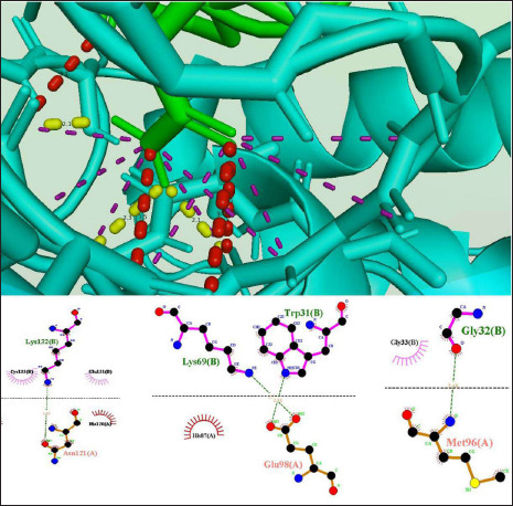
scores, Hydrophobic favored—Cluster scores, Balanced score—Cluster scores are −300.1, −993.4, −1150.8, and −900.5. As a result of this proteins interact with hydrogen bonds between LYS 132 B of B. asper toxin and the ASN 121 A of HIV-surface-glycoprotein with the shortest distance of 2.6 Å. (Fig. 1) (Table 1).
Fig. 1. HIV surface glycoprotein interaction with B. asper B.
HIV Surface-glycoprotein–toxin 2 (B. jararaca) complex molecular interactions with best-balanced cluster scores
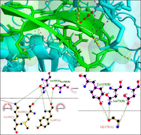
The HIV-surface-glycoprotein- B. jararaca toxin complex docked with significant binding affinities of Vander walls & Electrostatic force Cluster scores, Electrostatic favored—Cluster scores, Hydrophobic favored—Cluster scores, Balanced score—Cluster scores are −243.7, −1281.9, −1597, and −1312.6. As a result of this proteins interact with hydrogen bonds between TYR 387 B of B. jararaca toxin and ASN 389 A of HIV-surface-glycoprotein with the shortest distance of 2.7 Å. (Fig. 2) (Table 1).
Fig. 2. HIV surface glycoprotein interaction with B. jararaca B.
Details of interaction between HIV Surface-glycoprotein (PDB ID 6ES8) with toxin 3 (PDB ID 3S9B)
HIV Surface-glycoprotein–toxin 3 (Russell’s viper) complex
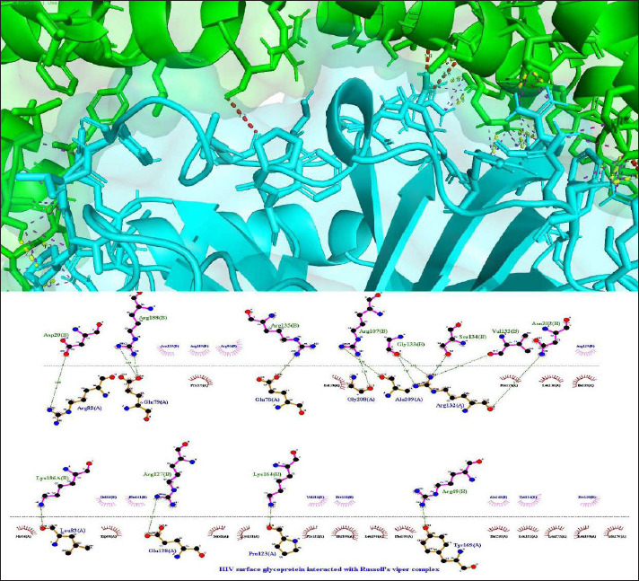
The HIV-surface-glycoprotein- Russell’s viper toxin complex docked with significant binding affinities of Vander walls & Electrostatic force Cluster scores, Electrostatic favored—Cluster scores, Hydrophobic favored—Cluster scores, Balanced score—Cluster scores are −219.4, −1112.2, −1325.6, and −1056.9. As a result of this proteins interaction with hydrogen bonds between ARG 107 B of Russell’s viper toxin and ALA 209 A of HIV-surface-glycoprotein with the shortest distance of 2.71 Å. (Fig. 3) (Table 1).
Fig. 3. HIV surface glycoprotein with Ryssell’s viper.
Details of interaction between HIV Surface-glycoprotein (PDB ID 6ES8) with toxin 4 (PDB ID 4E0V)
HIV Surface-glycoprotein–toxin 4 (B. jararacussu) complex
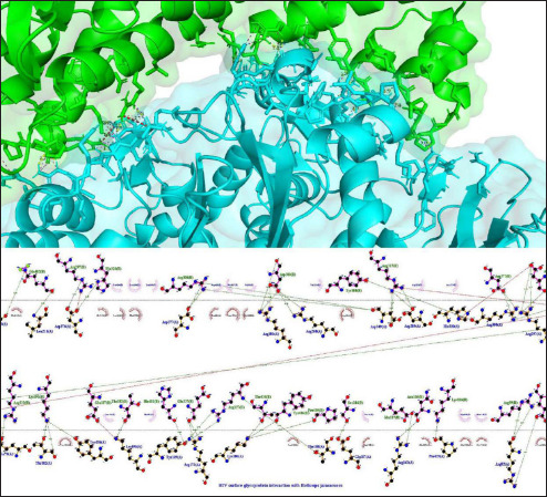
The HIV-surface-glycoprotein- B. jararacussu toxin complex docked with significant binding affinities of Vander walls & Electrostatic force Cluster scores, Electrostatic favored—Cluster scores, Hydrophobic favored—Cluster scores, Balanced score—Cluster scores are −251.9, −1049, −1167.7, and −1009.3. As a result of this proteins interaction with hydrogen bonds between LYS 191 B of B. jararacussu toxin and TYR 436 A of HIV-surface-glycoprotein with the shortest distance of 2.7 Å. (Fig. 4) (Table 1).
Fig. 4. HIV surface glycoprotein interaction with B. jararacussu ligplotv.
Details of interaction between HIV Surface-glycoprotein (PDB ID 6ES8) with toxin 5 (PDB ID 1WVR)
HIV Surface-glycoprotein–toxin 5 (P. flavoviridis) complex
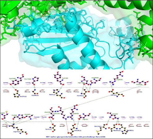
The HIV-surface-glycoprotein- P. flavoviridis toxin complex docked with significant binding affinities of Vander walls & Electrostatic force Cluster scores, Electrostatic favored—Cluster scores, Hydrophobic favored—Cluster scores, Balanced score—Cluster scores are −207.9, −951.2, −1239.9, and −919.3. As a result of this proteins interaction with hydrogen bonds between PRO 171 B of P. flavoviridis toxin and GLY 208 A of HIV-surface-glycoprotein with the shortest distance of 2.6 Å (Fig. 5) (Table 1).
Fig. 5. HIV surface glycoprotein interaction with P. flavoviridis ligplot.
Details of interaction between HIV Surface-glycoprotein (PDB ID 6ES8) with toxin 6 (PDB ID 1UOS)
HIV Surface-glycoprotein–toxin 6 (C. durissus terrificus) complex
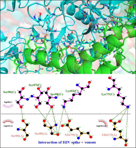
The HIV-surface-glycoprotein- C. durissus terrificus toxin complex docked with significant binding affinities of Vander walls & Electrostatic force Cluster scores, Electrostatic favored—Cluster scores, Hydrophobic favored—Cluster scores, Balanced score—Cluster scores are −222.7, −1151.9, −1299, and −1037.8. As a result of this proteins interaction with hydrogen bonds between GLU 94 B of C. durissus terrificus toxin and VAL 116 A of HIV-surface-glycoprotein with the shortest distance of 2.5 Å (Fig. 6) (Table 1).
Fig. 6. HIV surface glycoprotein interaction with C. durissus ligplot.
Details of interaction between HIV Surface-glycoprotein (PDB ID 6ES8) with toxin 7 (PDB ID 3UBU)
HIV Surface-glycoprotein–toxin 7 (D. acutus) complex
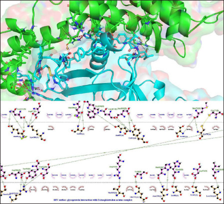
The HIV-surface-glycoprotein- D. acutus toxin complex docked with significant binding affinities of Vander walls & Electrostatic force Cluster scores, Electrostatic favored—Cluster scores, Hydrophobic favored—Cluster scores, Balanced score—Cluster scores are −244.1, −976.2, −1320.9, and −974.3. As a result of this proteins interaction with hydrogen bonds between THR 95 B of D. acutus toxin and ASN 116 A of HIV-surface-glycoprotein with the shortest distance of 2.5 Å (Fig. 7) (Table 1).
Fig. 7. HIV surface glycoprotein interactions with Deinagkistrodon cutus complex ligplot.
Details of interaction between HIV Surface-glycoprotein (PDB ID 6ES8) with toxin 8 (PDB ID 1RMR)
HIV Surface-glycoprotein–toxin 7 (E. carinatus) complex
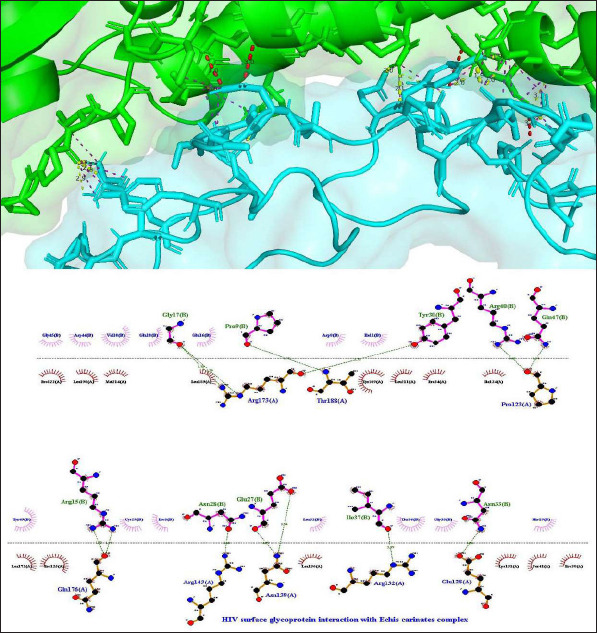
The HIV-surface-glycoprotein- E. carinatus toxin complex docked with significant binding affinities of Vander walls & Electrostatic force Cluster scores, Electrostatic favored—Cluster scores, Hydrophobic favored—Cluster scores, Balanced score—Cluster scores are −188.7, −1030.3, −1421.9, and −1015.5. As a result of this proteins interaction with hydrogen bonds between PRO 9 B of E. carinatus toxin and THR 188 A of HIV-surface-glycoprotein with the shortest distance of 2.6 Å (Fig. 8) (Table 1).
Fig. 8. HIV surface glycoprotein interaciton with E. carinatus ligplot.
Details of interaction between HIV Surface-glycoprotein (PDB ID 6ES8) with toxin 9 (PDB ID 4AA2)
HIV Surface-glycoprotein–toxin 7 (G. blomhoffii) complex
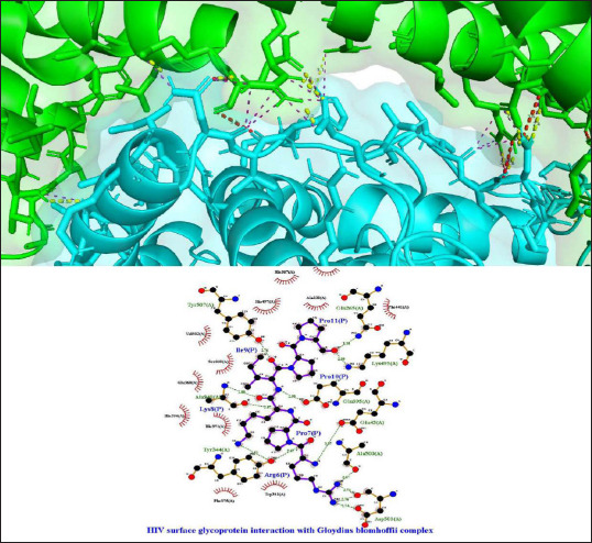
The HIV-surface-glycoprotein- G. blomhoffii toxin complex docked with significant binding affinities of Vander walls & Electrostatic force Cluster scores, Electrostatic favored—Cluster scores, Hydrophobic favored—Cluster scores, Balanced score—Cluster scores are −228.4, −917.3, −988.6, and −840.1. As a result of this proteins interact with hydrogen bonds between ILE 9 P of G. blomhoffii toxin and TYR 507 A of HIV-surface-glycoprotein with the shortest distance of 2.5 Å (Fig. 9) (Table 1).
Fig. 9. HIV surface glycoprotein interaction with G. blomhoffii complex ligplot.
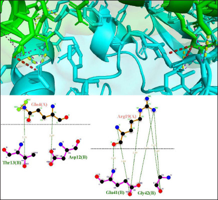
HIV Surface-glycoprotein–toxin 7 (P. textilis) complex with best electrostatic favored—cluster scores & hydrophobic favored—cluster scores
The HIV-surface-glycoprotein- P. textilis toxin complex docked with significant binding affinities of Vander walls & Electrostatic force Cluster scores, Electrostatic favored—Cluster scores, Hydrophobic favored—Cluster scores, Balanced score—Cluster scores are −228.4, −917.3, −988.6, and −840.1. As a result of this proteins interaction with hydrogen bonds between GLY 42 B and ASP 28 C of P. textilis toxin and ARG19 A and LYS 50 A of HIV-surface-glycoprotein with the shortest distance of 2.5 Å (Fig. 10) (Table 1).
Fig. 10. HIV surface glycoprotein interaction with P. textilis textilis B.
Details of interaction between HIV Surface-glycoprotein (PDB ID 6ES8) with toxin 11 (PDB ID 4GV5)
HIV Surface-glycoprotein–toxin 7 (C. durissus terrificus) complex
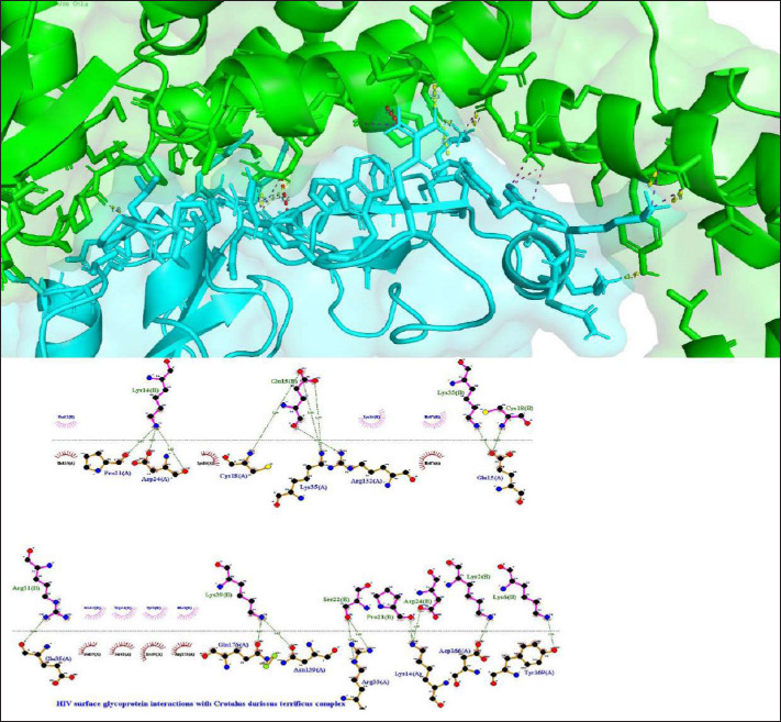
The HIV-surface-glycoprotein- C. durissus terrificus toxin complex docked with significant binding affinities of Vander walls & Electrostatic force Cluster scores, Electrostatic favored—Cluster scores, Hydrophobic favored—Cluster scores, Balanced score—Cluster scores are −217.5, −1115, −1501.7, and −217.5. As a result of this proteins interaction with hydrogen bonds between GLU 15 B of C. durissus terrificus toxin and LYS 35 A of HIV-surface-glycoprotein with the shortest distance of 2.4 Å (Fig. 11) (Table 1).
Fig. 11. HIV surface glycoprotein interaction with C. durissus complex ligplot.
Discussion
Strengthening human responses against the HIV virus is essential to ensuring human sustainability. A significant number of researchers globally are working on the development of new molecules or rethinking old ones in an attempt to control and slow the spread of HIV (Piscaglia et al., 2021). Parallelly, the present study aims to discover a new medication that might safeguard lives by analyzing the effects of snake poisons from the Elapidae and Viperidae families on the HIV surface spike protein. (Siniavin et al., 2022) Following a comprehensive analysis of the literature, eleven toxins from snakes in the Elapidae and Viperidae families were selected, as indicated in Table 1, and the HIV surface spike protein of the capsid with PDB ID: 6ES8 was selected for testing. Every toxin protein was selected to undergo processing using cluspro-2, (Kozakov et al., 2017) followed by an automated approach and PyMOL analysis and visualization.
The protein—protein interaction of all the toxins was the least for Hydrophobic favored—Cluster scores of −1611.2 were noted for the P. textilis toxin and the highest score was noted for G. blomhoffii −988.6. The lowest Electrostatic favored—Cluster scores of -1287.1 was noted for the P. textilis toxin and the highest score was noted for G. blomhoffii −917.3. The lowest Vander walls & Electrostatic force—Cluster scores of −300.1 was noted for the B. asper and the highest with −188.7was noted for E. carinatus and the lowest Balanced score—Cluster scores of −1312.6 was noted for the Bothrops jararaca and the highest with −840.1 was noted for G. blomhoffii. The Crotalus durissus terrificus toxin interacted with the target protein at a competitive H-bond distance of 2.4 Å. The above-given scores prove the fact that snake venom could be a good source of developing a drug which is helpful in the treatment of chronic and challenging disorders and infections like HIV (Meenakshisundaram et al., 2009; Uzair et al., 2018).
It is evident that the P. textilis toxin is a protease inhibitor named Textilinin-1, a Kunitz-type serine protease inhibitor including plasmin and trypsin. (Millers et al., 2009) Which was obtained from the Australian common brown snake venom was expressed with good docking scores and stood up as the most potent candidate for the new drug research and development as anti-HIV. Redesigning the protein in a favorable way may prove beneficial in limiting the spread of viral infections like HIV. An extensive literature review revealed that a few noticeable manuscripts are available reflecting the need of the current research, (Neri et al., 1990; Khusro et al., 2018; Oliveira et al., 2022); however, not concentrating on the toxins but on other antivirals, agent to improve and prepare the molecule with greater affinity (Alrajhi Abdulrahman and Almohaizeie, 2008). Hence, the current study is highly justifiable in this scenario. Interferons are one the desired candidates being proteinaceous in origin is highly applicable in the management of viral infections including HIV and this garners a chance for the molecules of the current investigation eleven of which are toxins but proteins to be a reliable source as anti-HIV in future. (Alrajhi Abdulrahman and Almohaizeie, 2008)
Furthermore, the lowest Vander walls & Electrostatic force—Cluster scores of −300.1 was noted for the B. asper and the lowest Balanced score—Cluster scores of −1312.6 was noted for the B. jararaca which can act as potential candidates for the novel drug development and research for preventing the vial entry into a healthy cell, like other entry inhibitors (Lobritz et al., 2010). Remodeling the amino acid sequence of the human cellular structures may result in the redesigning of the entry point of the for the virus into humans which may also can prevent the entry of the virus in healthy individuals (German Advisory Committee Blood 2016; Gutiérrez-Sevilla et al., 2021).
Conclusion
Snake venom can be a potential therapeutic source of anti-HIV/antiviral candidates that could potentially treat or reduce the incidences of HIV with acceptable to least side effects. 11 potential animal toxins from various families of snakes’ species were molecularly docked and revealed several binding interactions with the target protein. Pseudonaja textilis toxin a protease inhibitor named Textilinin-1, a Kunitz-type serine protease inhibitor including plasmin and trypsin exhibited favourable interactions with HIV capsid-glycoprotein with the highest affinities. These peptides could be used as HIV capsid-glycoprotein inhibitors because, according to molecular docking studies, they can prevent the interaction between human cells and the HIV capsid-glycoprotein which could be supported by the high-throughput screenings, cell line studies, preclinical studies, and human trials to concrete the evidence. Nonetheless, this research could be considered as the beginning of the new search for an anti-HIV entry inhibitor.
Acknowledgment
The authors extend their appreciation to the Deanship of Scientific Research at Jouf University, Saudi Arabia for funding this work through research grant number (DSR2022-RG-0118).
Conflict of interest
The authors declare no conflict of interest.
Funding
This work was funded by the Deanship of Scientific Research at Jouf University under Grant Number (DSR2022-RG-0118).
Authors’ contributions
MMA is the designer of the research. MJ, MF, MS, BA, DM, SA, and NG analyzed the data and wrote the manuscript. MOG, MMA, MS, AA, and MF revised the manuscript. MOG supervised the data analysis. MOG and MF applied for funding. All authors read, corrected, and approved the final manuscript.
Data availability
All data generated or analyzed during this study are included in this article.
References
- doi: 10.1111/febs.12038. Akif, M., Masuyer, G., Bingham, R.J., Sturrock, E.D., Isaac, R.E. and Acharya, K.R. 2012. Structural basis of peptide recognition by the angiotensin-1 converting enzyme homologue AnCE from Drosophila melanogaster. FEBS J. 279(24), 4525–4534. [DOI] [PMC free article] [PubMed] [Google Scholar]
- doi: 10.5144/0256-4947.2008.292. Alrajhi Abdulrahman, A. and Almohaizeie, A. 2008. Snake venom preparation for drug-resistant human immunodeficiency virus. Ann. Saudi Med.28(4), 292–293. [DOI] [PMC free article] [PubMed] [Google Scholar]
- doi: 10.2174/1381612822666151125000550. Baig, M. H., Ahmad, K., Roy, S., Ashraf, J., Adil, M., Siddiqui, M., Khan, S., Kamal, M., Provazník, V. and Choi, I. 2015. Computer aided drug design: success and limitations. Curr. Pharm. 22(5), 572–581. [DOI] [PubMed] [Google Scholar]
- doi: 10.1107/s0907444903021620. Batuwangala, T., Leduc, M., Gibbins, J.M., Bon, C. and Jones, E.Y. 2004. Structure of the snake-venom toxin convulxin. Acta Crystallogr. D. Biol. Crystallogr. 60(Pt 1), 46–53. [DOI] [PubMed] [Google Scholar]
- doi: 10.1093/nar/28.1.235. Berman, H.M., Westbrook, J., Feng, Z., Gilliland, G., Bhat, T.N., Weissig, H., Shindyalov, I. N. and Bourne, P.E. 2000. The protein data bank. Nucleic Acids. Res. 28(1), 235–242. [DOI] [PMC free article] [PubMed] [Google Scholar]
- doi: 10.1016/j.jmb.2004.06.048. Bilgrami, S., Tomar, S., Yadav, S., Kaur, P., Kumar, J., Jabeen, T., Sharma, S. and Singh, T.P. 2004. Crystal structure of schistatin, a disintegrin homodimer from saw-scaled viper (Echis carinatus) at 2.5Å resolution. J. Mol. Biol. 341(3), 829–837. [DOI] [PubMed] [Google Scholar]
- doi: 10.1007/s10930-021-10019-4. Chérifi, F. and Laraba-Djebari, F. 2021. Bioactive molecules derived from snake venoms with therapeutic potential for the treatment of thrombo-cardiovascular disorders associated with COVID-19. Protein J. 40(6), 799–841. [DOI] [PMC free article] [PubMed] [Google Scholar]
- doi: 10.1107/S0907444913018003. Coronado, M.A., Gabdulkhakov, A., Georgieva, D., Sankaran, B., Murakami, M.T., Arni, R. K. and Betzel, C. 2013. Structure of the polypeptide crotamine from the Brazilian rattlesnake \it Crotalus durissus terrificus. Acta Crystallogr. D. Biol. Crystallogr. 69(Pt 10), 1958–1964. [DOI] [PMC free article] [PubMed] [Google Scholar]
- doi: 10.1186/s40409-016-0089-0. da Mata É,C., Mourão, C.B., Rangel, M. and Schwartz, E.F. 2017. Antiviral activity of animal venom peptides and related compounds. J. Venom Anim. Toxins Incl. Trop. Dis. 23, 3. [DOI] [PMC free article] [PubMed] [Google Scholar]
- doi: 10.3390/molecules200713384. Ferreira, L.G., Dos Santos, R.N., Oliva, G. and Andricopulo, A.D. 2015. Molecular docking and structure-based drug design strategies. Molecules. 20(7), 13384–13421. [DOI] [PMC free article] [PubMed] [Google Scholar]
- doi: 10.1002/prot.24060. Gao, Y., Ge, H., Chen, H., Li, H., Liu, Y., Chen, L., Li, X., Liu, J., Niu, L. and Teng, M. 2012. Crystal structure of agkisacucetin, a Gpib-binding snake C-type lectin that inhibits platelet adhesion and aggregation. Proteins. 80(6), 1707–1711. [DOI] [PubMed] [Google Scholar]
- doi: 10.1159/000445852. German Advisory Committee Blood (Arbeitskreis Blut), Subgroup ‘Assessment of Pathogens Transmissible by Blood’. 2016. Human Immunodeficiency Virus (HIV). Transfus. Med. Hemother. 43(3), 203–222. [DOI] [PMC free article] [PubMed] [Google Scholar]
- doi: 10.1371/journal.pbio.1002140. Goodsell, D.S., Dutta, S., Zardecki, C., Voigt, M., Berman, H.M. and Burley, S.K. 2015. The RCSB PDB “Molecule of the Month”: Inspiring a Molecular View of Biology. PLoS Biol. 13(5), e1002140. [DOI] [PMC free article] [PubMed] [Google Scholar]
- doi: 10.1016/j.mrgentox.2021.503336. Gutiérrez-Sevilla, J.E., Cárdenas-Bedoya, J., Escoto-Delgadillo, M., Zúñiga-González, G.M., Pérez-Ríos, A.M., Gómez-Meda, B.C., González-Enríquez, G.V., Figarola-Centurión, I., Chavarría-Avila, E. and Torres-Mendoza, B.M. 2021. Genomic instability in people living with HIV. Mutat. Res. Genet. Toxicol. Environ. Mutagen. 865, 503336. [DOI] [PubMed] [Google Scholar]
- doi: 10.1016/j.micpath.2018.09.003. Khusro, A., Aarti, C., Barbabosa-Pliego, A., Rivas-Cáceres, R.R. and Cipriano-Salazar, M. 2018. Venom as therapeutic weapon to combat dreadful diseases of 21st century: a systematic review on cancer, TB, and HIV/AIDS. Microb. Pathog. 125, 96–107. [DOI] [PubMed] [Google Scholar]
- doi: 10.1038/nprot.2016.169. Kozakov, D., Hall, D.R., Xia, B., Porter, K.A., Padhorny, D., Yueh, C., Beglov, D. and Vajda, S. 2017. The ClusPro web server for protein–protein docking. Nat. Protoc. 12(2), 255–278. [DOI] [PMC free article] [PubMed] [Google Scholar]
- doi: 10.1021/ci200227u. Laskowski, R.A. and Swindells, M.B. 2011. LigPlot+: Multiple Ligand–protein interaction diagrams for drug discovery. J. Chem. Inf. Model. 51(10), 2778–2786. [DOI] [PubMed] [Google Scholar]
- doi: 10.3390/v2051069. Lobritz, M.A., Ratcliff, A.N. and Arts, E.J. 2010. HIV-1 Entry, inhibitors, and resistance. Viruses. (5), 1069–1105. [DOI] [PMC free article] [PubMed] [Google Scholar]
- doi: 10.7554/eLife.35335. Mallery, D.L., Márquez, C.L., McEwan, W.A., Dickson, C.F., Jacques, D.A., Anandapadamanaban, M., Bichel, K., Towers, G.J., Saiardi, A., Böcking, T. and James, L. C. 2018. IP6 is an HIV pocket factor that prevents capsid collapse and promotes DNA synthesis. Elife. 7, e35335. [DOI] [PMC free article] [PubMed] [Google Scholar]
- doi: 10.1186/1742-6405-6-25. Meenakshisundaram, R., Sweni, S. and Thirumalaikolundusubramanian, P. 2009. Hypothesis of snake and insect venoms against human immunodeficiency virus: a review. AIDS Res. Ther. 6, 25. [DOI] [PMC free article] [PubMed] [Google Scholar]
- doi: 10.1111/j.1742-4658.2009.07034.x. Millers, E.K.I., Trabi, M., Masci, P.P., Lavin, M.F., de Jersey, J. and Guddat, L.W. 2009. Crystal structure of textilinin-1, a Kunitz-type serine protease inhibitor from the venom of the Australian common brown snake (Pseudonaja textilis). FEBS J. 276(11), 3163–3175. [DOI] [PubMed] [Google Scholar]
- doi: 10.3390/toxins11100564. Mohamed Abd El-Aziz, T., Garcia Soares, A. and Stockand, J.D. 2019. Snake venoms in drug discovery: valuable therapeutic tools for life saving. Toxins (Basel). 11(10), 564. [DOI] [PMC free article] [PubMed] [Google Scholar]
- doi: 10.1016/j.toxicon.2008.08.021. Muniz, J.R.C., Ambrosio, A.L.B., Selistre-de-Araujo, H.S., Cominetti, M.R., Moura-da-Silva, A.M., Oliva, G., Garratt, R.C. and Souza, D.H.F. 2008. The three-dimensional structure of bothropasin, the main hemorrhagic factor from Bothrops jararaca venom: Insights for a new classification of snake venom metalloprotease subgroups. Toxicon 52, 807–816. [DOI] [PubMed] [Google Scholar]
- doi: 10.1016/j.febslet.2011.08.022. Nakayama, D., Ben Ammar, Y., Miyata, T. and Takeda, S. 2011. Structural basis of coagulation factor V recognition for cleavage by RVV-V. FEBS Lett. 585(19), 3020–3025. [DOI] [PubMed] [Google Scholar]
- doi: 10.1007/BF01310756. Neri, P., Bracci, L., Rustici, M. and Santucci, A. 1990. Sequence homology between HIV gp120, rabies virus glycoprotein, and snake venom neurotoxins. Arch. Virol. 114(3-4), 265–269. [DOI] [PubMed] [Google Scholar]
- doi: 10.1038/s41570-022-00393-7. Oliveira, A.L., Viegas, M.F., da Silva, S.L., Soares, A.M., Ramos, M.J. and Fernandes, P.A. 2022. The chemistry of snake venom and its medicinal potential. Nat Rev Chem 6, 451–469. [DOI] [PMC free article] [PubMed] [Google Scholar]
- doi: 10.1080/14728214.2021.1946036. Piscaglia, M., Cossu, M.V., Passerini, M., Petri, F., Gerbi, M., Fusetti, C., Capetti, A. and Rizzardini, G. 2021. Emerging drugs for the treatment of HIV/AIDS: a review of 2019/2020 phase II and III trials. Expert. Opin. Emerg. Drugs. 26(3), 219–230. [DOI] [PubMed] [Google Scholar]
- Rivero, J.V.R., de Castro, F.O.F., Stival, A.S., Magalhães, M.R., Carmo Filho, J.R. and Pfrimer, I.A.H. 2011. Mechanisms of virus resistance and antiviral activity of snake venoms. J. Venom. Anim. Toxins incl. Trop. Dis 17(4). 387–393. [Google Scholar]
- doi: 10.1016/j.biochi.2016.12.015. Salvador, G.H.M., dos Santos, J.I., Lomonte, B. and Fontes, M.R.M. 2017. Crystal structure of a phospholipase A2 from Bothrops asper venom: insights into a new putative “myotoxic cluster”. Biochimie. 133, 95–102. [DOI] [PubMed] [Google Scholar]
- Schrodinger, LLC. 2015. The PyMOL Molecular Graphics System, Version 1.8. Schrödinger, LLC, New York, NY, USA. [Google Scholar]
- doi: 10.1101/cshperspect.a006841. Sharp, P.M. and Hahn, B.H. 2011. Origins of HIV and the AIDS pandemic. Cold Spring Harb. Perspect. Med. 1(1), a006841. [DOI] [PMC free article] [PubMed] [Google Scholar]
- doi: 10.1016/j.jmb.2005.05.020. Shikamoto, Y., Suto, K., Yamazaki, Y., Morita, T. and Mizuno, H. 2005. Crystal structure of a CRISP family Ca2+-channel blocker derived from snake venom. J. Mol. Biol. 22(4), 735–743. [DOI] [PubMed] [Google Scholar]
- doi: 10.3390/ijms23031610. Siniavin, A., Grinkina, S., Osipov, A., Starkov, V., Tsetlin, V. and Utkin, Y. 2022. Anti-HIV activity of snake venom phospholipase A2s: updates for new enzymes and different virus strains. Int. J. Mol. Sci. 23(3), 1610. [DOI] [PMC free article] [PubMed] [Google Scholar]
- doi: 10.1016/j.bbrc.2012.03.129. Ullah, A., Souza, T.A.C.B., Abrego, J.R.B., Betzel, C., Murakami, M.T. and Arni, R.K. 2012. Structural insights into selectivity and cofactor binding in snake venom l-amino acid oxidases. Biochem. Biophys. Res. Commun. 27(1), 124–128. [DOI] [PubMed] [Google Scholar]
- doi: 10.2174/0929866525666180628161107. Uzair, B., Bushra, R., Khan, A.B., Zareen, S. and Fasim, F. 2018. Potential uses of venom proteins in treatment of HIV. Protein Pept. Lett. 25(7), 619–625. [DOI] [PubMed] [Google Scholar]
- doi: 10.1002/prot.25219. Vajda, S., Yueh, C., Beglov, D., Bohnuud, T., Mottarella, S.E., Xia, B., Hall, D.R. and Kozakov, D. 2017. New additions to the ClusPro server motivated by CAPRI. Proteins. 85(3), 435–444. [DOI] [PMC free article] [PubMed] [Google Scholar]
- WHO (2023). HIV—The Global Health Observatory. In “Global situation and trends”. WHO; Vol. 2023. Geneva, Switzerland. [Google Scholar]
- doi: 10.1080/17460441.2023.2246872. Xu, S., Sun, L., Liu, X. and Zhan, P. 2023. Opportunities and challenges in new HIV therapeutic discovery: what is the next step? Expert. Opin. Drug Discov. 18(11), 1195–1199. [DOI] [PubMed] [Google Scholar]
- Yuan, S., Chan, H.C.S. and Hu, Z. 2017. Using PyMOL as a platform for computational drug design. WIREs Comput. Mol. Sci. 7, e1298. [Google Scholar]
Associated Data
This section collects any data citations, data availability statements, or supplementary materials included in this article.
Data Availability Statement
All data generated or analyzed during this study are included in this article.


