

Abstract
In this work, cerium dioxide nanostructures were synthesized in an easy sonochemical way. CeO2 nanoparticles have received much attention in nanotechnology. CeO2NPs, exhibit biomimetic properties depending on their size, ratio of valency on their surface, and the ambient physico-chemical properties. Nanomedicine has emerged as a promising avenue for targeted cancer therapy, aiming to develop innovative approaches with improved efficacy and reduced side effects. Here, for the production of cerium dioxide nanostructures, a new and natural capping agent called Rosa Damascena extract was utilized, as well as ceric ammonium nitrate as a metal precursor. The results of the characterization of the oxide sample fabricated in the presence of Rosa Damascena extract demonstrated that nanostructures with a sponge-like morphology, which have a pure cubic crystal phase, were formed. The cytotoxicity effect of CeO2 NPs on glioblastoma and neuroblastoma cell lines (T98, SHSY5Y) was studied using the MTT test; cerium oxide nanoparticles exhibited cytotoxicity effects on T98 and SHSY5Y cell lines, compared to the control. The improved cytotoxic effects can be due to the plant secondary metabolites involved in the green synthesis of NPs. Consequently, synthesized CeO2 NPs have revealed an acceptable inhibitory impact upon cancer cell lines.
Keywords: Cerium dioxide, Sonochemistry, Nanostructures, Eco-friendly synthesis, Cancer cell lines, Cytotoxicity
Subject terms: Chemistry, Materials science, Nanoscience and technology
Introduction
Nanoscale compounds have attracted the attention of many researchers worldwide owing to their phenomenal optical, thermal, magnetic, mechanical, and electrical features1–5. In such a way preparing them and examining their utilization in various fields is a hot research issue6–10. Cerium dioxide (CeO2) is one of the remarkable members of the family of rare earth oxides. Owing to its unique f-electron configuration, this oxide compound can provide outstanding magnetic and catalytic attributes for versatile utilizations11,12. Cerium dioxide structures have been widely employed in sensors, catalytic processes, UV blockers, CO oxidation, fuel cells, environmental remediation, and other fields11,13–17. It has been accepted that the microscopic morphology of compounds can remarkably determine their attributes and applications11,18,19. Choosing the appropriate method for synthesis is known to be one of the influencing factors on their microscopic morphology11,20–22. Different methods for the synthesis of diverse structures of cerium dioxide have been utilized so far, including sol-gel, thermal decomposition, precipitation, hydrothermal, and template-assisted methods, etc23–29. The mentioned approaches are usually time-consuming and energy consuming and often comprise complex steps and, are costly. In recent years, applying procedures based on green chemistry to fabricate a variety of nanoscale compounds has been a noteworthy research topic for international scientists30–32. Utilizing natural and harmless combinations in the synthesis process of various compounds is one of the striking and valuable advantages of this kind of method9,33–36.
There is no report on the sono-synthesis of cerium dioxide nanostructures with the help of Rosa Damascena extract. Rosa Damascena extract contains phytochemicals like anthocyanins, glycosides, and flavonoids37–39, which can act as a capping agent for the green synthesis of diverse nanostructures, and can be a beneficial substitute for chemical surfactants. Also, owing to the presence of these phytochemicals, Rosa Damascena can exhibit medicinal features39.
Metal oxide (MO) nanoparticles are famous because of unique physicochemical properties like crystal structure diversity and the metal-oxygen bond nature. Investigators are involved in MO NPS studying for their various applications in medicine, biomedical, chemistry of materials, electronics, agriculture, environmental applications, etc39. Cell parameter variations like atom number increase at the surface and interface led to crystal structure disturbances, which cause changes in the size of particles. Variations in the nanoparticle size led to variations in the other nanoparticle properties39. CeO2 NPs have established more consideration than other MO NPs because of their exceptional applications in chemical and biomedical ones like sunblock, healing of the wounds, cancer therapy, antibiotics, as well as drug delivery40–44. Cancer is an illness in that cells increases wildly and can contaminate nearby tissues. Nowadays, numerous surgery approaches, radiation or, chemotherapy are used to treat of cancer. The significant disadvantage of these approaches is the ruin of healthy cells; therefore, scholars proceed with innovative treatments with lower side effects40,41. The technology of nano can allow doctors to use targeted drug delivery to cancerous cells. There are some nanoparticles for this purpose that indicate respectable potential for treating/ diagnosing in vitro42. Nanoparticles, after attaching to drugs, can be absorbed selectively by cancerous cells. Like this, healthy cells are safe and, side effects decrease43. Recently, CeO2 nanoparticles have shown cytotoxic impacts upon cancerous cell lines; consequently, additional investigations are significant to specify their side effects before utilization in cancer treatment41. Not many investigations reported the induction of oxidative stress produced by nanoceria in vitro/vivo where CeO2 NPs has operated with the role of antioxidant, as well as free extreme scavenger; these particles interact with radical of superoxide, radical of OH and peroxide radical, and accordingly caused to death in cell lines owing to oxidative stress44. Depending on the conditions, CeO2 nanoparticles can play a dual role as an antioxidant in normal cells or as an oxidant in cancer cells. This dual function depends on the pH of the environment. In acidic conditions, CeO2 nanoparticles act as oxidants, while in neutral conditions, these materials act as antioxidants45,46.
Subsequently, CeO2 nanoparticles can be applied as probable drug agents, and biological scaffolding.
Herein, pure CeO2 nanoparticles were synthesized in the presence of Rosa Damascena extract, and their cytotoxic effects were investigated on glioblastoma and neuroblastoma cell lines (T98, SHSY5Y). In the continue of our previous work, the aim of the study is to investigate and find effective nanoparticles in cancer therapy. For better understanding of readers, Fig. 1 indicated the graphical image of sono-synthesis as well as cytotoxic effect of nanoparticles on two various cell lines.
Fig. 1.

Graphical image of sono-synthesis as well as cytotoxic effect of nanoparticles on two various cell lines.
Experimental section
Preparation of Rosa Damascena extract
All methods were performed in accordance with the relevant guidelines/regulations/legislation. The same procedure as previously reported was utilized to prepare Rosa Damascena extract39. Rosa Damascena flowers were picked from the garden of our private villa (located in Qamsar, Kashan, Iran), rinsed with double distilled water, dried and ground into powder form. About 5 g of the obtained powder was added to 100 ml of DDW in an Erlenmeyer flask and the resulting mixture subjected to sonication at 70°C for 20 min39. The final product filtered, and the filtered solution was utilized as a new and natural capping agent to produce cerium dioxide nanostructures.
Sono-synthesis of cerium dioxide nanostructures
The two chemical reagents employed for the sono-synthesis of cerium dioxide nanostructures, including analytical grade of ceric ammonium nitrate and ammonia was applied as received. 4 ml of Rosa Damascena extract was added to a vessel containing ceric ammonium nitrate solution (1 mmol of cerium source in 15 ml H2O), and the resultant mixture was stirred for 6 min on a stirrer. After that, the resulting mixture was subjected to ultrasound waves (power of 80 watts for a quarter of an hour), its pH was adjusted to about 11 at the same time as ammonia gradually increased. The resulting gel was washed numerous times with distilled water and finally air-dried. Cerium dioxide nanostructures were obtained by calcining the residue at 500 °C for 110 min (Fig. 1).
Cell culture and treatment of cells by CeO2 NPs
Human glioblastoma and neuroblastoma cells were from the T98 and SH-SY5Y lines (Pasteur Institute, Tehran, Iran). T98 and SH-SY5Y lines without any treatment (not incubated with cerium dioxide NPs) considered as the control cell line. According to earlier reports47, the culture of SH-SY5Y as well as T98 cell lines was done. T98 glioblastoma cell is used in the study of cancer of brain, as well as the pharmaceuticals creation. SK-N-SH from a bone marrow biopsy was firstly described in 197348, while the SH-SY5Y line from a neuroblast-like subclone was initially pronounced in 197849. DMEM, dulbecco’s modified Eagle’s media (Gaithersburg, MD, USA) used for cultivating SH-SY5Y as well as T98 lines (Pasteur Institute, Tehran, Iran) in containers with 25 cm2 (Iwaki, Tokyo, Japan) with 4 × 10⁴ cells/ml density. The cultured cells were preserved at 37 ± 0.5 °C in a moistened environment with 5% carbon dioxide and supplemented with 1% (v/v) penicillin/streptomycin, and 10% (v/v) fetal calf inactivated serum. Daily, a diverse media is utilized. For investigation of 24 and 48 h, 4 × 10⁴ cells groups of SH-SY5Y and T98 cell line exposed to CeO2 NPs at variable quantities (0.05–0.8 m/mL). It is crucial that to prevent accumulation of suspended nanoparticles in DMEM medium, before cell treatment, suspensions dispersed within 10 min at forty watts. Following each treatment, biochemical and morphological features were explored.
Cell viability (MTT assay)
Cytotoxic analysis using the MTT test on the SHSYY and T98 cell lines was achieved to assess the biocompatibility of nanoparticles. Every cell line with the density of 10 × 103 per well is implanted in 96-well plates. The plates incubated in a carbon dioxide incubator at a 37 °C/24 h. Next that, numerous CeO2 NPs suspension quantities (0.05, 0.1, 0.2, 0.4, and 0.8 mg/mL) were added to each well, and finally incubated for 24, and 48 h. After the addition of a filtered solution of 10 µL of 5% MTT, the medium was substituted with a novel media, and incubated for 2 h. The following formula specifies the viable cells percent: Cell viability (%) = (Atest /Actrl) ×100, where Actrl, and Atest are the control wells, and absorbance quantities of the experiment, correspondingly.
Characterization
A diffractometer from Philips Company was applied to identify the crystalline nature of the as-fabricated nanostructure. Surface attributes like shape and particle size related to the as-produced oxide nanostructure were studied utilizing two apparatuses, FESEM (Zeiss Sigma 300) and TEM (Philips CM30 TEM). Also, the elemental composition of the cerium dioxide nanostructure surface was checked by EDS.
Statistical investigation
Each experiment was conducted a minimum of three times with new samples each time, and the standard deviations were computed from these results. The data was presented as the average ± standard deviation from the trio of experiments. Initially, statistical analysis was performed using a two-way ANOVA test. Subsequently, the significance of the results was assessed through post hoc Tukey’s test. A P-value of less than 0.05 was considered to indicate statistical significance.
Results and discussion
Investigating the characteristics of the CeO2 NPs
In the current paper, an easy and swift method for the production of cerium dioxide nanostructures was presented. Nanostructured cerium dioxide was created through a sonochemical process with the help of a new and environmentally friendly capping agent called Rosa Damascena extract. The XRD pattern of the sono-synthesized sample was recorded to identify its crystalline nature50, as depicted in Fig. 2. Based on the JCPDS database, all the signals appearing in the XRD pattern can be well ascribed to crystalline cerium dioxide (No. 00-034-0394). Thus, it can be said that the cubic phase of cerium dioxide can be successfully fabricated via sono-synthesis utilizing Rosa Damascena extract. The absence of any impurity signs in the diffraction pattern confirms the purity of the cerium dioxide nanostructure sample. In addition, the diffraction pattern includes sharp signals, which is owing to the excellent crystallinity of the nanostructure sample fabricated with Rosa Damascena extract. Utilizing Scherer’s Eqs22,51,52 and the corresponding XRD results for the sono-synthesized cerium oxide sample, its average crystallite size was calculated to be nearly 29 nm.
Fig. 2.

XRD pattern of the sono-synthesized cerium dioxide sample with the help of Rosa Damascena extract.
To examine the structural morphology of the cerium dioxide sample prepared with Rosa Damascena extract, FESEM micrographs were taken, which can be seen in Fig. 3. The nanostructured oxide sample has a sponge-like morphology. It is seen that a sponge-like nanostructure has been created from the assembly of spherical-like nanoparticles with good uniformity. It seems that phytochemicals like anthocyanins, glycosides, and flavonoids in Rosa Damascena extract37–39 can efficiently act as a green capping agent and lead to the fabrication of nanostructured cerium dioxide products. Since the phytochemicals in the Rosa Damascena extract can adhere to the surface of primary particles through their functional groups and prohibit their aggregation, they can also have a positive effect on their assembly in different directions37–39,53. Thus, it can be said that using of Rosa Damascena extract for the sono-synthesis of sponge-like cerium dioxide nanostructure can be a favorable option.
Fig. 3.
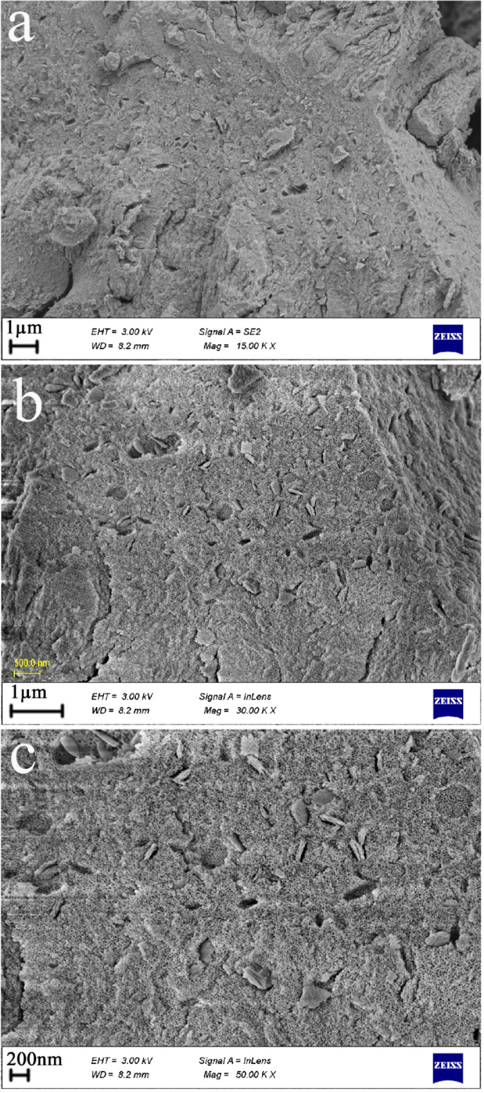
FESEM images of the sono-synthesized cerium dioxide sample with the help of Rosa Damascena extract.
EDS analysis was done to check the composition of elements of the oxide sample produced with Rosa Damascena extract and to check its purity further (see Fig. 4). The spectra obtained were good evidence for the existence of only oxygen and cerium elements in the sono-synthesized sample with the help of Rosa Damascena extract, which is in good agreement with the corresponding XRD outcomes regarding its purity.
Fig. 4.

EDS pattern of the sono-synthesized cerium dioxide sample with the help of Rosa Damascena extract.
Figure 5 exhibits the FTIR spectrum of the oxide sample produced with Rosa Damascena extract to seek the functional groups54. The absorption signal near 1627 cm− 1 may be allocated to the H-O-H bending vibration of the adsorbed or free H2O molecules55,56. Further, the absorption signal nearly 448 cm− 1 is ascribed to the Ce–O stretching vibration in the cerium dioxide sample57,58, demonstrating its formation.
Fig. 5.

FT-IR spectrum of the sono-synthesized cerium dioxide sample with the help of Rosa Damascena extract.
Furthermore, to further structural morphology investigations of the cerium dioxide sample sono-synthesized with Rosa Damascena extract, TEM micrographs (from different areas of the as-produced sample) were examined59 (see Fig. 6a-c). The images denote the creation of cerium dioxide nanostructures that are composed of spherical-like nanoparticles. These images are in good agreement with the FESEM micrographs.
Fig. 6.
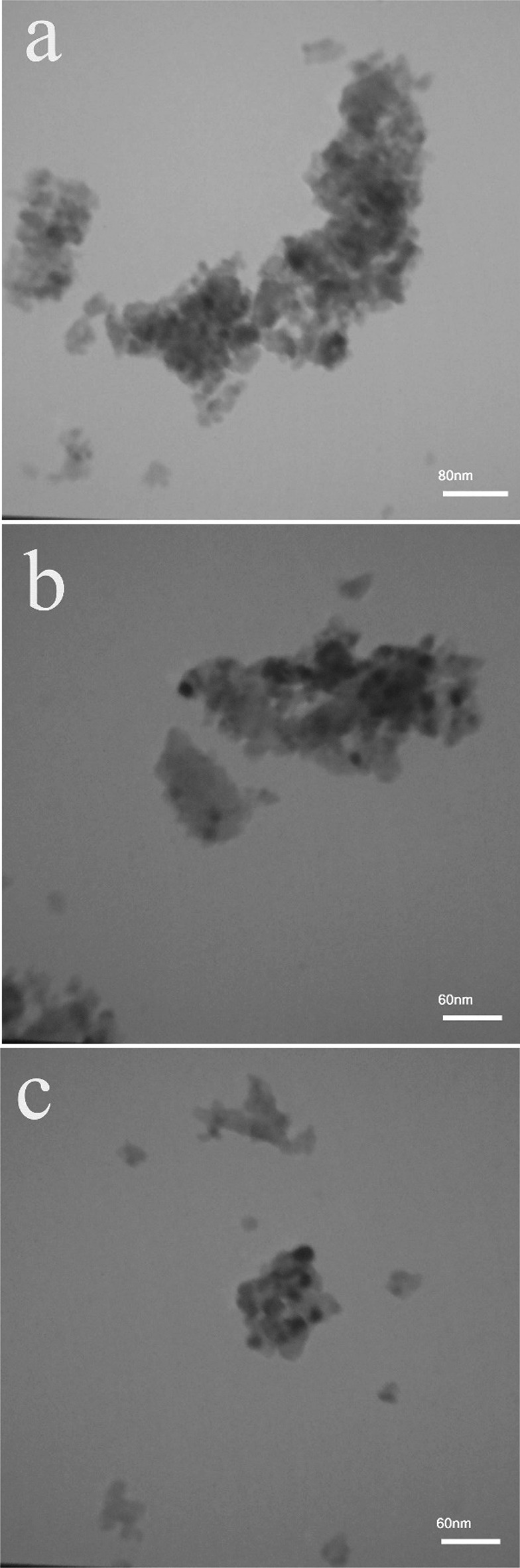
TEM images of the sono-synthesized cerium dioxide sample with the help of Rosa Damascena extract.
So, considering all the above findings, it can be concluded that utilizing the new capping agent, Rosa Damascena extract, through a rapid and simple sonochemical process, nanostructured cerium dioxide with sponge-like morphology can be produced.
Anticancer evaluation of the CeO2 NPs
The anti-cancer effect of CeO2 NPs against T98 and also SHSY5Y cell lines utilizing the various quantities of 0.0, 0.05, 0.1, 0.2, 0.4, and 0.8 mg/ml NP exposure within 24/ 48 h, was specified by MTT test. The T98 cell line survivability exposed to CeO2 NPs at several doses for 24 h (A), and 48 h, (B) is revealed in Fig. 7. Cell line viability markedly reduced in cultivated cells in the presence of various doses of NPs, indicating a dose-dependent procedure in 48 h. According to Fig. 7, the CeO2 NPs confirmed a significant cytotoxicity upon malignant cell lines. The glioblastoma features are probably the cause for the NP’s inhibitory effects upon this illness, since these cancer cells possess quick cell division, metabolism with great rate, and accordingly display improved NP internalization, which leads to cell death with a higher rate60. With the time passage, the toxicity level in diverse concentrations increases: nevertheless, the toxicity level in dissimilar doses becomes similar. Finally, all the nanoparticles presented comparable toxicity levels to this cell in 24, and 48 h. Figure 8. Indicated the optical microscope images of control and cell viability after 48 h of T98 cell lines incubated with 0.8 mg/ml cerium dioxide NPs. Furthermore, the in vitro cell survival experiment was done upon SHSY5Y cell lines utilizing CeO2 NPs at different doses within 24 h (A), and 48 h (B), and the outcomes are revealed in Fig. 9. Besides, the decrement in cellular ATP quantity and dehydrogenase performance, CeO2 NPs causes reactive oxygen species, which causes the mitochondrial respiratory chain harm and also other cellular components harm and eventually leads to death of cell. Consequently, before causing a general disruption in the functions of the cell membrane, NPs’ cytotoxicity is dependent upon the cell kind that displays an exceptional intracellular mechanism suppression growth. Moreover, the factors that determine nanoparticle cytotoxicity are 1- aggregation state, 2-interaction duration, 3-surface chemistry, 4-shape, and 5-size61. As a consequence, the level of toxicity of nanosized particles was greater in glioblastoma cells than in neuroblastoma cells.
Fig. 7.

In vitro cell viability assay, Cell viability of T98 cell lines incubated with cerium dioxide NPs at different concentrations for 24 h (a), and 48 h (b) (SD ± 2%).
Fig. 8.

The optical microscope images (magnification 200×) of control and cell viability after 48 h of T98 cell lines incubated with 0.8 mg/ml cerium dioxide NPs.
Fig. 9.

In vitro cell viability assay, Cell viability of SHSY5Y cell lines incubated with cerium dioxide NPs at different concentrations for 24 h (a), and 48 h (b) (SD ± 2%).
The basis for the anticancer activity of CeO2 NPs is the redox cycles among + 3 and + 4 states, and also their precise capability in oxygen the adsorption/release62,63. Though initially believed that the vacancy of oxygen and cycles of redox among these two cerium states is involved in antioxidant performance, now it has been recognized that redox process is mainly accountable for all the antioxidant features63; Ce+ 3 /Ce+ 4 surface ratio has a vital impact on the biological performance of CeO2 NPs mostly64. Figure 10. revealed the optical microscope images of control and cell viability after 48 h of SHSY5Y cell lines incubated with 0.8 mg/ml cerium dioxide NPs.
Fig. 10.

The optical microscope images (magnification 200×) of control and cell viability after 48 h of SHSY5Y cell lines incubated with 0.8 mg/ml cerium dioxide NPs.
Consequently, the most significant nanoparticles application is in response to ROS cell mechanisms which play a key role in the creation of free radicals. In biology of cancer, this mechanism involved in the creation of energy and regulation of intracellular activity. Meanwhile, cancer is known as an unplanned/uncontrolled cell proliferation; this, path may interrupt cancer correspondingly65. High ROS levels ruin the inner/outer membranes of mitochondria, cause the cytochrome release, and finally leads apoptotic cascade activation66. microenvironment around the tumor due to the Warburg effect has much more acidic conditions than normal tissues. The Warburg effect is a predominant metabolic characteristic of cancer cells that cancer cells prefer to ferment glucose to lactate, even in the presence of sufficient oxygen. This type of metabolism causes lactate to accumulate around cancer cells and the environment becomes acidic. In acidic pH, ceria nanoparticles show oxidant effects as toxic agents. So, Effects of catalase and superoxide dismutase of ceria nanoparticles.
inhibited and nanoparticles act as pro-oxidants46. The improved cytotoxic effects can be due to the plant secondary metabolites involved in the green synthesis of NPs. Consequently, the fabricated nanoparticles may be employed for the treatment of cancer but more investigations on other cell lines is recommended.
Conclusions
In summary, we demonstrated the sono-synthesis of cerium dioxide sponge-like nanostructures via an easy and quick method utilizing a new and natural capping agent called Rosa Damascena extract. These sponge-shaped cerium dioxide nanostructures have high purity, desirable crystallinity, and a cubic crystal structure. The NPs presented a striking cytotoxic, as well as dose-dependent influence upon the cancer cell lines T98, and SHSY5Y. An ideal and appropriate chemotherapy agent should have the most lethal effects on cancer cells without causing toxicity to healthy cells. Due to the pH-dependent function of seria and the different pH levels around cancer cells and healthy cells, serias can specifically target only cancer cells and do not harm healthy tissues. The improved cytotoxic effects can be due to the plant secondary metabolites involved in the green synthesis of NPs. More pharmacological investigation is undoubtedly required; however, the in vitro outcomes displayed that CeO2 NPs are favorable chemotherapeutic agents against invasive cancer cells, and demonstrated great potential for anticancer uses.
Acknowledgements
Authors are grateful to University of Bonab for supporting this work.
Author contributions
M. A.: Methodology, Validation, Writing—original draft, Writing—review & editing, Supervision, Conceptualization, Validation, Investigation, Resources. S. Z.A.: Methodology, Validation, writing—original draft, Writing—review & editing, Supervision, Conceptualization, Validation, Investigation, Resources. M. A.Z.: Methodology, Validation, Conceptualization, Validation, Investigation, Resources. F. Sh.: Validation, Conceptualization, Investigation, Resources, Investigation.
Data availability
The datasets used and analyzed during the current study available from the corresponding author on reasonable request.
Declarations
Competing interests
The authors declare no competing interests.
Footnotes
Publisher’s note
Springer Nature remains neutral with regard to jurisdictional claims in published maps and institutional affiliations.
Contributor Information
Sahar Zinatloo-Ajabshir, Email: s.zinatloo@ubonab.ac.ir, Email: s.zinatloo@gmail.com.
Meysam Ahmadi-Zeidabadi, Email: meysamcell@yahoo.com.
References
- 1.Yi, S. et al. Green and facile synthesis of nanostructured Co3O4/CeO2 catalysts via a glucose-urea method for NO oxidation. Appl. Surf. Sci.626, 157180 (2023). [Google Scholar]
- 2.Kumar, S. et al. Controlled synthesis and magnetic properties of monodispersed ceria nanoparticles. AIP Adv.5 (2015).
- 3.Xiao, R., Jia, J., Wang, R., Feng, Y. & Chen, H. Strong ligand control for noble metal nanostructures. Acc. Chem. Res.56, 1539–1552 (2023). [DOI] [PubMed] [Google Scholar]
- 4.Kushwah, N. et al. Molecular precursor-driven synthesis of copper telluride nanostructures for LIB anode application. Inorg. Chem.62, 8823–8834 (2023). [DOI] [PubMed] [Google Scholar]
- 5.Chitara, B., Shringi, A. K., Roy, B., Wu, M. H. & Yan, F. Facile synthesis and morphology-induced photoconductivity modulation of Bi2O2S nanostructures. Mater. Lett.346, 134545 (2023). [Google Scholar]
- 6.Goswami, M. et al. Morphology Variations in Copper Sulfide Nanostructures as Anode Materials for Na-Ion Capacitors (ACS Applied Nano Materials, 2023).
- 7.Gupta, S., Jain, P., Jagadeesan, D. & Vinod, C. P. Morphology-dependent catalysis by Co3O4 nanostructures in Atmospheric pressure Carbon Dioxide Hydrogenation. J. Phys. Chem. C127, 13055–13064 (2023). [Google Scholar]
- 8.Jamal, F. et al. Review of metal sulfide nanostructures and their applications. ACS Appl. Nano Mater.6 7077–7106 (2023).
- 9.Haque, S. et al. Green synthesis of nanostructures from rice straw food waste to improve the antimicrobial efficiency: New insight. Int. J. Food Microbiol.386, 110016 (2023). [DOI] [PubMed] [Google Scholar]
- 10.Scheid, C. M. et al. Glycerol carbonate synthesis over nanostructured titanate catalysts: Effect of morphology and structure of catalyst. Chem. Eng. Res. Des.197, 392–404 (2023). [Google Scholar]
- 11.Li, H. L. et al. Synthesis and characterization of novel coral-like hollow CeO2 nanostructures and their potential as peroxidase mimics. Solid State Sci.97, 106011 (2019). [Google Scholar]
- 12.Marques, T. M. F. et al. Viana, homogeneously dispersed CeO2 nanoparticles on exfoliated hexaniobate nanosheets. J. Phys. Chem. Solids111, 335–342 (2017). [Google Scholar]
- 13.Slavinskaya, E. M. et al. Low-temperature CO oxidation by Pd/CeO2 catalysts synthesized using the coprecipitation method. Appl. Catal. B 166–167 91–103 (2015).
- 14.Laguna, O. H. et al. Gold supported on CuOx/CeO2 catalyst for the purification of hydrogen by the CO preferential oxidation reaction (PROX). Fuel 118 176–185 (2014).
- 15.Thakur, S. & Patil, P. Rapid synthesis of cerium oxide nanoparticles with superior humidity-sensing performance. Sens. Actuators B194, 260–268 (2014). [Google Scholar]
- 16.Zhang, W. & Chen, D. Preparation and performance of CeO2 hollow spheres and nanoparticles. J. Rare Earths34, 295–299 (2016). [Google Scholar]
- 17.Rajan, A. R., Vilas, V., Rajan, A., John, A. & Philip, D. Synthesis of nanostructured CeO2 by chemical and biogenic methods: optical properties and bioactivity. Ceram. Int.46, 14048–14055 (2020). [Google Scholar]
- 18.Arul, N. S., Mangalaraj, D. & Kim, T. W. Photocatalytic degradation mechanisms of CeO2/Tb2O3 nanotubes. Appl. Surf. Sci.349, 459–464 (2015). [Google Scholar]
- 19.Xu, J., Wang, W., Wang, J. & Liang, Y. Controlled fabrication and enhanced photocatalystic performance of BiVO4@CeO2 hollow microspheres for the visible-light-driven degradation of rhodamine B. Appl. Surf. Sci.349, 529–537 (2015). [Google Scholar]
- 20.Mu, J., Zhao, X., Li, J., Yang, E. C. & Zhao, X. J. Coral-like CeO2/NiO nanocomposites with efficient enzyme-mimetic activity for biosensing application. Mater. Sci. Eng. C74, 434–442 (2017). [DOI] [PubMed] [Google Scholar]
- 21.Rada, E., Lima, E., Ruiz, F. & Sergio Moreno, M. Synthesis of LiMn2O4 nanostructures with controlled morphology. Mater. Sci. Eng. B292, 116410 (2023). [Google Scholar]
- 22.Beshkar, F., Zinatloo-Ajabshir, S. & Salavati-Niasari, M. Simple morphology-controlled fabrication of nickel chromite nanostructures via a novel route. Chem. Eng. J.279, 605–614 (2015). [Google Scholar]
- 23.Manoj, D., Manigandan, R., Rajendran, S., Cornejo, L. & Ponce Self-assembled dendrite-like 3D-CeO2 nanostructures for non-enzymatic vitamin B2 sensor. Mater. Lett.295, 129834 (2021). [Google Scholar]
- 24.González-Souto, L., González-Rovira, L., Botana, F. J., Calvino, J. J. & Cauqui, M. Á. J.C. Hernández-Garrido, The Role of Gold-Alumina Template in the Electrochemical Deposition of CeO2Nanotubes, 361900168 (Particle & Particle Systems Characterization, 2019).
- 25.Parlak, O. & Demir, M. M. Toward transparent nanocomposites based on polystyrene matrix and PMMA-Grafted CeO2 nanoparticles. ACS Appl. Mater. Interfaces. 3, 4306–4314 (2011). [DOI] [PubMed] [Google Scholar]
- 26.Xiao, H., Ai, Z. & Zhang, L. Nonaqueous Sol – Gel Synthesized hierarchical CeO2 Nanocrystal microspheres as Novel adsorbents for Wastewater Treatment. J. Phys. Chem. C113, 16625–16630 (2009). [Google Scholar]
- 27.Devaraju, M. K., Yin, S. & Sato, T. Morphology control of cerium oxide particles synthesized via a supercritical solvothermal method. ACS Appl. Mater. Interfaces1, 2694–2698 (2009). [DOI] [PubMed] [Google Scholar]
- 28.Mock, S. et al. CeO2 Nanorods-supported transition metal catalysts for CO oxidation. J. Colloid Interface Sci., 466 (2015). [DOI] [PubMed]
- 29.Shen, Q. et al. Hollow MnOx-CeO2 mixed oxides as highly efficient catalysts in NO oxidation. Chem. Eng. J.322, 46–55 (2017). [Google Scholar]
- 30.Gupta, D., Boora, A., Thakur, A. & Gupta, D. T. Green and sustainable synthesis of nanomaterials: recent advancements and limitations. Environ. Res., 231 (2023). [DOI] [PubMed]
- 31.Thongsai, N. et al. Real-time detection of alcohol vapors and volatile organic compounds via optical electronic nose using carbon dots prepared from rice husk and density functional theory calculation. Colloids Surf. A560, 278–287 (2019). [Google Scholar]
- 32.Davar, F., Majedi, A. & Mirzaei, A. Green Synthesis of ZnO nanoparticles and its application in the degradation of some dyes. J. Am. Ceram. Soc.98, 1739–1746 (2015). [Google Scholar]
- 33.Singh, V. et al. Chickpea peel waste as sustainable precursor for synthesis of fluorescent carbon nanotubes for bioimaging application. Carbon Lett.31, 117–123 (2021). [Google Scholar]
- 34.Shi, H. et al. Green synthesis of Fe3O4 nanoparticles with controlled morphologies using urease and their application in dye adsorption. Dalton Trans.43, 12474–12479 (2014). [DOI] [PubMed] [Google Scholar]
- 35.Hudlikar, M., Joglekar, S., Dhaygude, M. & Kodam, K. Green synthesis of TiO2 nanoparticles by using aqueous extract of Jatropha curcas L. latex. Mater. Lett.75, 196–199 (2012). [Google Scholar]
- 36.Mdlovu, N. V., Juang, R. S., Weng, M. T. & Lin, K. S. Green synthesis and characterization of silicate nanostructures coated with Pluronic F127/gelatin for triggered drug delivery in tumor microenvironments. Int. J. Biol. Macromol.251, 126337 (2023). [DOI] [PubMed] [Google Scholar]
- 37.Boskabady, M. H., Shafei, M. N., Saberi, Z. & Amini, S. Pharmacological effects of rosa damascena. Iran. J. Basic. Med. Sci.14, 295–307 (2011). [PMC free article] [PubMed] [Google Scholar]
- 38.Chopra, H. et al. Green metallic nanoparticles: Biosynthesis to applications. Front. Bioeng. Biotechnol. 10 874742 (2022). [DOI] [PMC free article] [PubMed]
- 39.Ebrahimian, J. et al. Rosa Damascena mediated ZnO-Red ochre nanocomposite for the electrochemical determination of 5-Fluorouracil. Arab. J. Chem.16, 104586 (2023). [Google Scholar]
- 40.Colon, J. et al. Cerium Oxide Nanoparticles Protect Gastrointestinal Epithelium from radiation-induced Damage by Reduction of Reactive Oxygen Species and Upregulation of Superoxide Dismutase 2, 6698–705 (Nanotechnology, Biology and Medicine, 2010). [DOI] [PubMed]
- 41.Clark, A., Zhu, A., Sun, K. & Petty, H. R. Cerium oxide and platinum nanoparticles protect cells from oxidant-mediated apoptosis. J. Nanopart. Res.13, 5547–5555 (2011). [DOI] [PMC free article] [PubMed] [Google Scholar]
- 42.Baumber, J., Ball, B. A., GRAVANCE, C. G., Medina, V. & DAVIES-MOREL, M. C. The effect of reactive oxygen species on equine sperm motility, viability, acrosomal integrity, mitochondrial membrane potential, and membrane lipid peroxidation. J. Androl.21, 895–902 (2000). [PubMed] [Google Scholar]
- 43.Patra, J. K. et al. Nano based drug delivery systems: recent developments and future prospects. J. Nanobiotechnol.16, 1–33 (2018). [DOI] [PMC free article] [PubMed] [Google Scholar]
- 44.Sabir, F. et al. Onco-receptors targeting in lung cancer via application of surface-modified and hybrid nanoparticles: a cross-disciplinary review. Processes9, 621 (2021). [Google Scholar]
- 45.Pezzini, I. et al. Cerium oxide nanoparticles: the regenerative redox machine in bioenergetic imbalance. Nanomedicine12, 403–416 (2017). [DOI] [PubMed] [Google Scholar]
- 46.Perez, J. M., Asati, A., Nath, S. & Kaittanis, C. Synthesis of biocompatible dextran-coated nanoceria with pH-dependent antioxidant properties. Small4, 552–556 (2008). [DOI] [PubMed] [Google Scholar]
- 47.Razavi, R., Amiri, M., Alshamsi, H. A., Eslaminejad, T. & Salavati-Niasari, M. Green synthesis of Ag nanoparticles in oil-in-water nano-emulsion and evaluation of their antibacterial and cytotoxic properties as well as molecular docking. Arab. J. Chem.14, 103323 (2021). [Google Scholar]
- 48.Biedler, J. L., Helson, L. & Spengler, B. A. Morphology and growth, tumorigenicity, and cytogenetics of human neuroblastoma cells in continuous culture. Cancer Res.33, 2643–2652 (1973). [PubMed] [Google Scholar]
- 49.Biedler, J. L., Roffler-Tarlov, S., Schachner, M. & Freedman, L. S. Multiple neurotransmitter synthesis by human neuroblastoma cell lines and clones. Cancer Res.38, 3751–3757 (1978). [PubMed] [Google Scholar]
- 50.Hublikar, L. V., Ganachari, S. V. & Patil, V. B. Zn and Co ferrite nanoparticles: towards the applications of sensing and adsorption studies. Environ. Sci. Pollut. Res.30, 66994–67007 (2023). [DOI] [PubMed] [Google Scholar]
- 51.Jenkins, R. & Snyder, R. L. Introduction to X-ray Powder Diffractometry 138 (Wiley Online Library, 1996).
- 52.Raghavendra, N. et al. Evaluation of PANI-Averraoha bilimbi leaves activated carbon nanocomposite for Cd2 + and Pb2 + removal from wastewater. J. Indian Chem. Soc.100, 100872 (2023). [Google Scholar]
- 53.Valian, M., Masjedi-Arani, M. & Salavati-Niasari, M. Sol-gel synthesis of DyFeO3/CuO nanocomposite using Capsicum Annuum extract: fabrication, structural analysis, and assessing the impacts of g-C3N4 on electrochemical hydrogen storage behavior. Fuel306, 121638 (2021). [Google Scholar]
- 54.Patil, S. B., Hublikar, L. V., Raghavendra, N., Shanbhog, C. & Kamble, A. Synthesis and exploration of anticancer activity of silver nanoparticles using Pandanus amaryllifolius Roxb. Leaf extract: promising approach against lung cancer and breast cancer cell lines. Biologia76, 3533–3545 (2021). [Google Scholar]
- 55.Rahim Pouran, S. et al. Comprehensive study on the influence of molybdenum substitution on characteristics and catalytic performance of magnetite nanoparticles. Res. Chem. Intermed.44, 883–900 (2018). [Google Scholar]
- 56.Mousavi-Kamazani, M. & Ashrafi, S. Single-step sonochemical synthesis of Cu2O-CeO2 nanocomposites with enhanced photocatalytic oxidative desulfurization. Ultrason. Sonochem.63, 104948 (2020). [DOI] [PubMed] [Google Scholar]
- 57.Liu, I. T., Hon, M. H. & Teoh, L. G. Structure and Optical properties of CeO2 nanoparticles synthesized by precipitation. J. Electron. Mater.42, 2536–2541 (2013). [Google Scholar]
- 58.Ishfaq, M. et al. Synthesis of binary metal doped CeO2 via the subcritical hydrothermal method for photo-mineralizing methyl orange dye. J. Alloys Compd.960, 170661 (2023). [Google Scholar]
- 59.Hublikar, L. V., Ganachari, S. V. & Patil, V. B. Phytofabrication of silver nanoparticles using Averrhoa bilimbi leaf extract for anticancer activity. Nanoscale Adv.5, 4149–4157 (2023). [DOI] [PMC free article] [PubMed] [Google Scholar]
- 60.Goudarzi, M., Alshamsi, H. A., Amiri, M. & Salavati-Niasari, M. ZnCo2O4/ZnO nanocomposite: facile one-step green solid-state thermal decomposition synthesis using Dactylopius Coccus as capping agent, characterization and its 4T1 cells cytotoxicity investigation and anticancer activity. Arab. J. Chem.14, 103316 (2021). [Google Scholar]
- 61.Amiri, M., Pardakhti, A., Ahmadi-Zeidabadi, M., Akbari, A. & Salavati-Niasari, M. Magnetic nickel ferrite nanoparticles: Green synthesis by Urtica and therapeutic effect of frequency magnetic field on creating cytotoxic response in neural cell lines. Colloids Surf., B. 172, 244–253 (2018). [DOI] [PubMed] [Google Scholar]
- 62.Deshpande, S., Patil, S., Kuchibhatla, S. V. & Seal, S. Size dependency variation in lattice parameter and valency states in nanocrystalline cerium oxide. Appl. Phys. Lett. 87 (2005).
- 63.Korsvik, C., Patil, S., Seal, S. & Self, W. T. Superoxide dismutase mimetic properties exhibited by vacancy engineered ceria nanoparticles. Chem. Commun., 1056–1058 (2007). [DOI] [PubMed]
- 64.Celardo, I. et al. Ce3+ ions determine redox-dependent anti-apoptotic effect of cerium oxide nanoparticles. ACS Nano5, 4537–4549 (2011). [DOI] [PubMed] [Google Scholar]
- 65.Park, E. J., Choi, J., Park, Y. K. & Park, K. Oxidative stress induced by cerium oxide nanoparticles in cultured BEAS-2B cells. Toxicology245, 90–100 (2008). [DOI] [PubMed] [Google Scholar]
- 66.Ghaznavi, H. et al. Neuro-protective effects of cerium and yttrium oxide nanoparticles on high glucose-induced oxidative stress and apoptosis in undifferentiated PC12 cells. Neurol. Res.37, 624–632 (2015). [DOI] [PubMed] [Google Scholar]
Associated Data
This section collects any data citations, data availability statements, or supplementary materials included in this article.
Data Availability Statement
The datasets used and analyzed during the current study available from the corresponding author on reasonable request.