

Abstract
This case highlights an unusually rapid onset and extensive presentation of eruptive cutaneous sarcoidosis in a 61-year-old woman who developed a sudden, widespread pruritic rash overnight after ingesting a supplement containing black seed oil and vitamins D3, K2, and E. She responded well to corticosteroid treatment. We compared the findings with two other cases found in the literature. This type of presentation has not been previously documented.
Keywords: sarcoidosis, granuloma, eruptive sarcoidosis
Introduction
Cutaneous manifestations of sarcoidosis are observed in 20 to 35 percent of patients and can serve as the primary presentation of the disease (1). Skin findings encompass a broad spectrum of manifestations, including papules, plaques, cutaneous and subcutaneous nodules, ulcers, erythema nodosum, lupus pernio, ichthyosiform sarcoidosis, and scar sarcoidosis (2). The occurrence of abrupt diffuse lesions, as observed in this case, is uncommon and is sparsely documented in the literature.
Case report
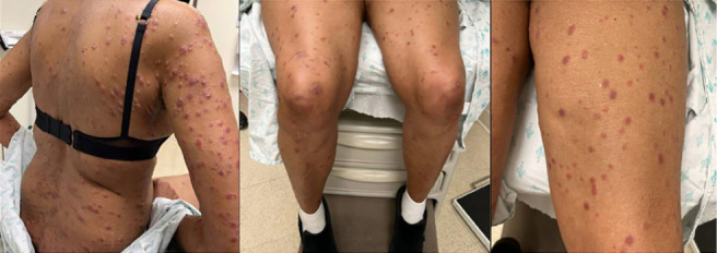
A 61-year-old female with past medical history significant for hypertension, chronic back pain, and a thrombotic event presented with a diffuse pruritic rash that started suddenly two weeks prior to presentation. She reported that she first noticed the lesions that involved nearly her entire body upon awakening, and it remained stable until she presented for evaluation. The patient noted that this occurred one day after taking a single dose of a new supplement containing black seed oil and vitamins D3, K2, and E, but otherwise she denied other new medications, exposures, and travel. She never took the supplement again after this. She also reported changes in her vision and shortness of breath, both of which started around the time of the rash onset. She denied experiencing fevers or chills. This was her first occurrence of such a rash. The patient’s medications included trazodone, risperidone, hydrocodone, amlodipine, and a calcium supplement. She reported an allergy to penicillin. Her family history revealed a cousin with sarcoidosis. On physical exam her vital signs were normal and widespread, tender, edematous, erythematous papules and nodules, some with scale and excoriation, were present (Figure 1). Two 4mm punch biopsies were performed on her upper back, one for Hematoxylin and eosin (H&E) stain and one for direct immunofluorescence (DIF). Biopsies showed superficial and mid-dermal noncaseating granulomatous inflammation, suggestive of cutaneous sarcoidosis (CS). Negative, non-specific findings were seen on DIF. She refused blood testing. The patient was prescribed triamcinolone 0.1% ointment to be used twice per day, hydroxyzine 25 mg nightly as needed for itching, and prednisone, which was started at 40mg per day and tapered over 21 days. She responded well to this therapy. She was also referred to rheumatology for further workup and consideration of starting methotrexate, as well as ophthalmology to evaluate for possible ocular involvement.
Figure 1.
Discussion
Eruptive CS is characterized by the sudden onset of numerous skin lesions, such as papules or nodules, which are widely distributed across the skin, representing a distinctive and less common clinical presentation of sarcoidosis (3). In a prior report of eruptive CS from 1978, a previously healthy 54-year-old male developed a progressive, widespread eruption that started on the shoulder and rapidly spread throughout his body over three months. Small, firm, smooth, dome-shaped, pinkish-brown papules approximately 4 mm in diameter were noted. Over the trunk, these formed confluent sheets with a 'cobblestone' surface. There were no inciting triggers or new medications prior to onset of skin lesions (3). Another report from 1978 detailed a 60-year-old man who experienced a broad outbreak of shiny, non-itchy, small, firm, waxy papules ranging from 1 to 5 mm in diameter affecting his trunk and extremities. This occurred one week after treatment with oral phenylbutazone 100 mg three times daily. Topical corticosteroids provided limited improvement, and a two-month course of oral prednisone (starting at 30 mg daily) resulted in significant improvement. However, the skin eruption recurred when the prednisone dose was reduced to 5 mg daily (3). In the case under consideration, the skin examination revealed similar findings observed in earlier instances of eruptive CS. Remarkably, unlike previous cases of eruptive CS, the lesions in this patient were reported to develop rapidly overnight. To the best of our knowledge, this swift and extensive presentation of CS has not been previously reported. Immunohistochemistry studies and single-cell RNA sequencing analyses may elucidate distinctive patterns of immune cell activation within granulomas, along with the upregulation of unique immune pathways.
Consent Statement:
The authors obtained written consent from patients for their photographs and medical information to be published in print and online and with the understanding that this information may be publicly available. Patient consent forms were not provided to the journal but are retained by the authors.
Conflict of Interest:
Each author declares that he or she has no commercial associations (e.g. consultancies, stock ownership, equity interest, patent/licensing arrangement etc.) that might pose a conflict of interest in connection with the submitted article.
Author Contributions:
All authors contributed directly to the intellectual content of the paper. The authors conceived and planned the work that led to the article, played important roles in interpreting the results, and contributed to writing the manuscript or providing substantive suggestions for revision. All authors approved the final version and accepted public responsibility for its content.
Funding Source:
None
References
- Yanardag H, Tetikkurt C, Bilir M, Demirci S, Iscimen A. Diagnosis of cutaneous sarcoidosis; clinical and the prognostic significance of skin lesions. Multidiscip Respir Med. Mar 22 2013;8(1):26. doi: 10.1186/2049-6958-8-26. doi:10.1186/2049-6958-8-26. [DOI] [PMC free article] [PubMed] [Google Scholar]
- Williams JR, Frey C, Cohen GF. Cutaneous Sarcoidosis in Skin of Color. J Drugs Dermatol. Jul 1 2023;22(7):695–7. doi: 10.36849/JDD.7008. doi:10.36849/JDD.7008. [DOI] [PubMed] [Google Scholar]
- Gange RW, Smith NP, Fox ED. Eruptive cutaneous sarcoidosis of unusual type. Report of two cases without radiologically demonstrable lung involvement. Clin Exp Dermatol. Sep 1978;3(3):299–306. doi: 10.1111/j.1365-2230.1978.tb01502.x. doi:10.1111/j.1365-2230.1978.tb01502.x. [DOI] [PubMed] [Google Scholar]