Abstract
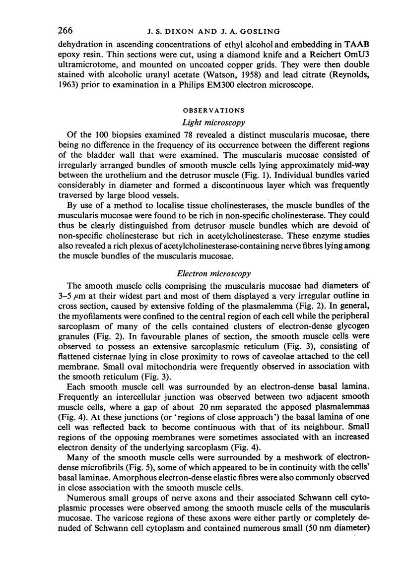
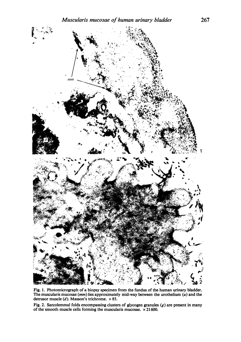
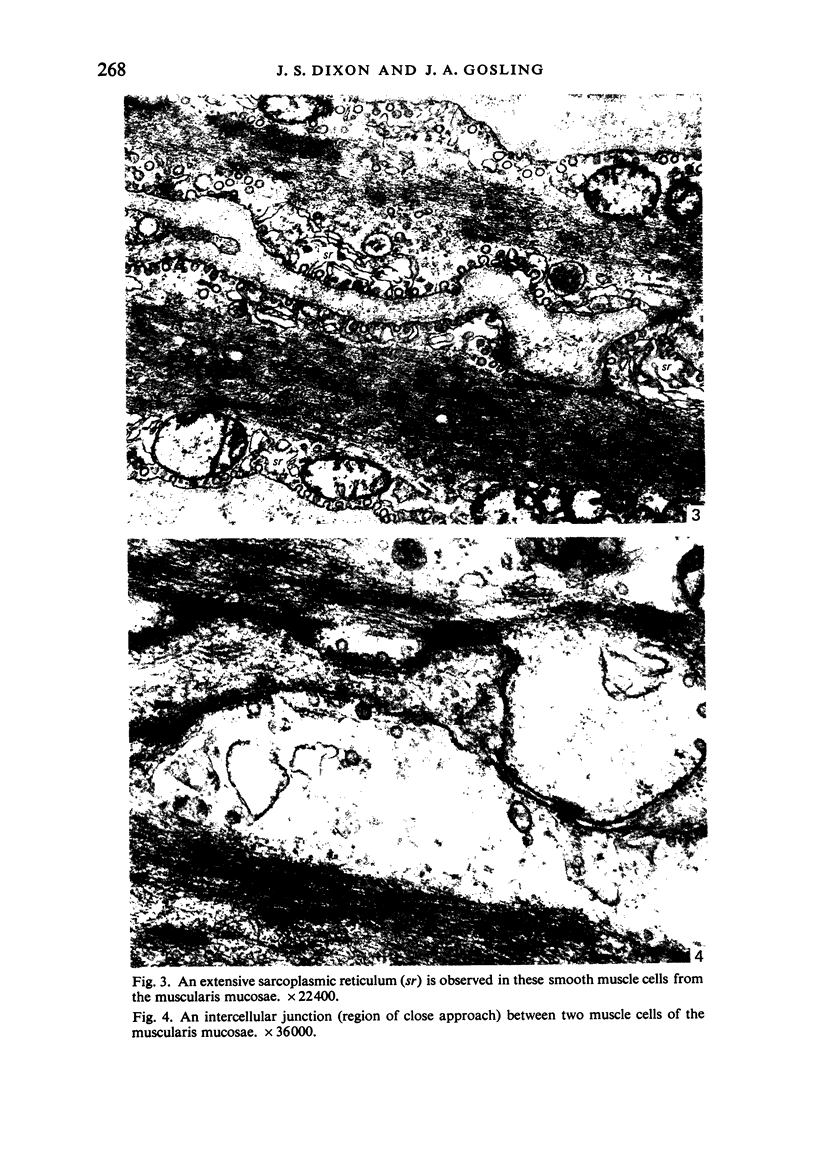
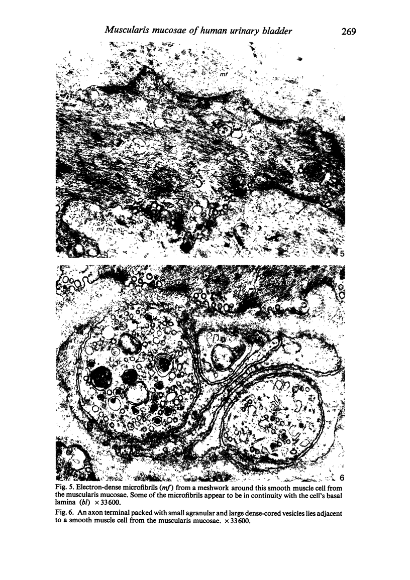
Study of biopsy specimens has revealed the presence of a muscularis mucosae in all regions of the human urinary bladder. The muscularis mucosae is discontinuous and consists of irregularly-arranged muscle bundles composed of relatively small-diameter smooth muscle cells. These cells are both morphologically and histochemically distinct from those forming the detrusor muscle, being rich in non-specific cholinesterase and glycogen. However, like detrusor muscle, the muscularis mucosae is richly supplied with acetylcholinesterase-positive nerve fibres. In the electron microscope, the constituent smooth muscle cells possess an extensive sarcoplasmic reticulum and large, peripheral clusters of dense glycogen granules; the myofilaments are confined to the central regions of the cells. Numerous intercellular junctions occur between adjacent cells while presumptive cholinergic nerve terminals containing small agranular and large granulated vesicles lie in close proximity to the muscle cells' surface.
Full text
PDF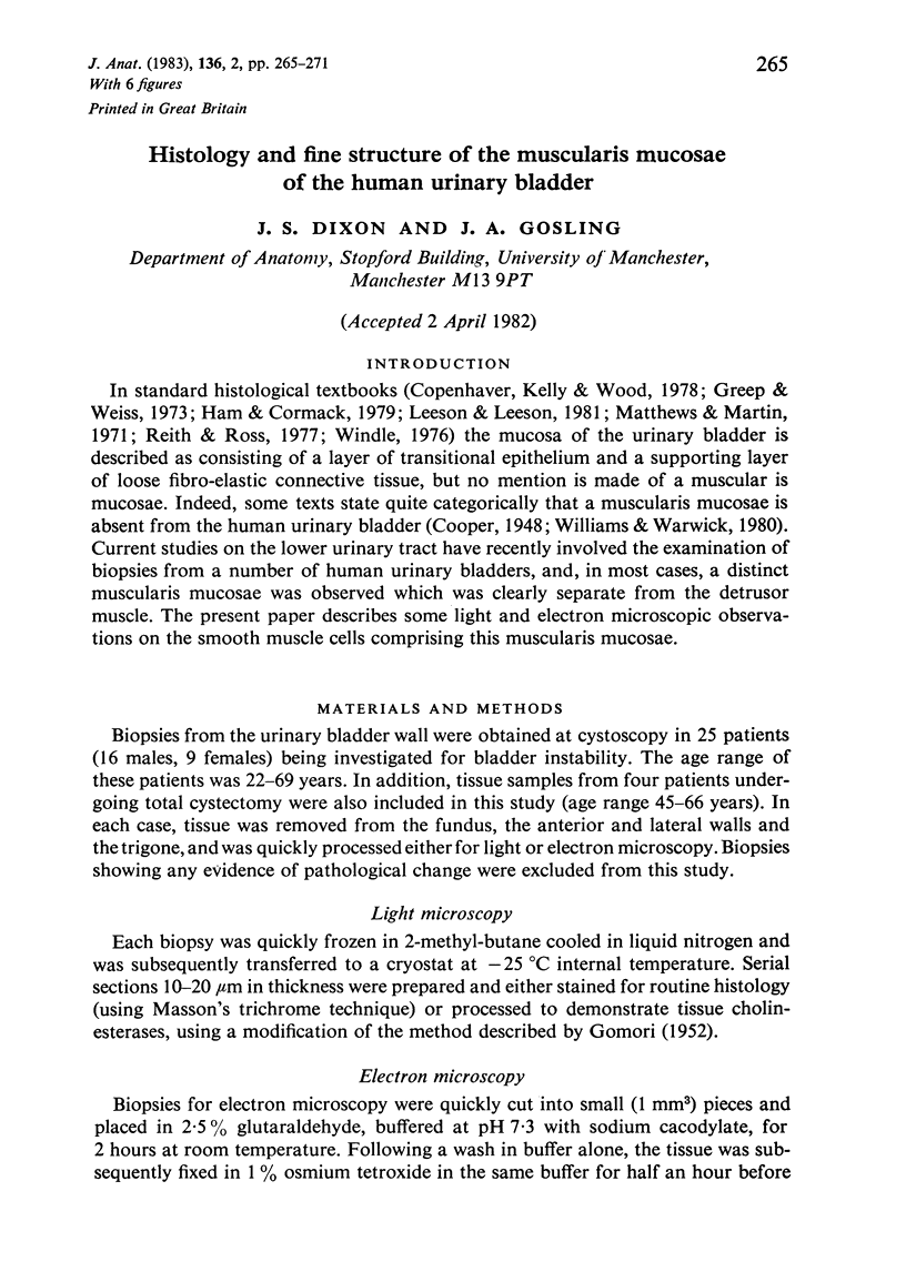
Images in this article
Selected References
These references are in PubMed. This may not be the complete list of references from this article.
- Gerrity R. G., Adams E. P., Cliff W. J. The aortic tunica media of the developing rat. II. Incorporation by medial cells 3-H-proline into collagen and elastin: autoradiographic and chemical studies. Lab Invest. 1975 May;32(5):601–609. [PubMed] [Google Scholar]
- REYNOLDS E. S. The use of lead citrate at high pH as an electron-opaque stain in electron microscopy. J Cell Biol. 1963 Apr;17:208–212. doi: 10.1083/jcb.17.1.208. [DOI] [PMC free article] [PubMed] [Google Scholar]
- WATSON M. L. Staining of tissue sections for electron microscopy with heavy metals. J Biophys Biochem Cytol. 1958 Jul 25;4(4):475–478. doi: 10.1083/jcb.4.4.475. [DOI] [PMC free article] [PubMed] [Google Scholar]


