

Abstract
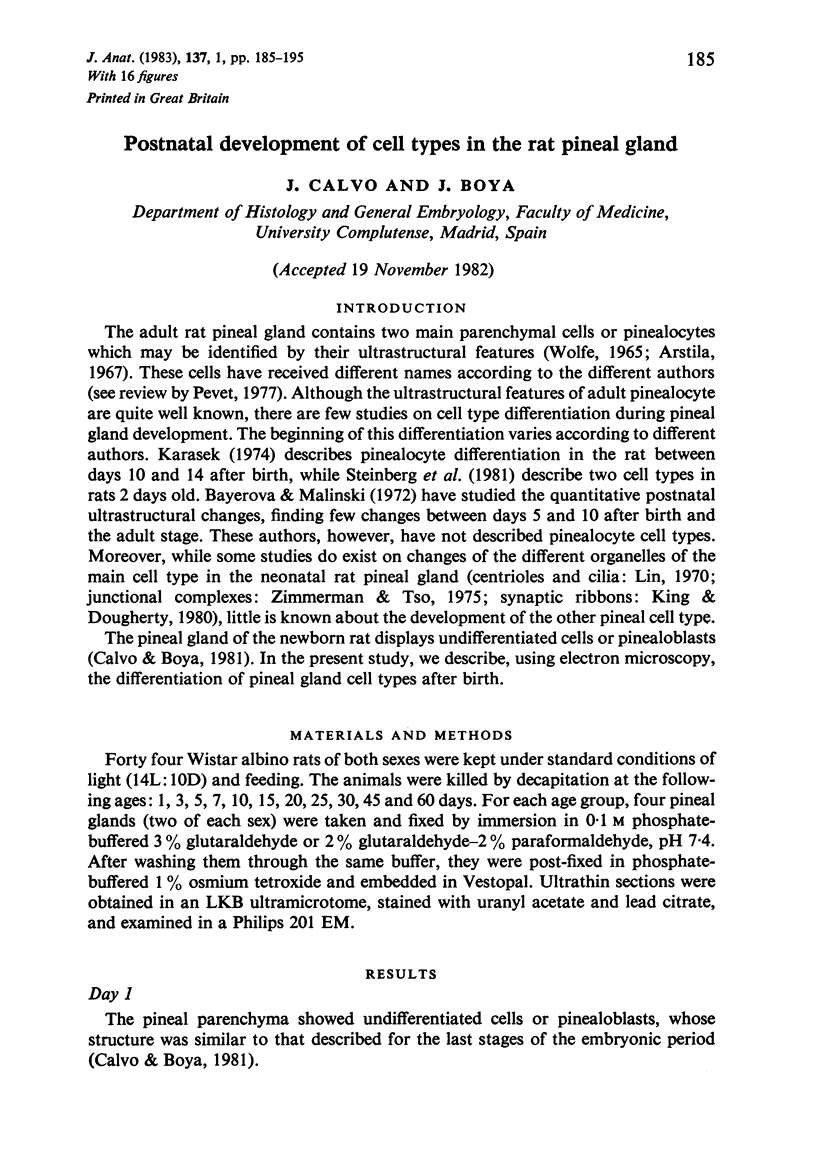
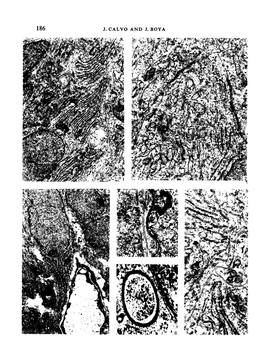
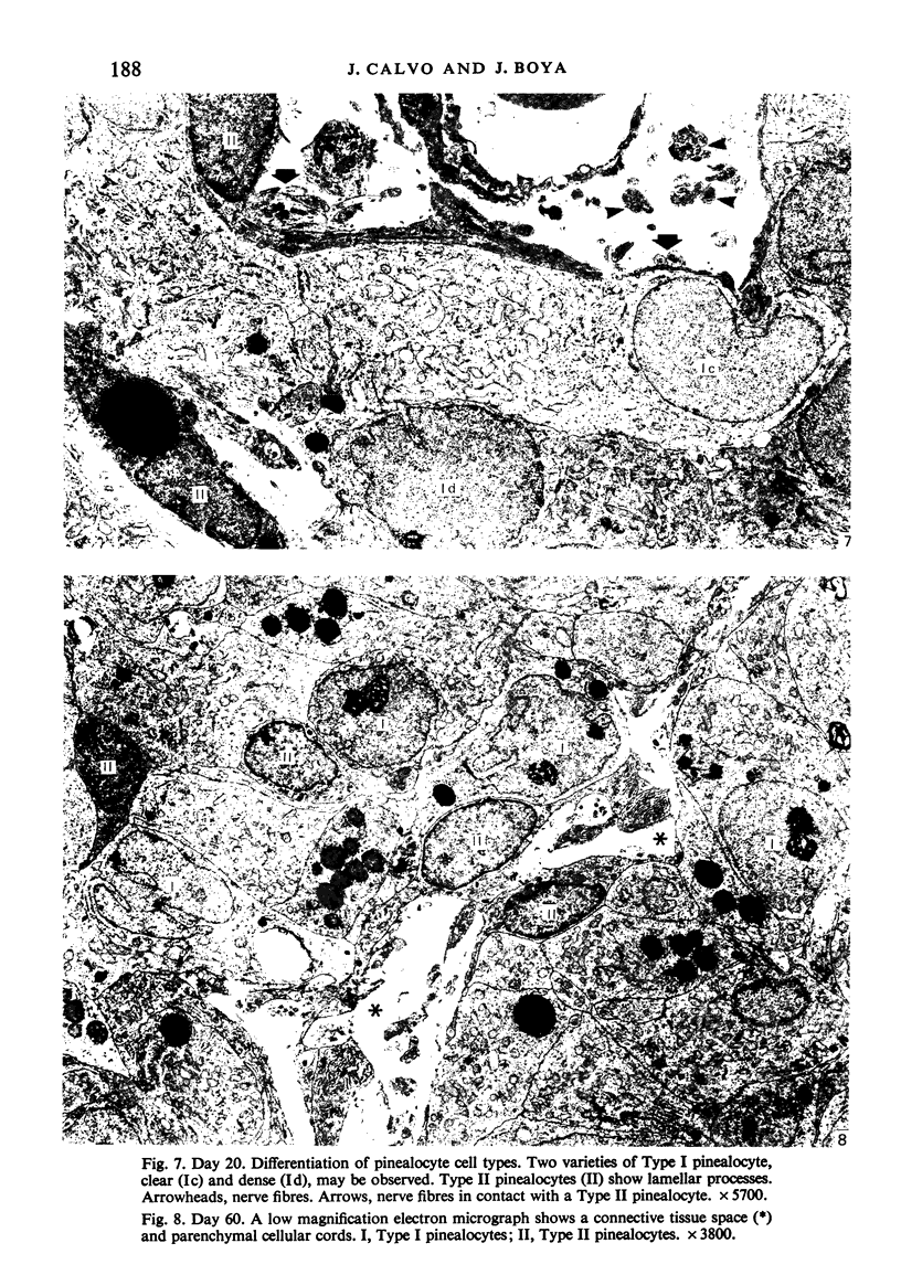
The morphological development of the rat pineal gland has been studied from 1 to 60 days of age. During the first days, undifferentiated cells (pinealoblasts) with scanty cytoplasm and frequent mitotic figures were observed. The differentiation of cell types (Types I and II pinealocytes) began on the third day after birth and was completed by days 15-20. At 3 days of age, nerve fibres were first observed, both in the connective spaces and in the parenchyma. After 5 days, an important hypertrophy of pinealocytes began, mostly Type I, which continued until 60 days of age. After 45 days, all the ultrastructural features described in the adult pineal gland were already present. The findings are discussed.
Full text
PDF
Images in this article
Selected References
These references are in PubMed. This may not be the complete list of references from this article.
- Bayerová G., Malínský J. Quantitative ultrastructural study of rat pinealocytes during postnatal development. Folia Morphol (Praha) 1972;20(1):56–59. [PubMed] [Google Scholar]
- Calvo J., Boya J. Oxytalan fibres in the rat pineal gland. J Anat. 1983 Mar;136(Pt 2):363–366. [PMC free article] [PubMed] [Google Scholar]
- Calvo J., Boya J. Ultrastructural study of the embryonic development in the rat pineal gland. Anat Rec. 1981 Apr;199(4):543–553. doi: 10.1002/ar.1091990410. [DOI] [PubMed] [Google Scholar]
- Ellison N., Weller J. L., Klein D. C. Development of a circadian rhythm in the activity of pineal serotonin N-acetyltransferase. J Neurochem. 1972 May;19(5):1335–1341. doi: 10.1111/j.1471-4159.1972.tb01458.x. [DOI] [PubMed] [Google Scholar]
- Håkanson R., Lombard des Gouttes M. N., Owman C. Activities of tryptophan hydroxylase, dopa decarboxylase, and monoamine oxidase as correlated with the appearance of monoamines in developing rat pineal gland. Life Sci. 1967 Dec 15;6(24):2577–2585. doi: 10.1016/0024-3205(67)90107-5. [DOI] [PubMed] [Google Scholar]
- King T. S., Dougherty W. J. Neonatal development of circadian rhythm in "synaptic" ribbon numbers in the rat pinealocyte. Am J Anat. 1980 Apr;157(4):335–343. doi: 10.1002/aja.1001570403. [DOI] [PubMed] [Google Scholar]
- Klein D. C., Namboodiri M. A., Auerbach D. A. The melatonin rhythm generating system: developmental aspects. Life Sci. 1981 May 4;28(18):1975–1986. doi: 10.1016/0024-3205(81)90644-5. [DOI] [PubMed] [Google Scholar]
- Pevet P. On the presence of different populations of pinealocytes in the mammalian pineal gland. J Neural Transm. 1977;40(4):289–304. doi: 10.1007/BF01257021. [DOI] [PubMed] [Google Scholar]
- Steinberg V. I., Rowe V., Watanabe I., Parr J., Degenhardt M. Morphologic development of neonatal rat pinealocytes in monolayer culture. Cell Tissue Res. 1981;220(2):337–347. doi: 10.1007/BF00210513. [DOI] [PubMed] [Google Scholar]
- WOLFE D. E. THE EPIPHYSEAL CELL: AN ELECTRON-MICROSCOPIC STUDY OF ITS INTERCELLULAR RELATIONSHIPS AND INTRACELLULAR MORPHOLOGY IN THE PINEAL BODY OF THE ALBINO RAT. Prog Brain Res. 1965;10:332–386. doi: 10.1016/s0079-6123(08)63460-3. [DOI] [PubMed] [Google Scholar]
- Zimmerman B. L., Tso M. O. Morphologic evidence of photoreceptor differentiation of pinealocytes in the neonatal rat. J Cell Biol. 1975 Jul;66(1):60–75. doi: 10.1083/jcb.66.1.60. [DOI] [PMC free article] [PubMed] [Google Scholar]