Abstract
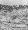
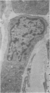
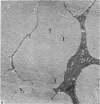
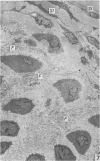
The ultrastructure of the marginal transitional zone of femoral articular cartilage has been studied in the rabbit knee. There is an abrupt boundary between the convex margin of the cartilage and the synovial membrane. This is due to the arrangement and amount of collagen and of cells, because cell ultrastructure changes gradually from synovium to cartilage. The densely fibrous marginal synovium contains scattered fibrocytic cells with sparse cytoplasm and long filopodia. Near the synovium/cartilage interface, oval boundary cells containing more abundant cytoplasm abut on the cartilage matrix. In the periphery of the cartilage, an edge-belt of collagen fibrils runs obliquely from articular surface to subchondral bone. Chondrocytes near the edge-belt, whatever their depth from the articular surface, ultrastructurally resemble middle zone (zone II) cells of articular cartilage generally. The synovial surface of the marginal zone is smooth and resembles articular cartilage surfaces. Most intimal cells contain plentiful granular endoplasmic reticulum and Golgi membranes and hence are intermediate between A and B synoviocytes commonly found elsewhere. Non-fenestrated (type I) capillaries lie in a superficial stratum beneath the synovial surface and in a deep stratum near the synovium/cartilage boundary, and are surrounded by pericytes. No mast cells, macrophages, lymph vessels or nerves could be identified in the marginal zone. Contrary to earlier accounts of collagen orientation in this zone, most of the fibrils in the marginal synovium appear to run around the perimeter of the cartilage and only a few bundles run radially from the synovium towards the cartilage. It is suggested that the circumferential collagen both contains the marginal cartilage and prevents displacement of synovial tissue on to the articular surface. The radial strata of collagen serve to anchor the circumferential collagen to the cartilage edge-belt. In agreement with earlier investigators, it is considered that the edge-belt withstands tensile stresses arising from deformation of the articular surface. The role of the marginal synovium is also discussed in relation to synovial fluid formation and cartilage nutrition.
Full text
PDF
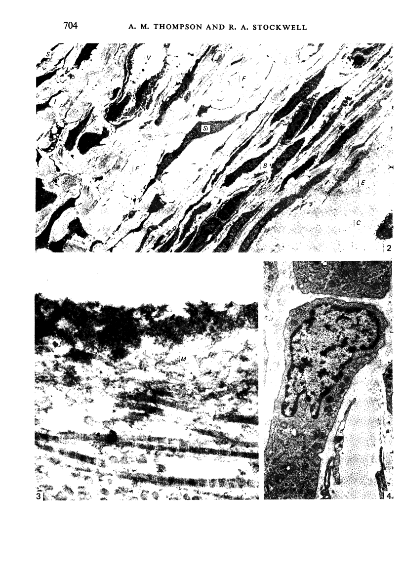
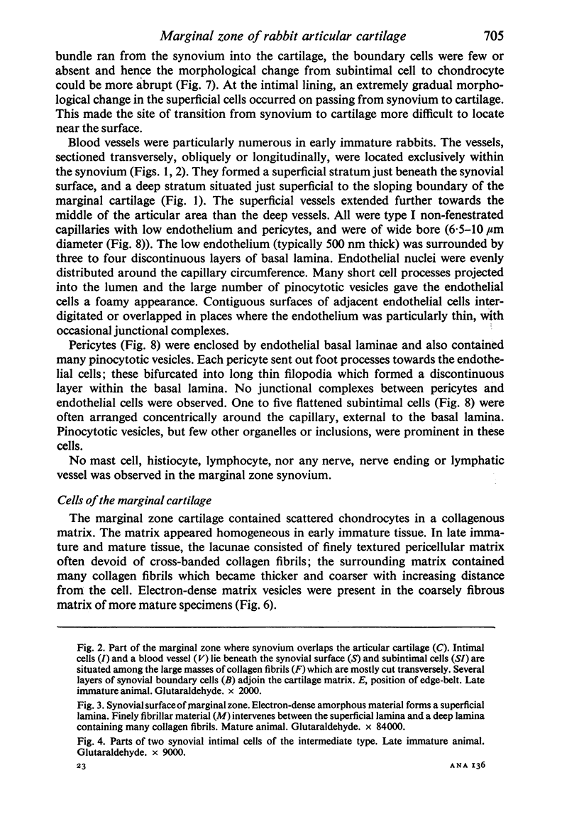
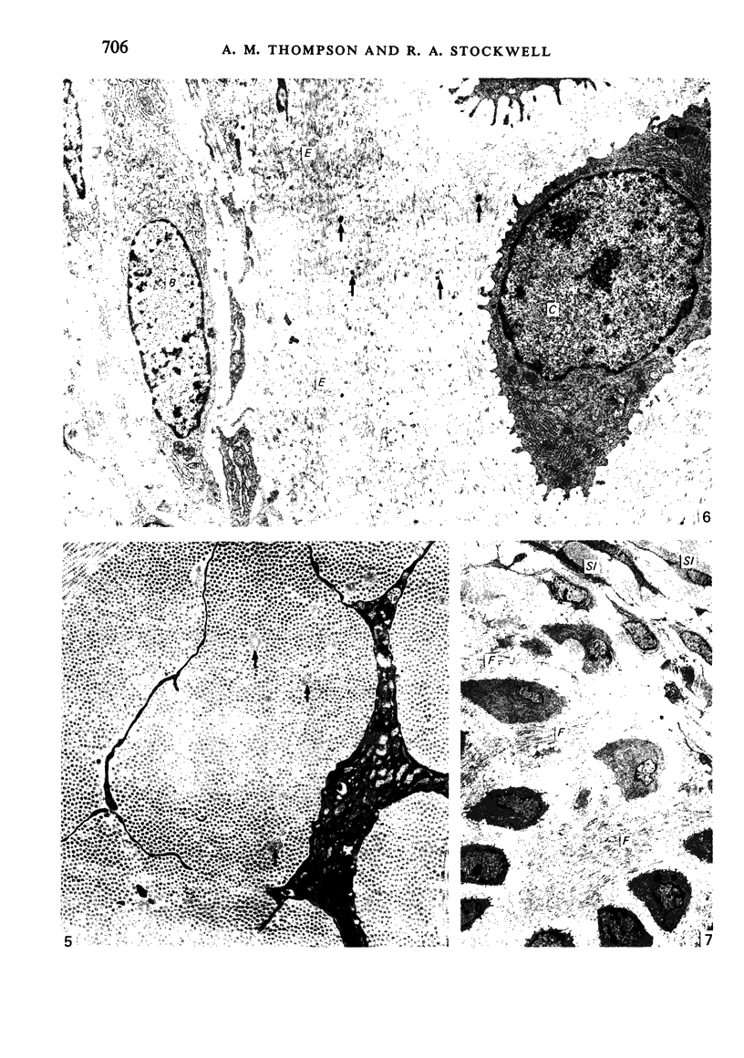
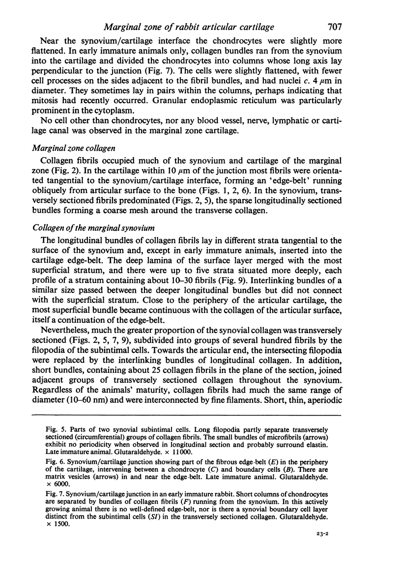
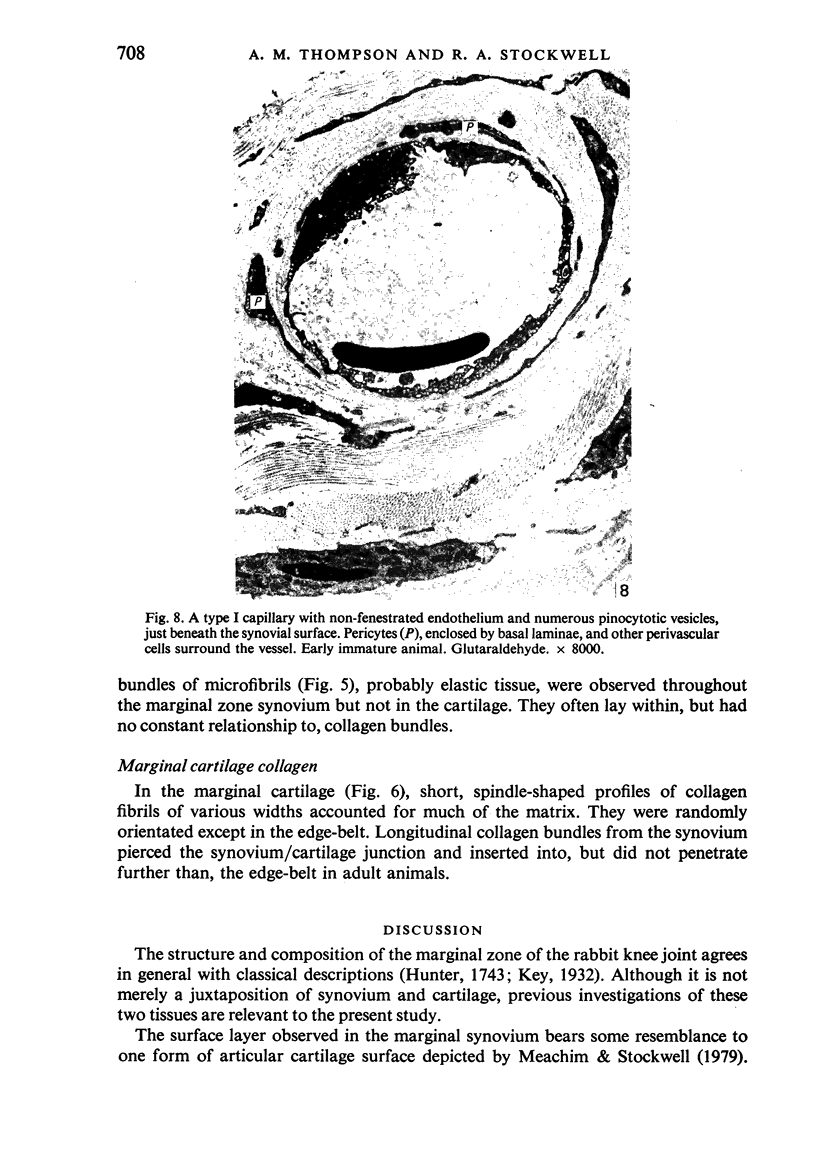
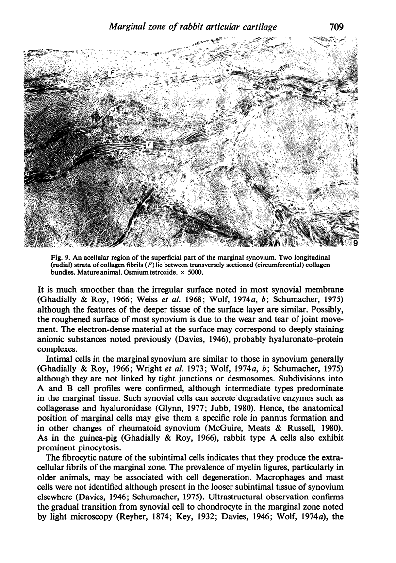
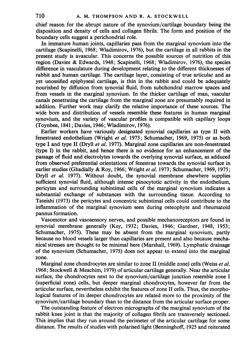
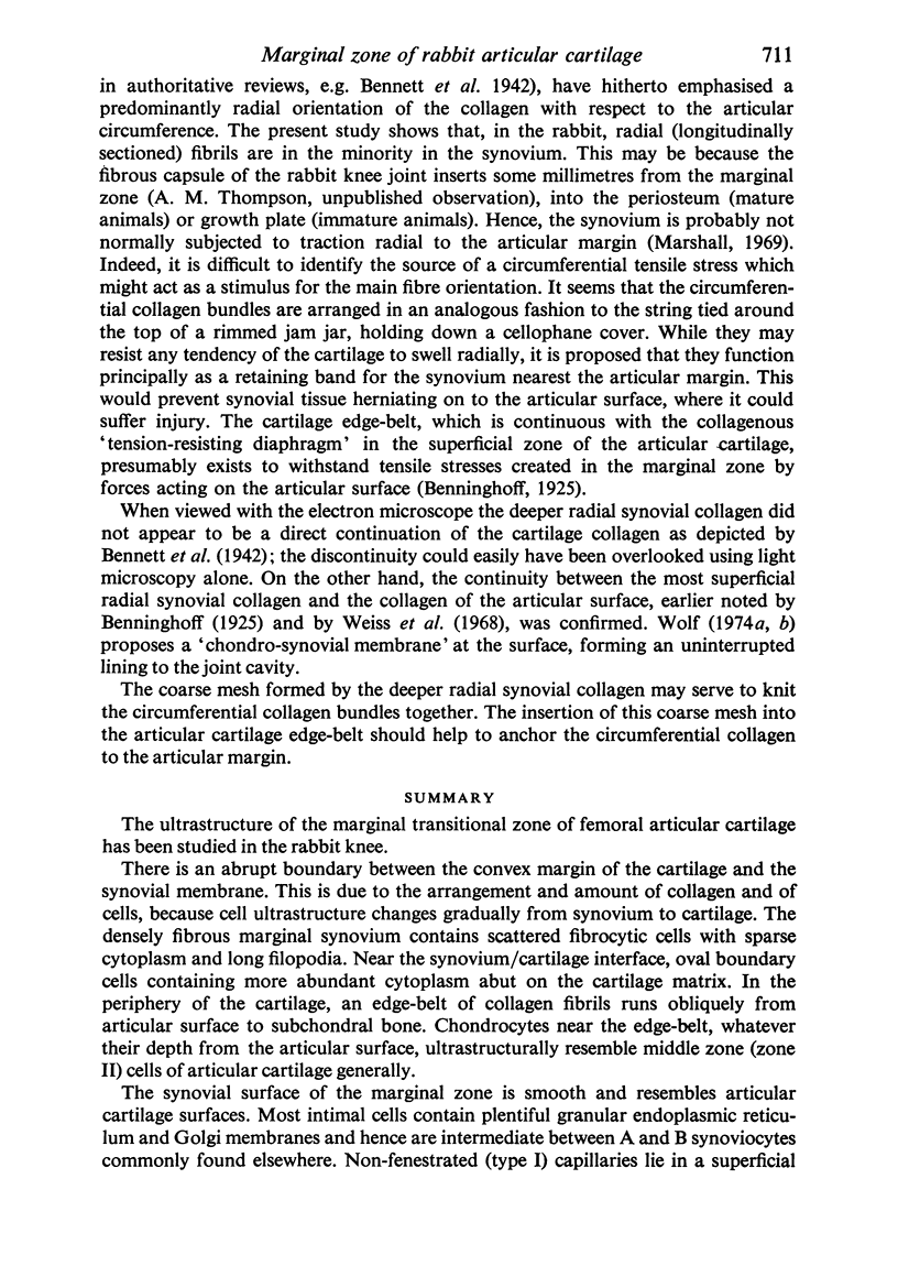
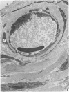
Images in this article
Selected References
These references are in PubMed. This may not be the complete list of references from this article.
- BARLAND P., NOVIKOFF A. B., HAMERMAN D. Electron microscopy of the human synovial membrane. J Cell Biol. 1962 Aug;14:207–220. doi: 10.1083/jcb.14.2.207. [DOI] [PMC free article] [PubMed] [Google Scholar]
- DAVIES D. V., BARNETT C. H., COCHRANE W., PALFREY A. J. Electron microscopy of articular cartilage in the young adult rabbit. Ann Rheum Dis. 1962 Mar;21:11–22. doi: 10.1136/ard.21.1.11. [DOI] [PMC free article] [PubMed] [Google Scholar]
- Davies D. V. The staining reactions of normal synovial membrane with special reference to the origin of synovial mucin. J Anat. 1943 Jan;77(Pt 2):160–169. [PMC free article] [PubMed] [Google Scholar]
- Dryll A., Lansaman J., Cazalis P., Peltier A. P., De Seze S. Light and electron microscopy study of capillaries in normal and inflammatory human synovial membrane. J Clin Pathol. 1977 Jun;30(6):556–562. doi: 10.1136/jcp.30.6.556. [DOI] [PMC free article] [PubMed] [Google Scholar]
- GARDNER E. Blood and nerve supply of joints. Stanford Med Bull. 1953 Nov;11(4):203–209. [PubMed] [Google Scholar]
- Ghadially F. N., Roy S. Ultrastructure of rabbit synovial membrane. Ann Rheum Dis. 1966 Jul;25(4):318–326. doi: 10.1136/ard.25.4.318. [DOI] [PMC free article] [PubMed] [Google Scholar]
- Glynn L. E. Primary lesion in osteoarthrosis. Lancet. 1977 Mar 12;1(8011):574–575. doi: 10.1016/s0140-6736(77)92003-7. [DOI] [PubMed] [Google Scholar]
- ITO S., WINCHESTER R. J. The fine structure of the gastric mucosa in the bat. J Cell Biol. 1963 Mar;16:541–577. doi: 10.1083/jcb.16.3.541. [DOI] [PMC free article] [PubMed] [Google Scholar]
- Marshall J. L. Periarticular osteophytes. Initiation and formation in the knee of the dog. Clin Orthop Relat Res. 1969 Jan-Feb;62:37–47. [PubMed] [Google Scholar]
- McDevitt C., Gilbertson E., Muir H. An experimental model of osteoarthritis; early morphological and biochemical changes. J Bone Joint Surg Br. 1977 Feb;59(1):24–35. doi: 10.1302/0301-620X.59B1.576611. [DOI] [PubMed] [Google Scholar]
- McGuire M. K., Meats J. E., Russell R. G. Cell-cell interactions in the rheumatoid joint. Agents Actions. 1980 Dec;10(6):486–490. doi: 10.1007/BF02024146. [DOI] [PubMed] [Google Scholar]
- Mills K. Pathology of the knee joint in rheumatoid arthritis. A contribution to the understanding of synovectomy. J Bone Joint Surg Br. 1970 Nov;52(4):746–756. [PubMed] [Google Scholar]
- Ogata K., Whiteside L. A., Lesker P. A. Subchondral route for nutrition to articular cartilage in the rabbit. Measurement of diffusion with hydrogen gas in vivo. J Bone Joint Surg Am. 1978 Oct;60(7):905–910. [PubMed] [Google Scholar]
- Reÿher C. The Cartilages and Synovial Membranes of the Joints. J Anat Physiol. 1874 May;8(Pt 2):261–273. [PMC free article] [PubMed] [Google Scholar]
- Scapinelli R. Studies on the vasculature of the human knee joint. Acta Anat (Basel) 1968;70(3):305–331. doi: 10.1159/000143133. [DOI] [PubMed] [Google Scholar]
- Schumacher H. R., Jr Ultrastructure of the synovial membrane. Ann Clin Lab Sci. 1975 Nov-Dec;5(6):489–498. [PubMed] [Google Scholar]
- Schumacher H. R. The microvasculature of the synovial membrane of the monkey: ultrastructural studies. Arthritis Rheum. 1969 Aug;12(4):387–404. doi: 10.1002/art.1780120406. [DOI] [PubMed] [Google Scholar]
- Shively J. A., Van Sickle D. C. Scanning electron microscopy of equine synovial membrane. Am J Vet Res. 1977 May;38(5):681–684. [PubMed] [Google Scholar]
- Tateishi H. Ultrastructure of synovio-cartilage junction in rheumatoid arthritis. Kobe J Med Sci. 1973 Jun;19(2):51–66. [PubMed] [Google Scholar]
- Thompson R. C., Jr, Bassett C. A. Histological observations on experimentally induced degeneration of articular cartilage. J Bone Joint Surg Am. 1970 Apr;52(3):435–443. [PubMed] [Google Scholar]
- Weiss C., Rosenberg L., Helfet A. J. An ultrastructural study of normal young adult human articular cartilage. J Bone Joint Surg Am. 1968 Jun;50(4):663–674. doi: 10.2106/00004623-196850040-00002. [DOI] [PubMed] [Google Scholar]
- Wladimirov B. Uber die Blutversorgung des Kneigelenkknorpels beim Menschen. Anat Anz. 1976;140(5):469–476. [PubMed] [Google Scholar]
- Wolf J. Reinforcement of transitional zone between chondral and synovial membranes. Folia Morphol (Praha) 1974;22(2):161–164. [PubMed] [Google Scholar]
- Wolf J. Transition from synovial into chondral membrane. Folia Morphol (Praha) 1974;22(2):151–160. [PubMed] [Google Scholar]
- Wright V., Dowson D., Kerr J. The structure of joints. Int Rev Connect Tissue Res. 1973;6:105–125. doi: 10.1016/b978-0-12-363706-2.50009-x. [DOI] [PubMed] [Google Scholar]


