

Abstract
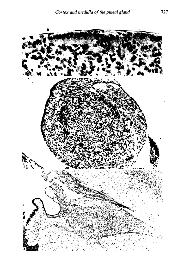
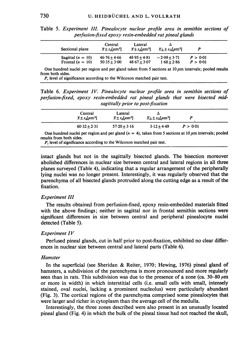
Because, in previous investigations on the rat pineal gland, karyometric studies of pinealocytes from cortical and medullary regions had yielded contradictory results, experiments were carried out to resolve this problem. In immersion-fixed, paraffin-embedded pineal glands, nuclear size of cortical regions was invariably larger than that in the medulla, the nuclear size clearly depending on the plane of sectioning. The differences between cortex and medulla were abolished in (a) pineal glands sagittally bisected prior to immersion fixation and (b) perfusion-fixed, epoxy resin-embedded pineal tissue, suggesting that unequal pinealocyte nuclear size in cortex and medulla is artefactual. In the hamster pineal gland, cortical and medullary regions are separated by a narrow band of interstitial cells, indicating that different structural features account for this subdivision in different species.
Full text
PDF


Images in this article

Selected References
These references are in PubMed. This may not be the complete list of references from this article.
- Bubenik G. A., Brown G. M., Uhlir I., Grota L. J. Immunohistological localization of N-acetylindolealkylamines in pineal gland, retina and cerebellum. Brain Res. 1974 Dec 6;81(2):233–242. doi: 10.1016/0006-8993(74)90938-x. [DOI] [PubMed] [Google Scholar]
- Cuello A. C., Tramezzani J. H. The epiphysis cerebri of the Weddell seal: its remarkable size and glandular pattern. Gen Comp Endocrinol. 1969 Feb;12(1):154–164. doi: 10.1016/0016-6480(69)90148-8. [DOI] [PubMed] [Google Scholar]
- Deutsch K., Hillman H. The effect of six fixatives on the areas of rabbit neurons and rabbit and rat cerebral slices. J Microsc. 1977 Apr;109(3):303–309. doi: 10.1111/j.1365-2818.1977.tb01143.x. [DOI] [PubMed] [Google Scholar]
- Diehl B. J. Time-related changes in size of nuclei of pinealocytes in rats. Cell Tissue Res. 1981;218(2):427–438. doi: 10.1007/BF00210355. [DOI] [PubMed] [Google Scholar]
- Fleischhauer K., Vossel A. Cell densities in the various layers of the rabbit's striate area. Anat Embryol (Berl) 1979 Jul 26;156(3):269–281. doi: 10.1007/BF00299627. [DOI] [PubMed] [Google Scholar]
- Freund D., Arendt J., Vollrath L. Tentative immunohistochemical demonstration of melatonin in the rat pineal gland. Cell Tissue Res. 1977 Jul 11;181(2):239–244. doi: 10.1007/BF00219983. [DOI] [PubMed] [Google Scholar]
- Ganote C. E., Moses H. L. Light and dark cells as artifacts of liver fixation. Lab Invest. 1968 Jun;18(6):740–745. [PubMed] [Google Scholar]
- Greeley D., Crapo J. D., Vollmer R. T. Estimation of the mean caliper diameter of cell nuclei. I. Serial section reconstriction method and endothelial nuclei from human lung. J Microsc. 1978 Sep;114(1):31–39. doi: 10.1111/j.1365-2818.1978.tb00114.x. [DOI] [PubMed] [Google Scholar]
- Gusek W., Buss H. Morphologische und histochemische Veränderungen der Zirbeldrüse unter dem Einfluss von Prolan und nach Ovarektomie. Frankf Z Pathol. 1966;75(2):172–186. [PubMed] [Google Scholar]
- Hewing M. Die postnatale Entwicklung der Epiphysis cerebri beim Goldhamster. Verh Anat Ges. 1976;(70 Pt 1):85–92. [PubMed] [Google Scholar]
- Hewing M. Synaptic ribbons during postnatal development of the pineal gland in the golden hamster (Mesocricetus auratus). Cell Tissue Res. 1979 Jul 17;199(3):473–482. doi: 10.1007/BF00236083. [DOI] [PubMed] [Google Scholar]
- Hildebrand R. Nuclear volume and cellular metabolism. Adv Anat Embryol Cell Biol. 1980;60:1–54. [PubMed] [Google Scholar]
- ITO S., WINCHESTER R. J. The fine structure of the gastric mucosa in the bat. J Cell Biol. 1963 Mar;16:541–577. doi: 10.1083/jcb.16.3.541. [DOI] [PMC free article] [PubMed] [Google Scholar]
- Ito T., Matsushima S. Effects of gonadectomy and hypophysectomy on the pineal body in the mouse: a quantitative morphological study. Anat Rec. 1968 Dec;162(4):479–482. doi: 10.1002/ar.1091620409. [DOI] [PubMed] [Google Scholar]
- QUAY W. B. Cytologic and metabolic parameters of pineal inhibition by continuous light in the rat (Rattus norvegicus). Z Zellforsch Mikrosk Anat. 1963;60:479–490. doi: 10.1007/BF00336620. [DOI] [PubMed] [Google Scholar]
- Quay W. B., Renzoni A. Twenty-four-hour rhythms in pineal mitotic activity and nuclear and nucleolar dimensions. Growth. 1966 Sep;30(3):315–324. [PubMed] [Google Scholar]
- RICHARDSON K. C., JARETT L., FINKE E. H. Embedding in epoxy resins for ultrathin sectioning in electron microscopy. Stain Technol. 1960 Nov;35:313–323. doi: 10.3109/10520296009114754. [DOI] [PubMed] [Google Scholar]
- Sailinger-Holle R., Vollrath L. Pineal structure and wheel-running activity in rats. Physiol Behav. 1980 Dec;25(6):985–987. doi: 10.1016/0031-9384(80)90322-4. [DOI] [PubMed] [Google Scholar]
- Sheridan M. N., Reiter R. J. Observations on the pineal system in the hamster. I. Relations of the superficial and deep pineal to the epithalamus. J Morphol. 1970 Jun;131(2):153–161. doi: 10.1002/jmor.1051310203. [DOI] [PubMed] [Google Scholar]
- Vivien-Roels B., Pévet P., Dubois M. P., Arendt J., Brown G. M. Immunohistochemical evidence for the presence of melatonin in the pineal gland, the retina and the Harderian gland. Cell Tissue Res. 1981;217(1):105–115. doi: 10.1007/BF00233830. [DOI] [PubMed] [Google Scholar]
- Vollrath L. Comparative morphology of the vertebrate pineal complex. Prog Brain Res. 1979;52:25–38. doi: 10.1016/S0079-6123(08)62909-X. [DOI] [PubMed] [Google Scholar]